. 2023 Jan 24:10:1111598.
doi: 10.3389/fmolb.2023.1111598. eCollection 2023.

Quinate-based ligands for irreversible inactivation of the bacterial virulence factor DHQ1 enzyme-A molecular insight

Affiliations

Quinate-based ligands for irreversible inactivation of the bacterial virulence factor DHQ1 enzyme-A molecular insight

Ángela Rodríguez et al. Front Mol Biosci. .

Abstract

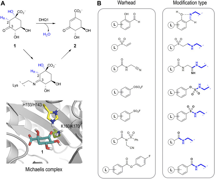
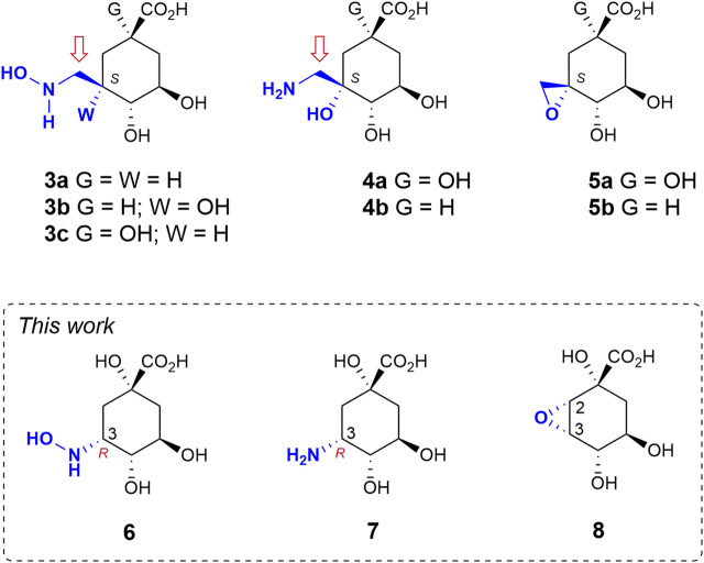
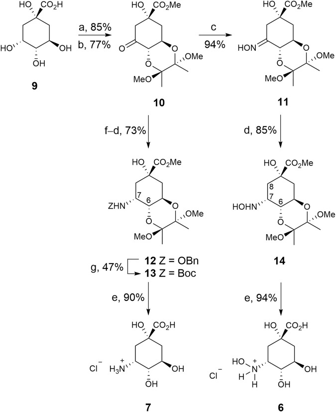
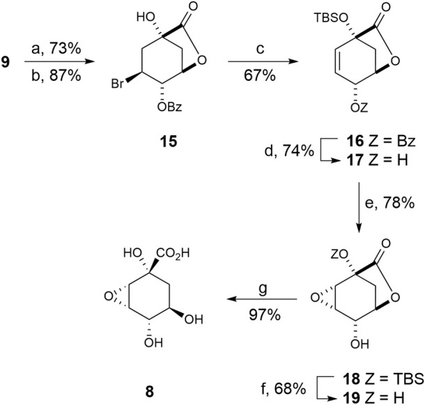
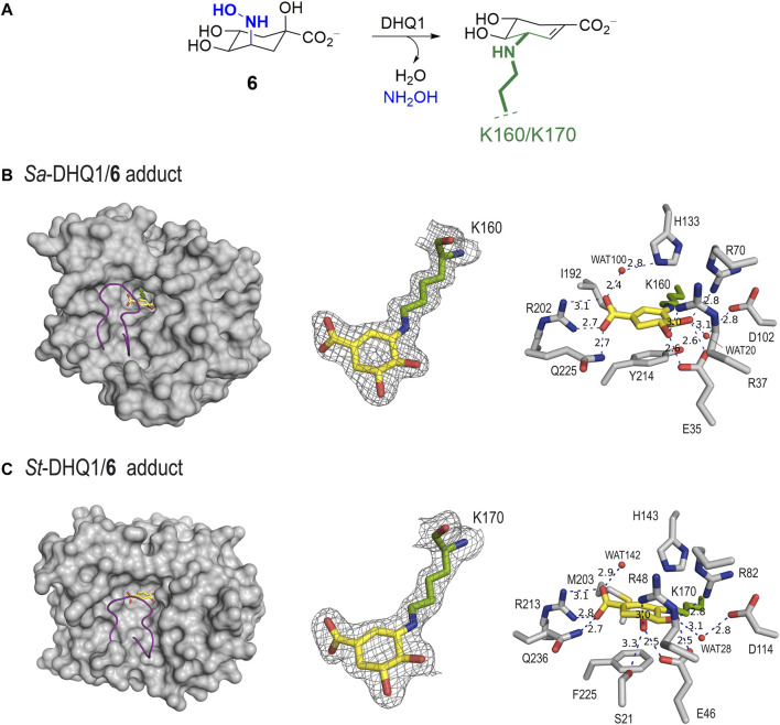
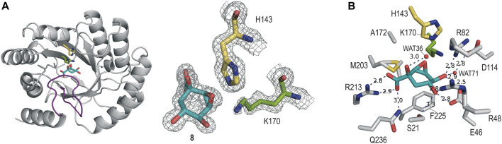
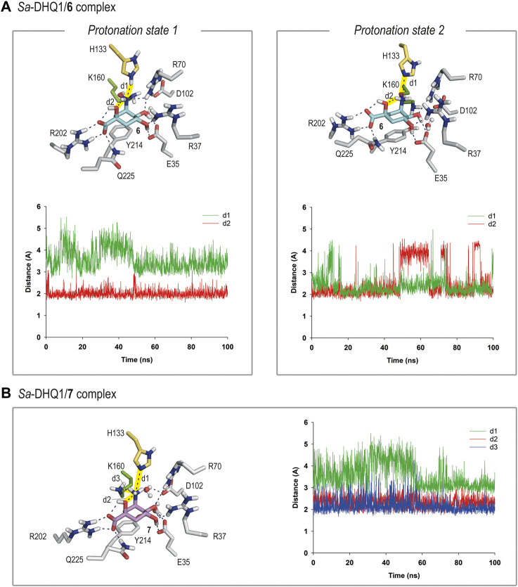
Irreversible inhibition of the enzyme type I dehydroquinase (DHQ1), a promising target for anti-virulence drug development, has been explored by enhancing the electrophilicity of specific positions of the ligand towards covalent lysine modification. For ligand design, we made use of the advantages offered by the intrinsic acid-base properties of the amino substituents introduced in the quinate scaffold, namely compounds 6-7 (R configuration at C3), to generate a potential leaving group, as well as the recognition pattern of the enzyme. The reactivity of the C2-C3 bond (Re face) in the scaffold was also explored using compound 8. The results of the present study show that replacement of the C3 hydroxy group of (-)-quinic acid by a hydroxyamino substituent (compound 6) provides a time-dependent irreversible inhibitor, while compound 7, in which the latter functionality was substituted by an amino group, and the introduction of an oxirane ring at C2-C3 bond, compound 8, do not allow covalent modification of the enzyme. These outcomes were supported by resolution of the crystal structures of DHQ1 from Staphylococcus aureus (Sa-DHQ1) and Salmonella typhi (St-DHQ1) chemically modified by 6 at a resolution of 1.65 and 1.90 Å, respectively, and of St-DHQ1 in the complex with 8 (1.55 Å). The combination of these structural studies with extensive molecular dynamics simulation studies allowed us to understand the molecular basis of the type of inhibition observed. This study is a good example of the importance of achieving the correct geometry between the reactive center of the ligand (electrophile) and the enzyme nucleophile (lysine residue) to allow selective covalent modification. The outcomes obtained with the hydroxyamino derivative 6 also open up new possibilities in the design of irreversible inhibitors based on the use of amino substituents.

Keywords: anti-virulence agents; binding mode; biomacromolecule simulations; enzyme recognition; irreversible inhibition; ligand activation; organic synthesis; type I dehydroquinase.


Conflict of interest statement

The authors declare that the research was conducted in the absence of any commercial or financial relationships that could be construed as a potential conflict of interest.

Figures

FIGURE 1
FIGURE 1
(A) Overall syn elimination of water from 3-dehydroquinic acid (1) catalyzed by DHQ1. The enzymatic process involves the formation of diverse Schiff base species via a catalytic lysine residue. Magnified view of the Michaelis complex obtained by MD simulation studies. Only the catalytic side-chain residues are shown. (B) Relevant reported electrophilic warheads for reversible/irreversible targeting of lysine residues and the covalent modification obtained.
FIGURE 2
FIGURE 2
Relevant reported irreversible inhibitors and targeted compounds. The methylene group undergoing lysine modification is indicated with an arrow. The numbering corresponds to the natural substrate.
SCHEME 1
SCHEME 1
Synthesis of compounds 67. Reagents and conditions. (a) (MeCO)2, CH(OMe)3, MeOH, camphorsulfonic acid, 65°C. (b) PDC, MS 4 Å, CH2Cl2, RT. (c) NH2OH.HCl, NaOAc, MeCN, H2O, RT. (d) NaBH3CN, MeOH, HCl, RT. (e) HCl (0.3 M), 100°C. (f) NH2OBn.HCl, NaOAc, MeOH, MS 4 Å, RT. (g) 1. H2(g), Pd/C (10%), AcOH, MeOH, RT. 2. Boc2O, Et3N, DMF, RT.
SCHEME 2
SCHEME 2
Synthesis of compound 8. Reagents and conditions. (a) PhCHO, DMF, PhMe, p-TsOH (cat), ∆. (b) NBS, AIBN (cat), PhH, ∆. (c) DBU, ClTBS, CH3CN, ∆. (d) 1. KCN, MeOH, RT. 2. NaH, THF, 0°C. (e) MCPBA, NaHCO3, CH2Cl2, ∆. (f) (20:1) TFA/H2O, RT. (g) 1. LiOH, RT, 2. Amberlite IR-120.
FIGURE 3
FIGURE 3
(A) Schematic representation of the chemical modification to the catalytic lysine residue of Sa-DHQ1 and St-DHQ1, as identified by X-ray crystallography. (B,C) Crystal structures of Sa-DHQ1 (B, PDB ID 8B2A, 1.65 Å, chain A) and St-DHQ1 (C, PDB ID 8B2B, 1.90 Å, chain A) covalently modified by hydroxylamine 6. Overall views of the enzyme adducts and interactions of the modified inhibitor (yellow) are shown. The substrate-covering loop (purple) is shown as a cartoon to visualize the active site. Hydrogen-bonding and electrostatic interactions between the ligands and both homologous enzymes are shown as dashed lines (blue). Relevant residues are shown and labeled. Unbiased electron density for inhibitor 6 and its covalent attachment to residues K160/K170 of Sa-DHQ1 and St-DHQ1, respectively. From the model obtained by molecular replacement, and before inclusion of the inhibitor molecule, refinement was performed to obtain unbiased density for the inhibitor molecule and other model changes. A maximum-likelihood weighted 2Fo − Fc map contoured at 1σ is shown up to 1.6 Å around the inhibitor molecule (yellow) and the catalytic lysine residue (green). The final model, including the inhibitor molecule, is superimposed onto the map. The distances are in angstroms.
FIGURE 4
FIGURE 4
(A) Crystal structure of St-DHQ1 in complex with epoxide 8 (PDB ID 8B2C, 1.55 Å). Unbiased electron density for inhibitor 8 and the catalytic residues K170 and H143 of St-DHQ1. The substrate-covering loop is highlighted in purple. From the model obtained by molecular replacement and before inclusion of the inhibitor molecule, refinement was performed to obtain unbiased density for the inhibitor molecule and other model changes. A maximum-likelihood weighted 2Fo − Fc map contoured at 1σ is shown up to 1.6 Å around the inhibitor molecule (cyan) and residues K170 (green) and H143 (yellow). (B) Main contacts of epoxide 8 with St-DHQ1. Hydrogen-bonding and electrostatic interactions are shown as blue dashed lines. The distances are shown in angstrom. Only relevant residues are shown and labeled. Note how residue K170 shows a bent conformation.
FIGURE 5
FIGURE 5
(A) Binding mode of compound 6 in the active site of Sa-DHQ1, as obtained by MD simulation studies, in which the two plausible protonation states of the NHOH moiety in 6 and residue H133 were considered. Snapshots taken after 80 (left) and 100 ns (right) are provided. Hydrogen-bonding and electrostatic interactions are shown as blue dashed lines. Only relevant residues are shown and labeled. Variation of the distances between H133 (HE2 or NE2 atoms) and the nitrogen atom in 6 (d1) and the NH group in 6 and its C1 hydroxyl group (oxygen atom, d2) in the Sa-DHQ1/6 binary complex during the whole simulation considering protonation states 1 and 2. The distances studied are highlighted with yellow shading in the protein figure. (B) Binding mode of compound 7 (ammonium salt) in the active site of Sa-DHQ1 obtained by MD simulation studies. Residue H133 was considered in its neutral form. Snapshot taken after 100 ns of simulation. Variation of the distances between the ammonium group in 7 (H or N atoms) and H133 (NE2 atom, d1), its tertiary hydroxyl group (oxygen atom, d2) and K170 (NZ atom, d3) in the Sa-DHQ1/7 binary complex during the whole simulation. The distances studied are highlighted with yellow shading in the protein figure. Note how the position of the catalytic lysine residue is frozen close to the C2 pocket by a strong hydrogen bond with the ammonium group in 7, with an average distance of 2.2 Å during the simulation.

Similar articles

References

    1. Anscombe E., Meschini E., Mora-Vidal R., Martin M. P., Staunton D., Geitmann M., et al. (2015). Identification and characterization of an irreversible inhibitor of CDK2. Chem. Biol. 22 (9), 1159–1164. 10.1016/j.chembiol.2015.07.018 - DOI - PMC - PubMed
    1. Bartlett P. A., Maitra U., Chouinard P. M. (1986). Synthesis of "iso-EPSP" and evaluation of its interaction with chorismate synthase. J. Am. Chem. Soc. 108 (25), 8068–8071. 10.1021/ja00285a031 - DOI
    1. Bauer R. A. (2015). Covalent inhibitors in drug discovery: From accidental discoveries to avoided liabilities and designed therapies. Drug Discov. Today 20 (9), 1061–1073. 10.1016/j.drudis.2015.05.005 - DOI - PubMed
    1. Boulet P., Gilardoni F., Weber J., Chermette H., Ellinger Y. (1999). Theoretical study of interstellar hydroxylamine chemistry: Protonation and proton transfer mediated by H3+. Chem. Phys. 244 (2), 163–174. 10.1016/s0301-0104(99)00151-2 - DOI
    1. Brünger A. T. (1997). Free R value: Cross-validation in crystallography. Methods Enzymol. 277, 366–396. 10.1016/s0076-6879(97)77021-6 - DOI - PubMed