. 2022 Jun 14:200:1-423.
doi: 10.3897/phytokeys.200.71667. eCollection 2022.

Monograph of wild and cultivated chili peppers (Capsicum L., Solanaceae)

Affiliations

Monograph of wild and cultivated chili peppers (Capsicum L., Solanaceae)

Gloria E Barboza et al. PhytoKeys. .

Abstract

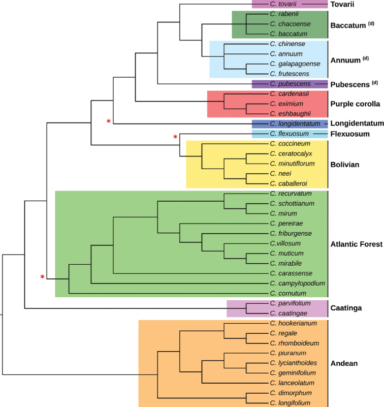
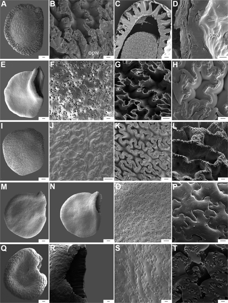
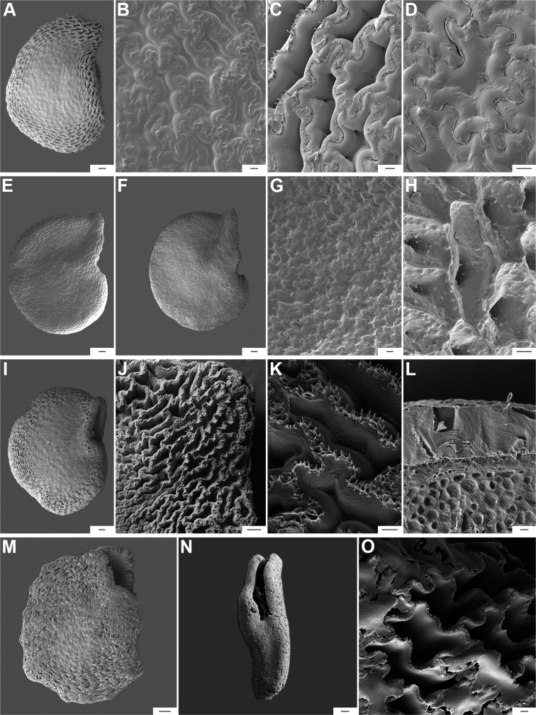
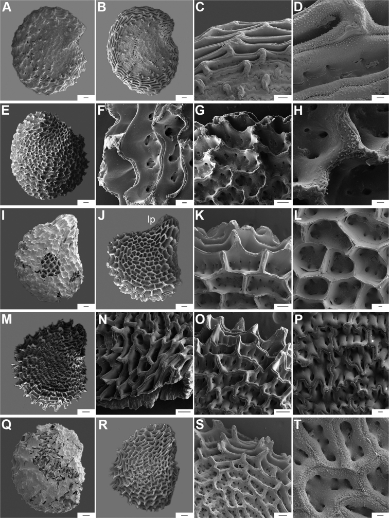
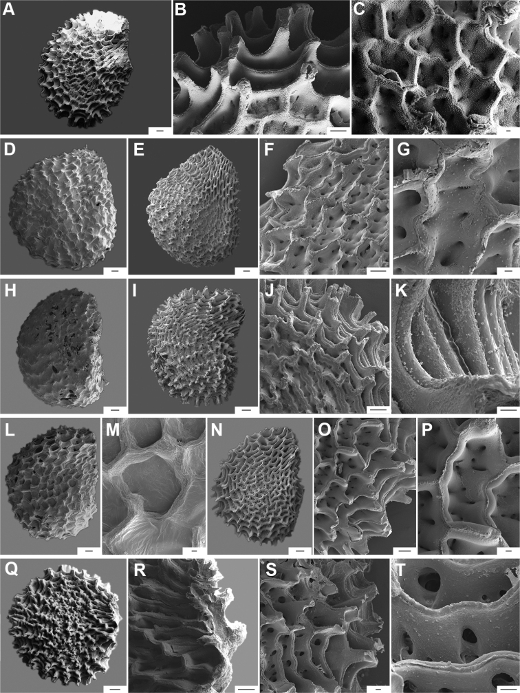
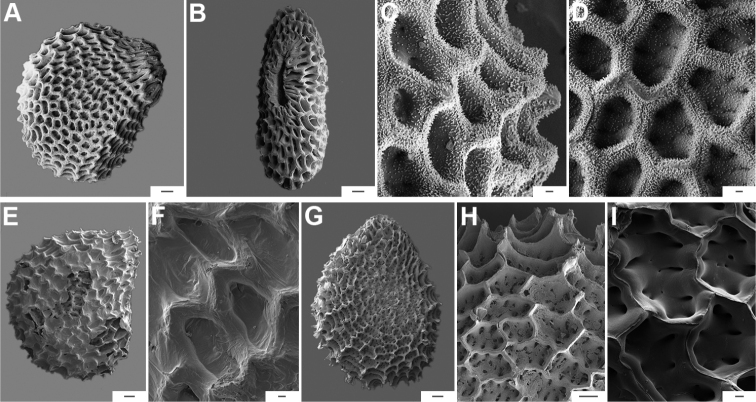
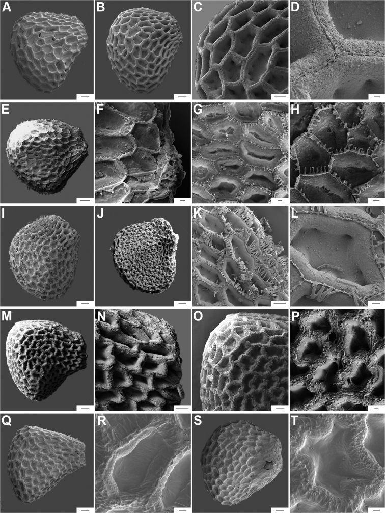
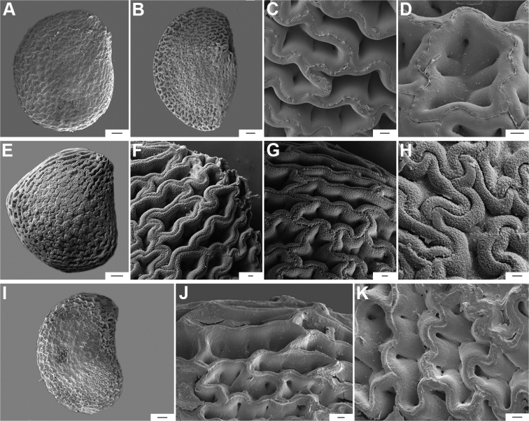
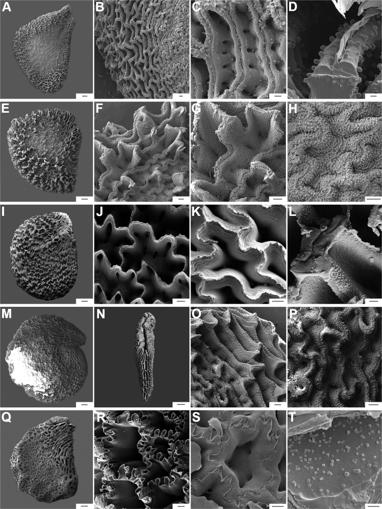
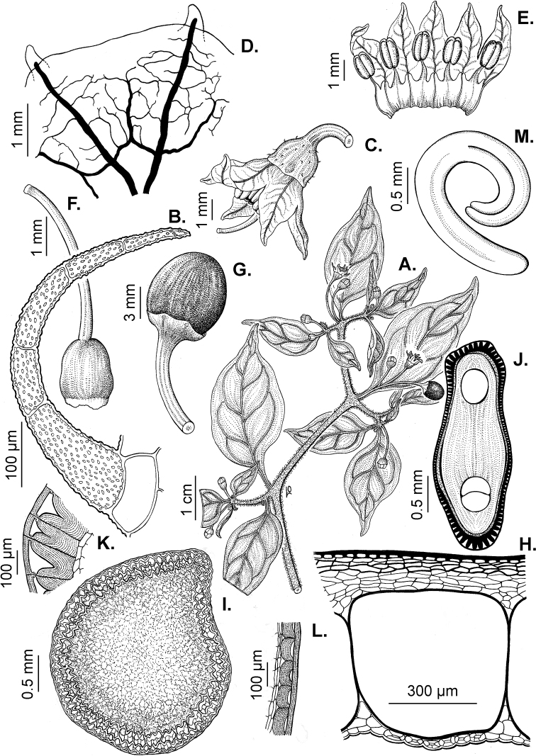
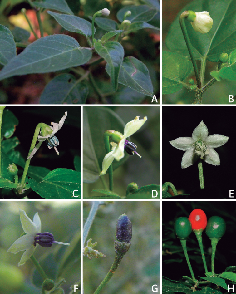
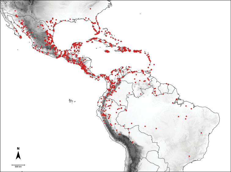
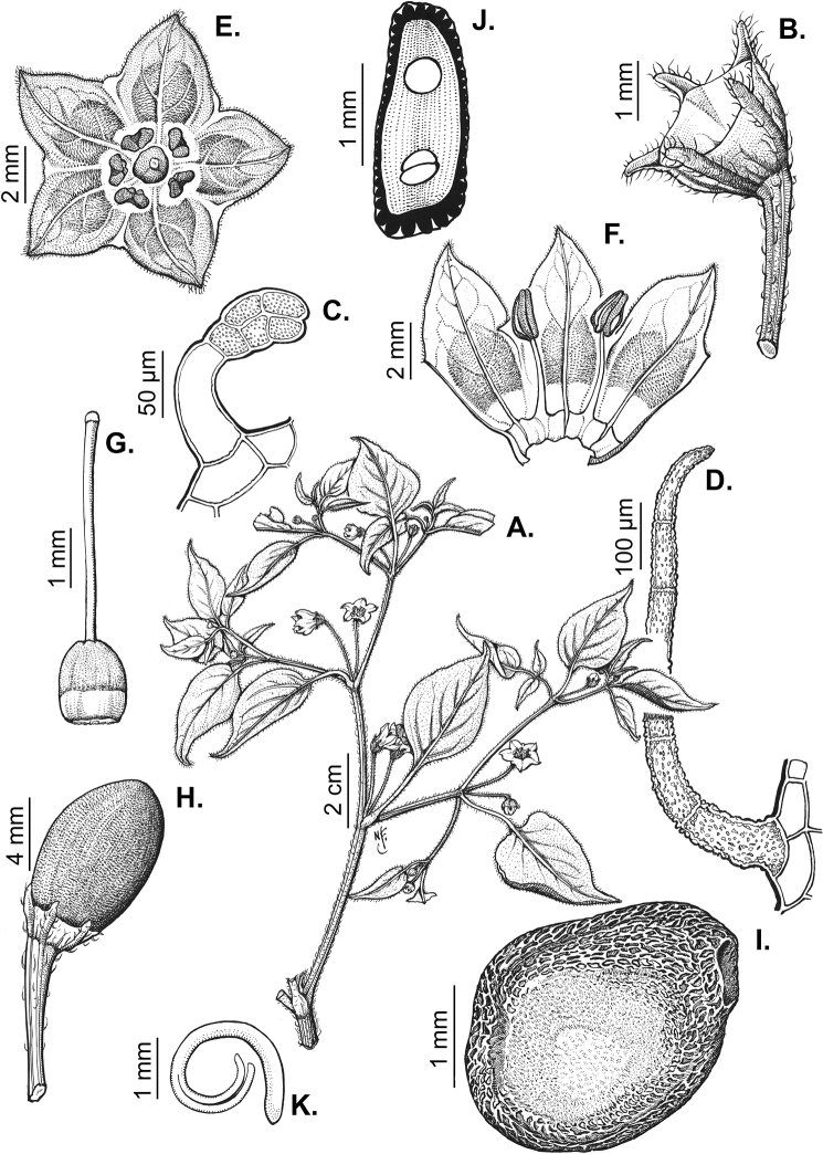
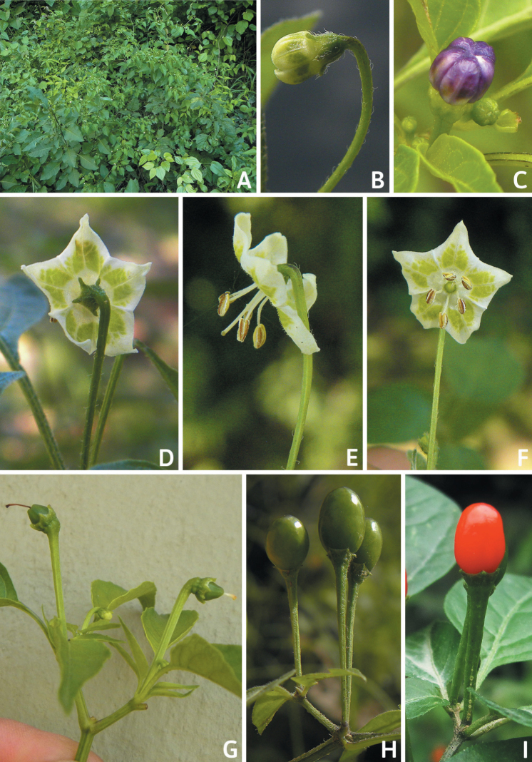
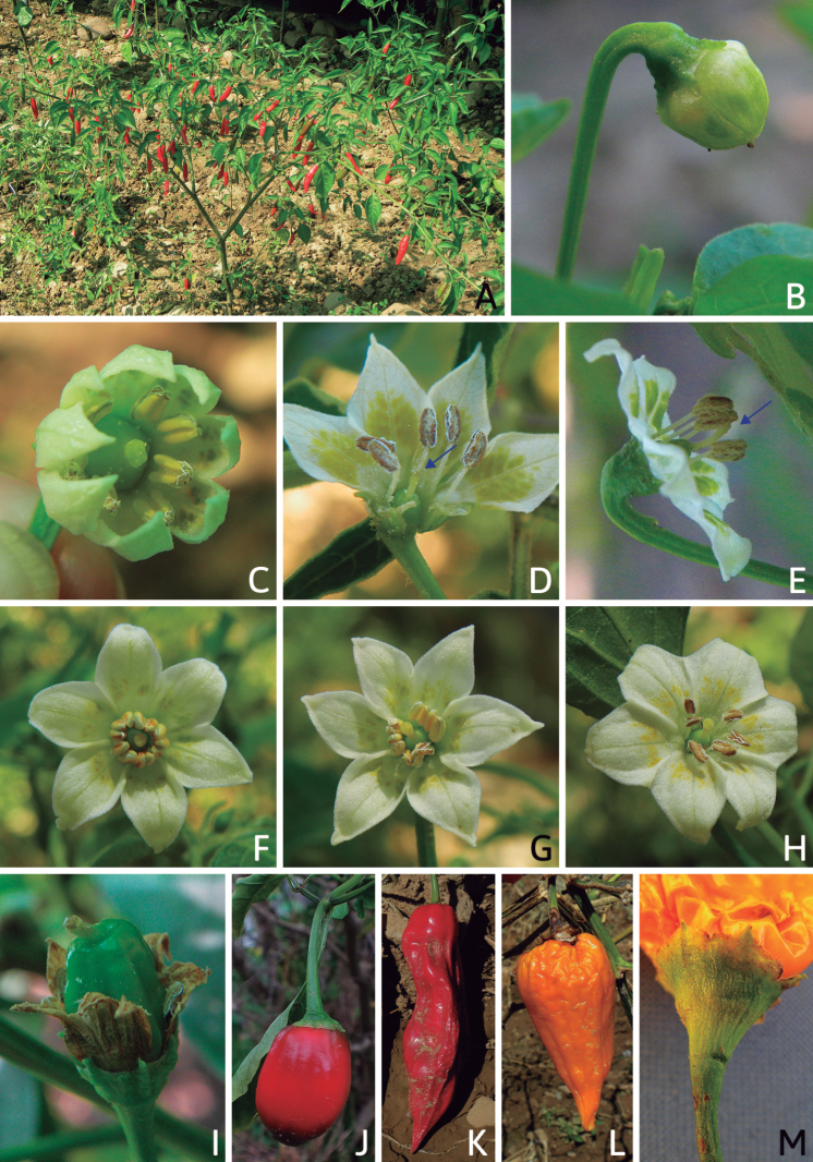
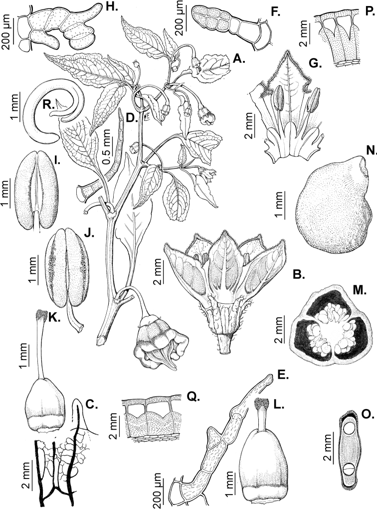
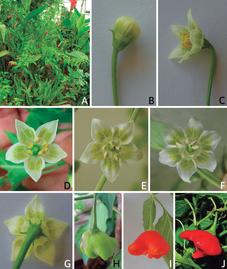
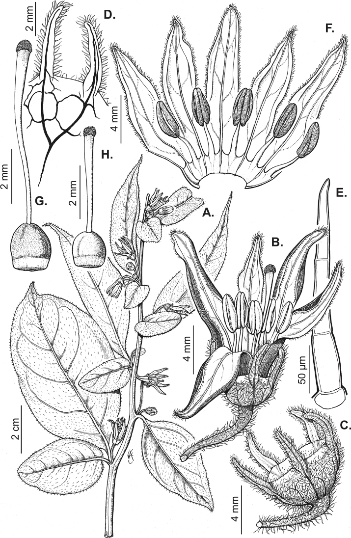
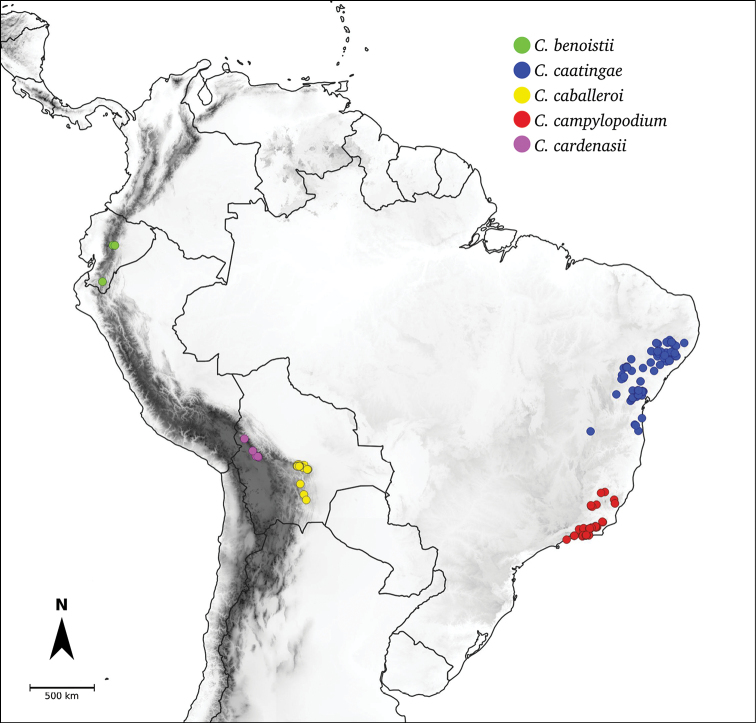
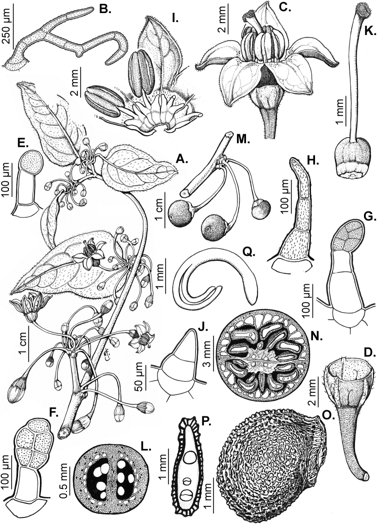
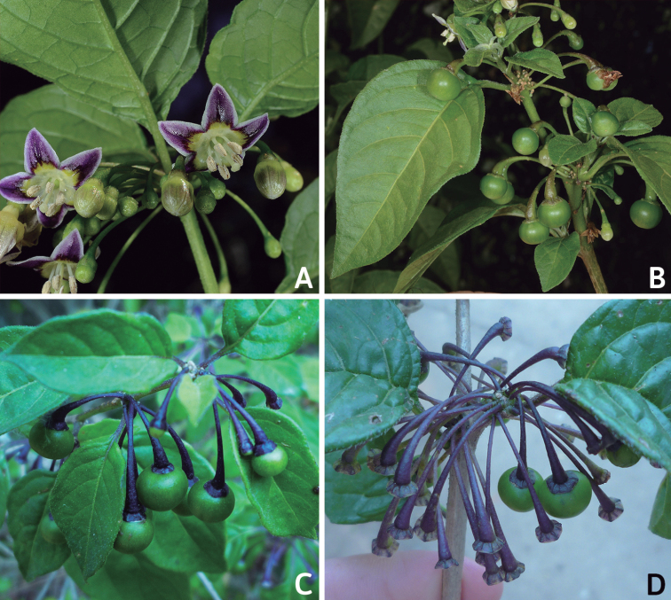
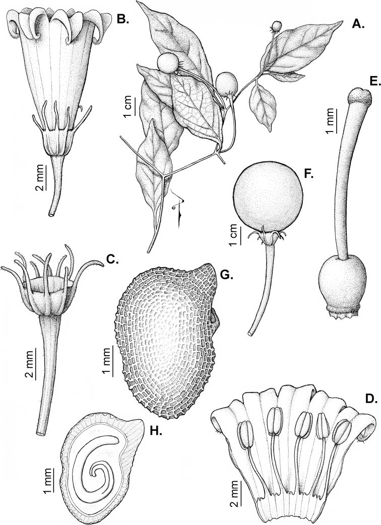
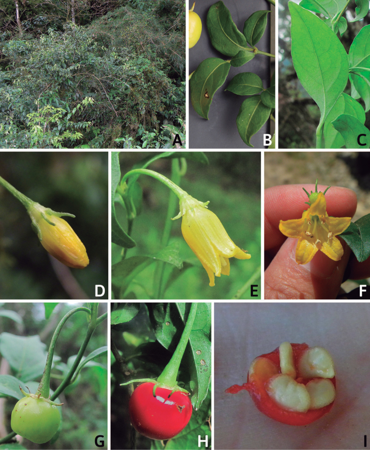
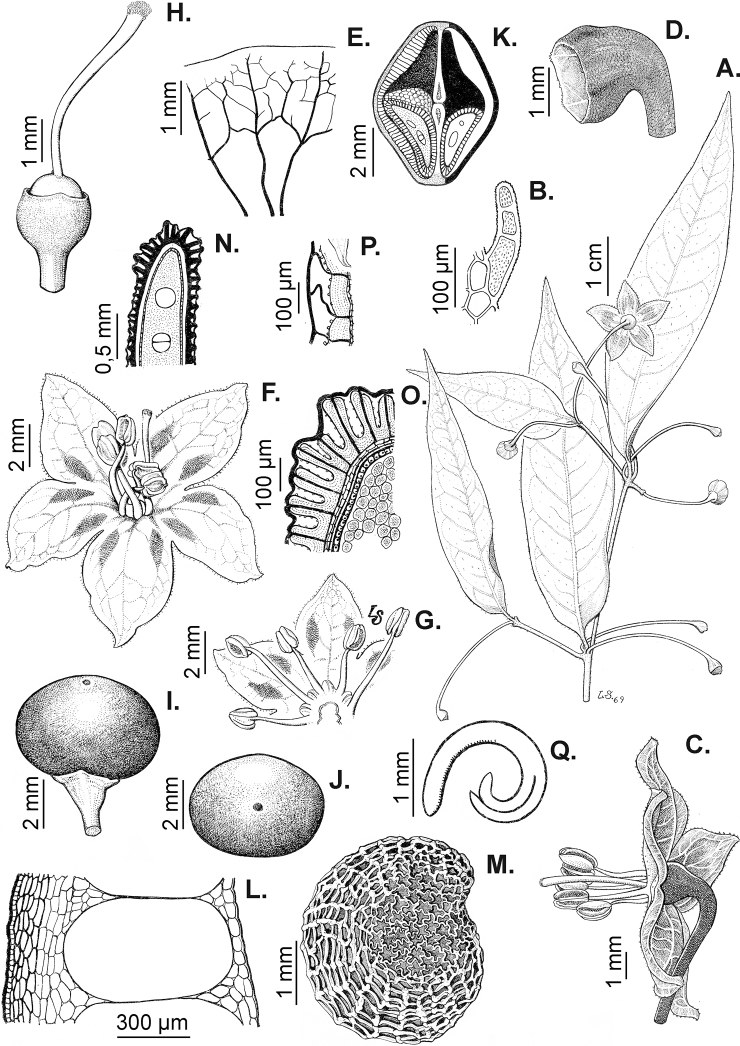
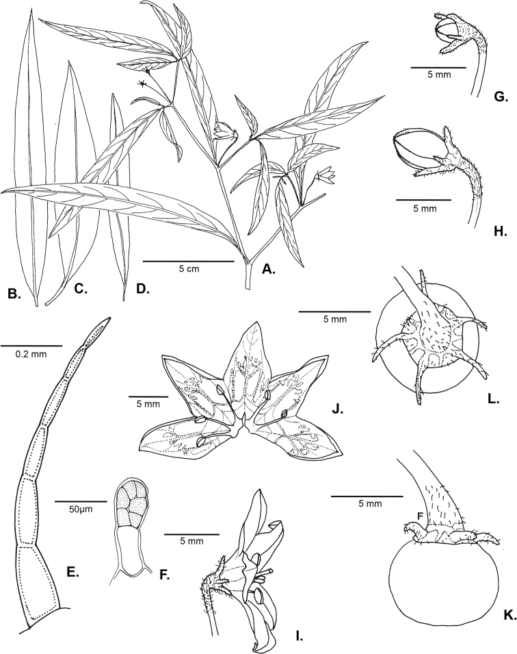
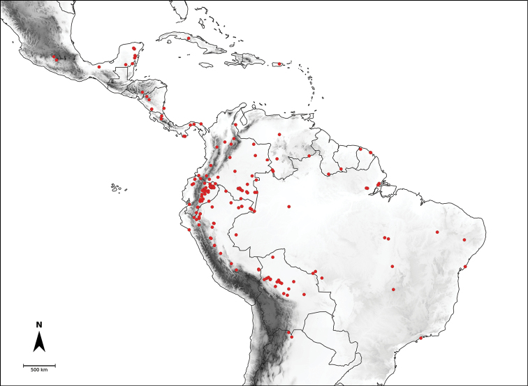
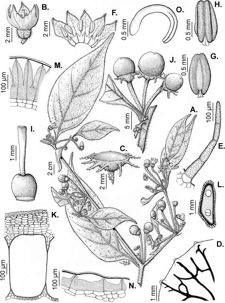
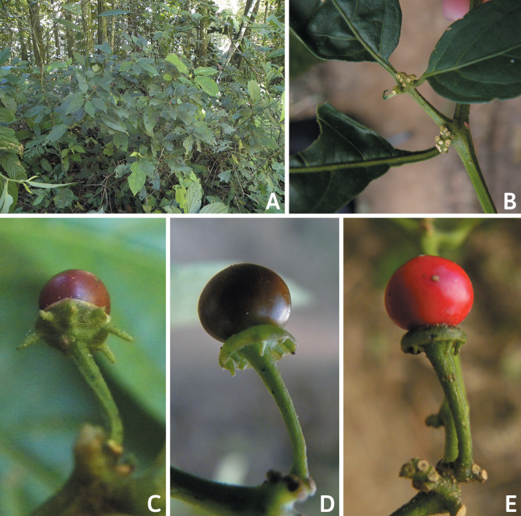
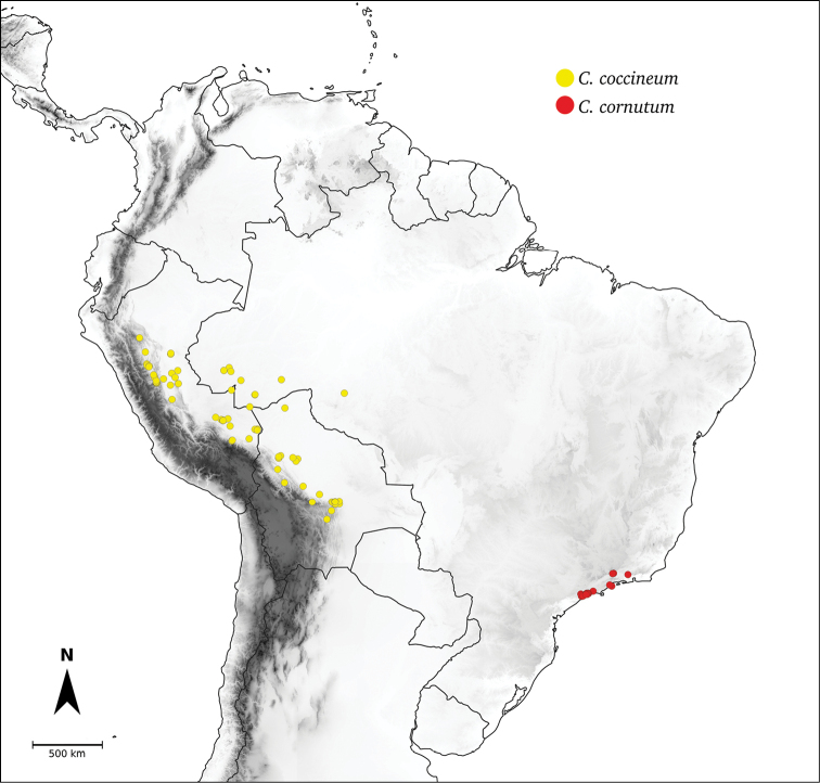
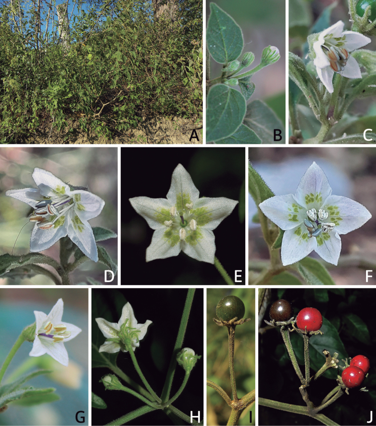
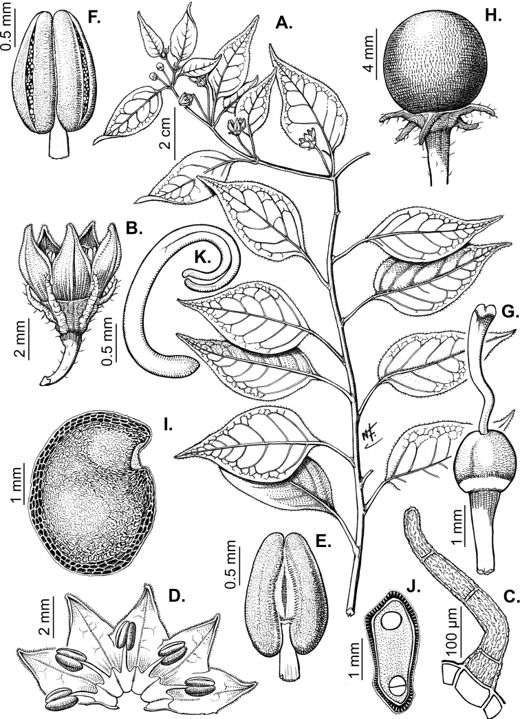
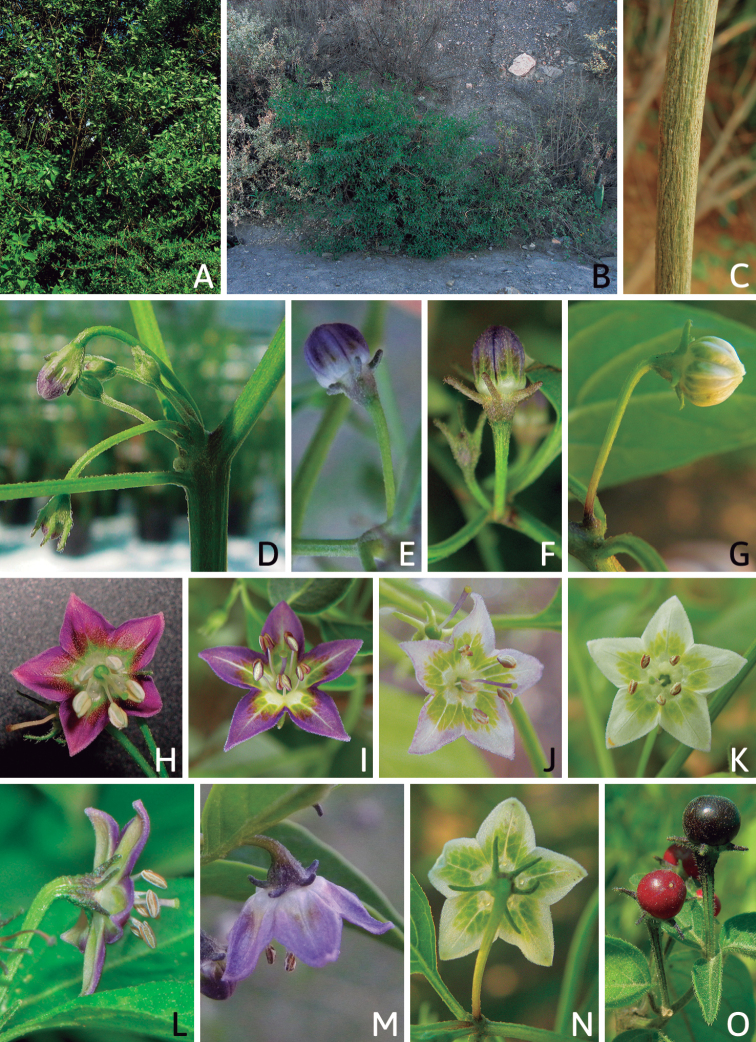
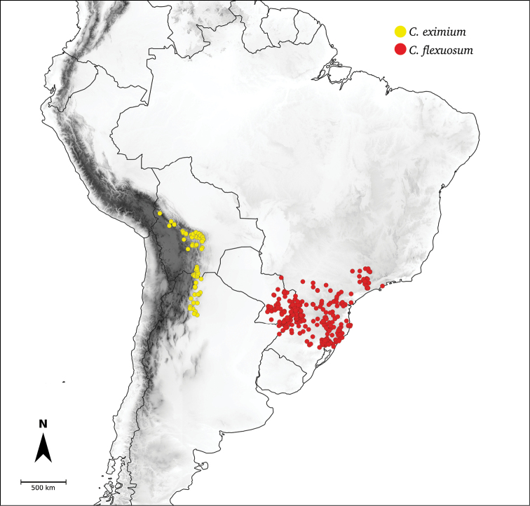
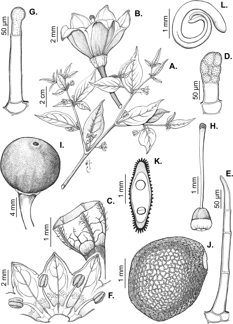
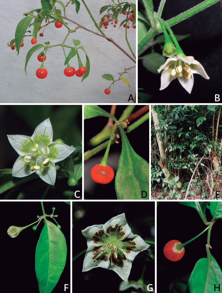
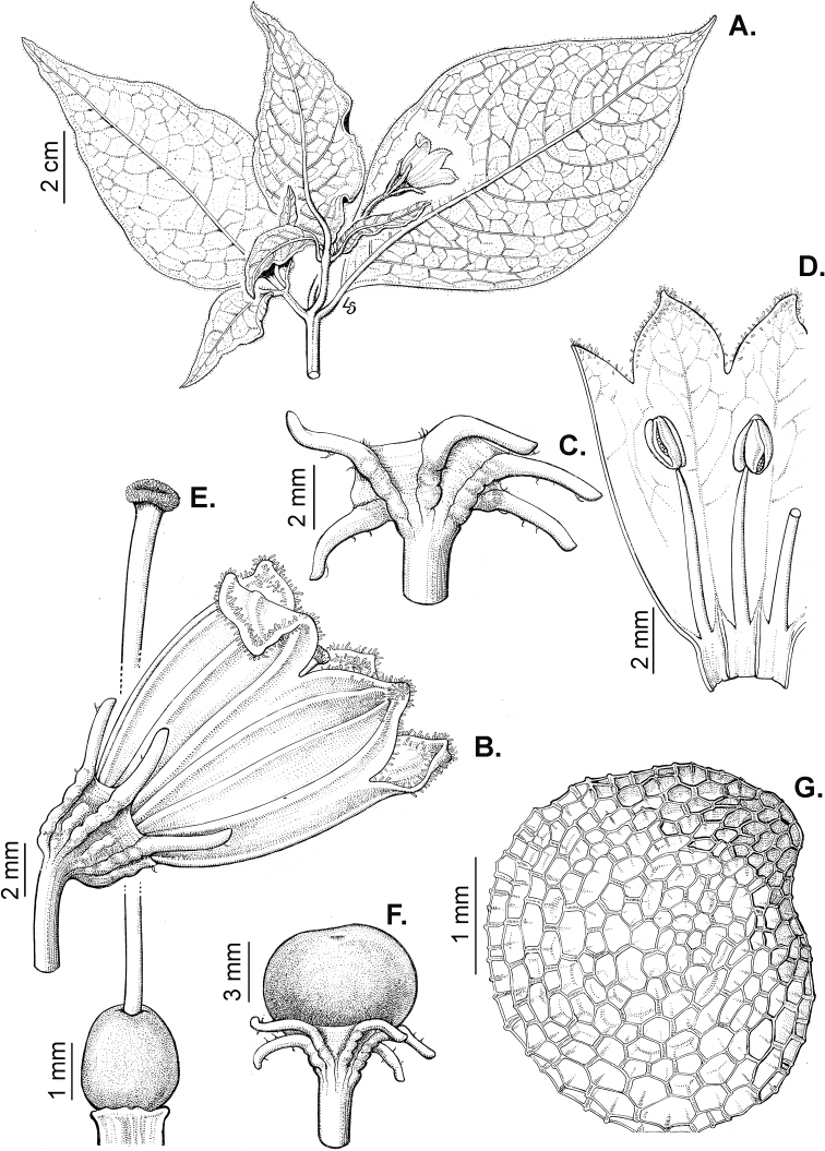
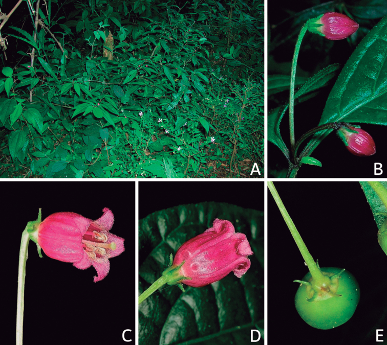
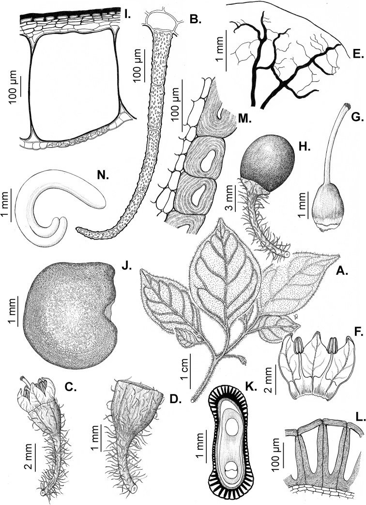
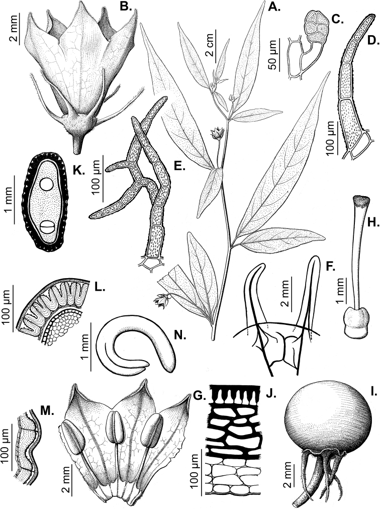
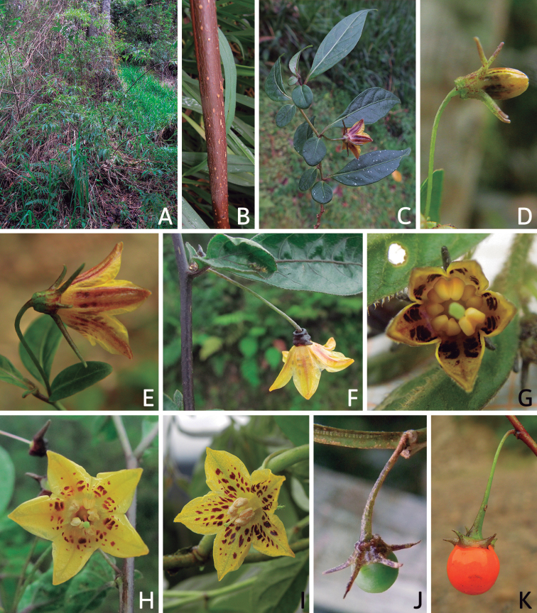
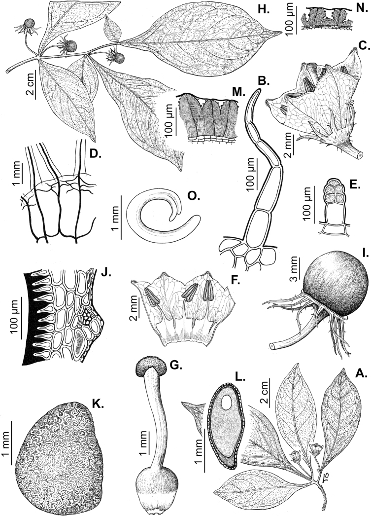
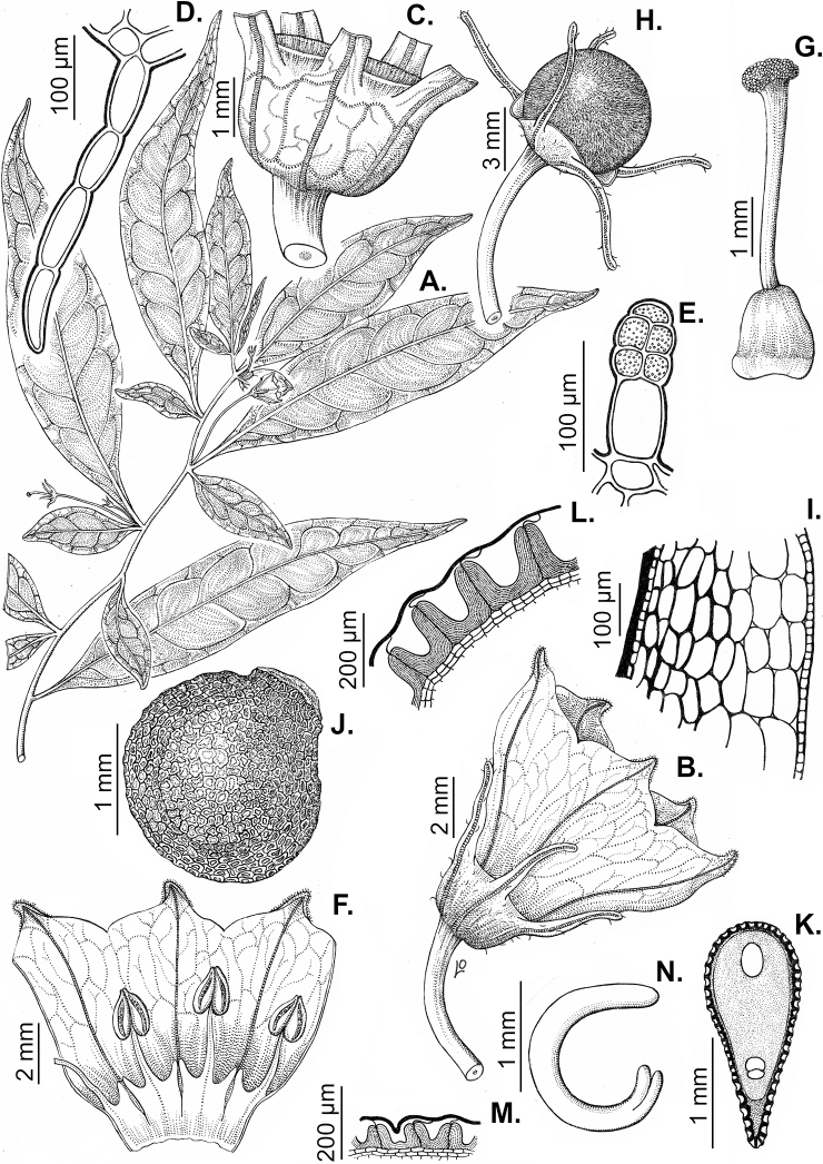
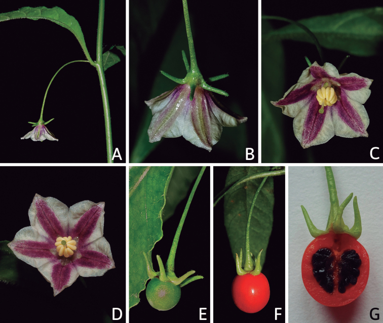
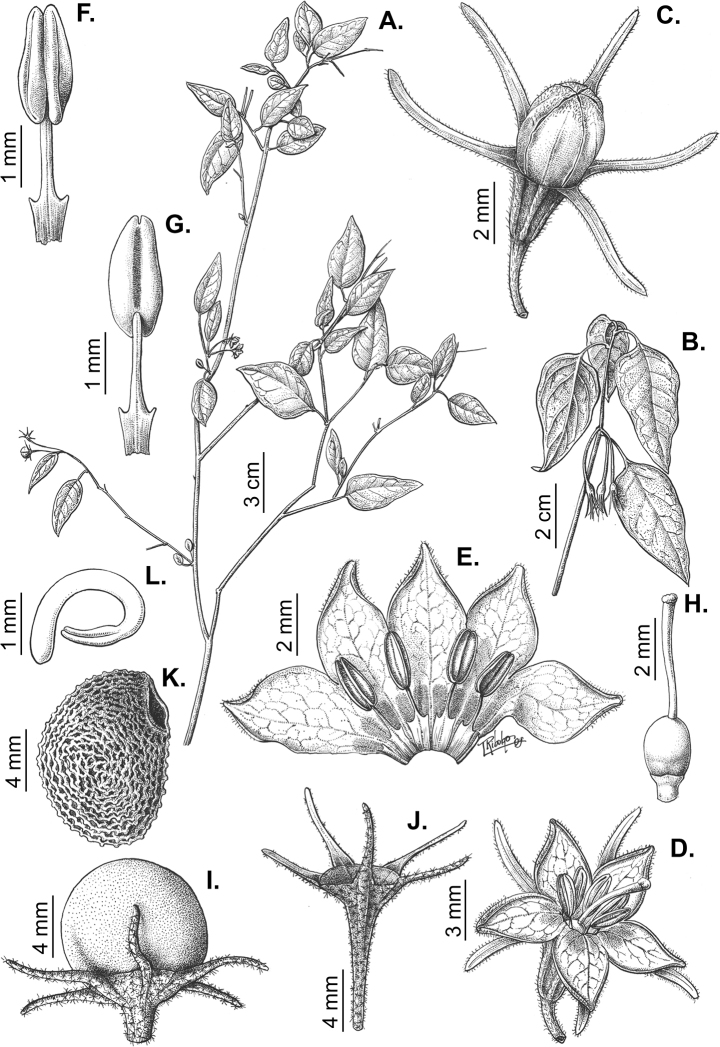
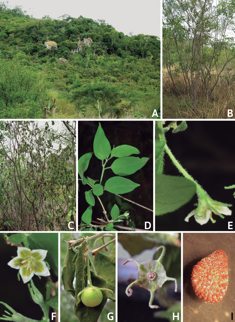
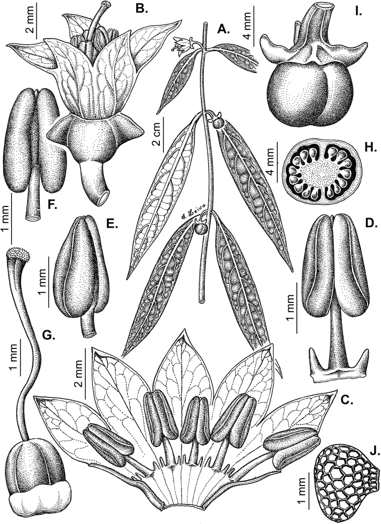
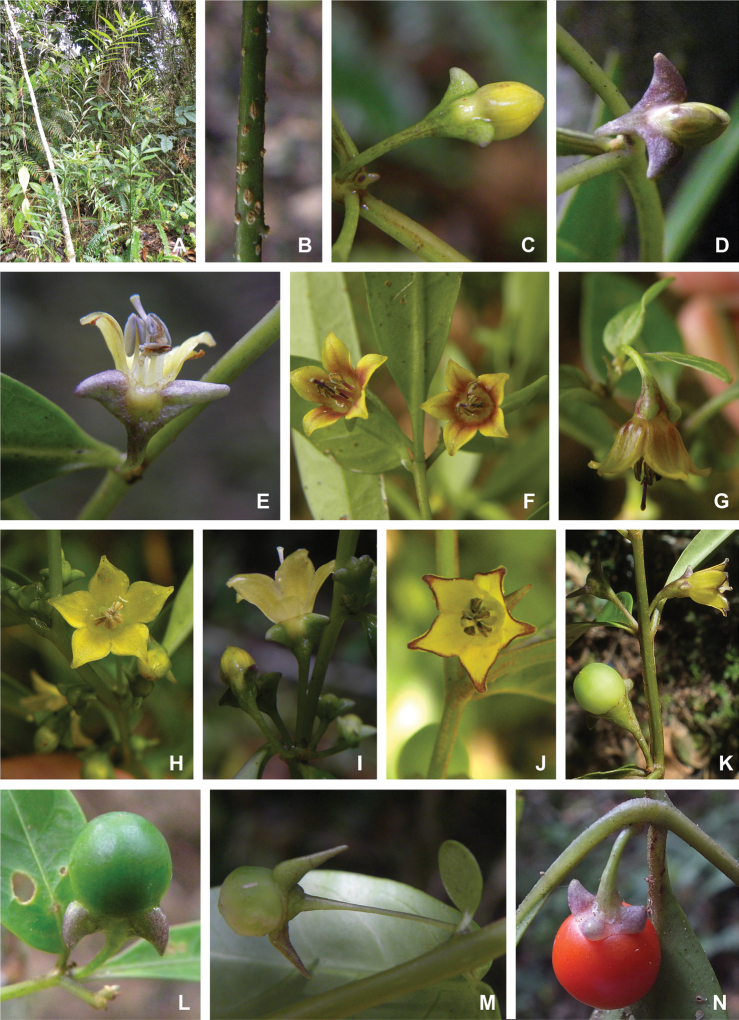
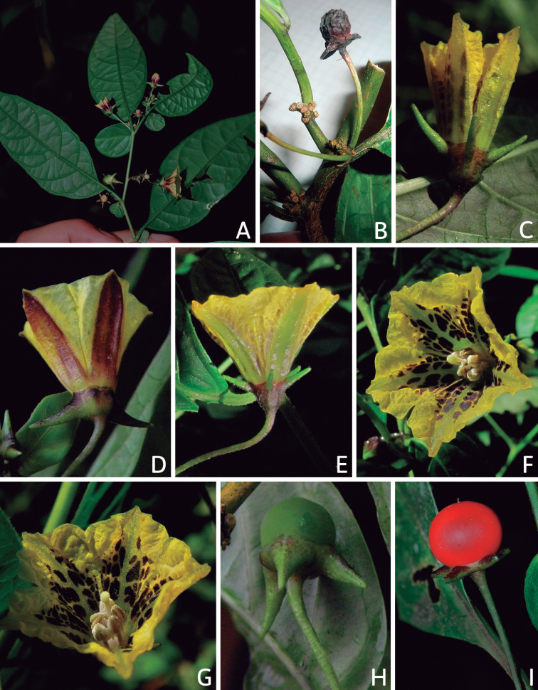
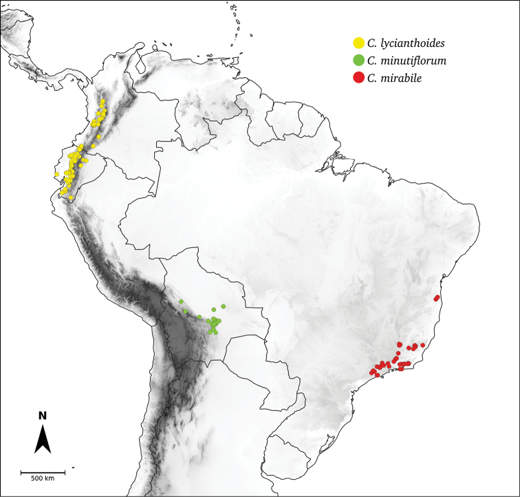
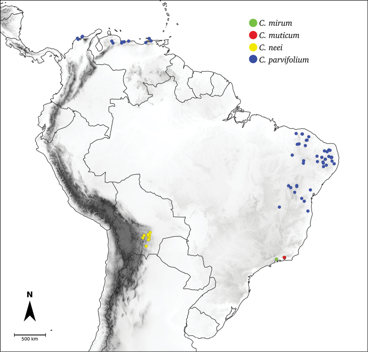
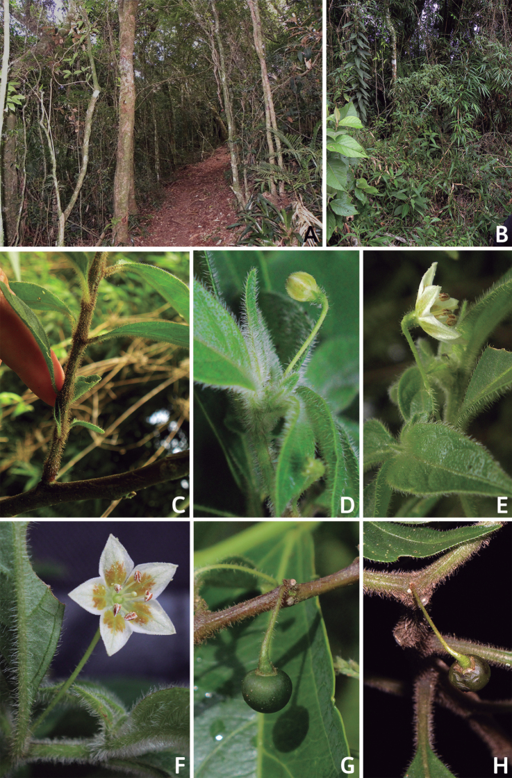
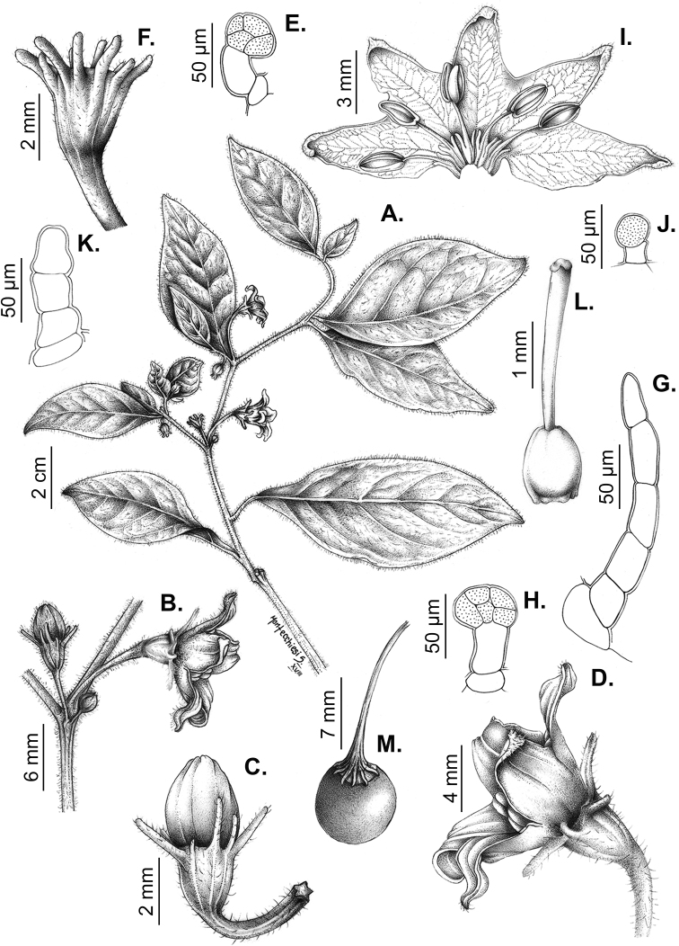
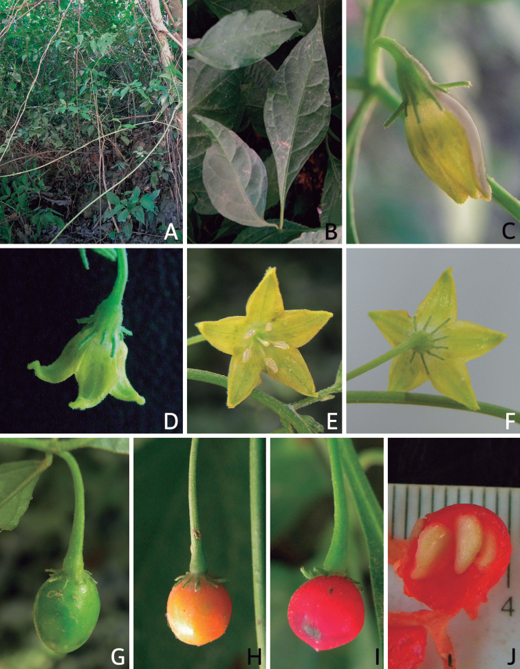
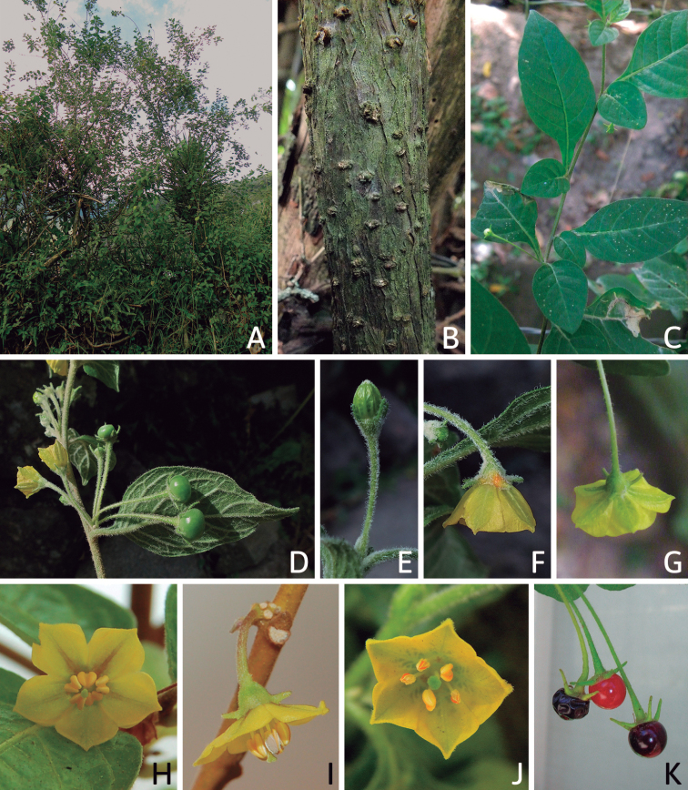
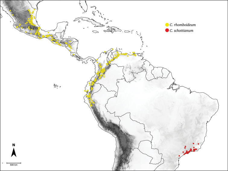
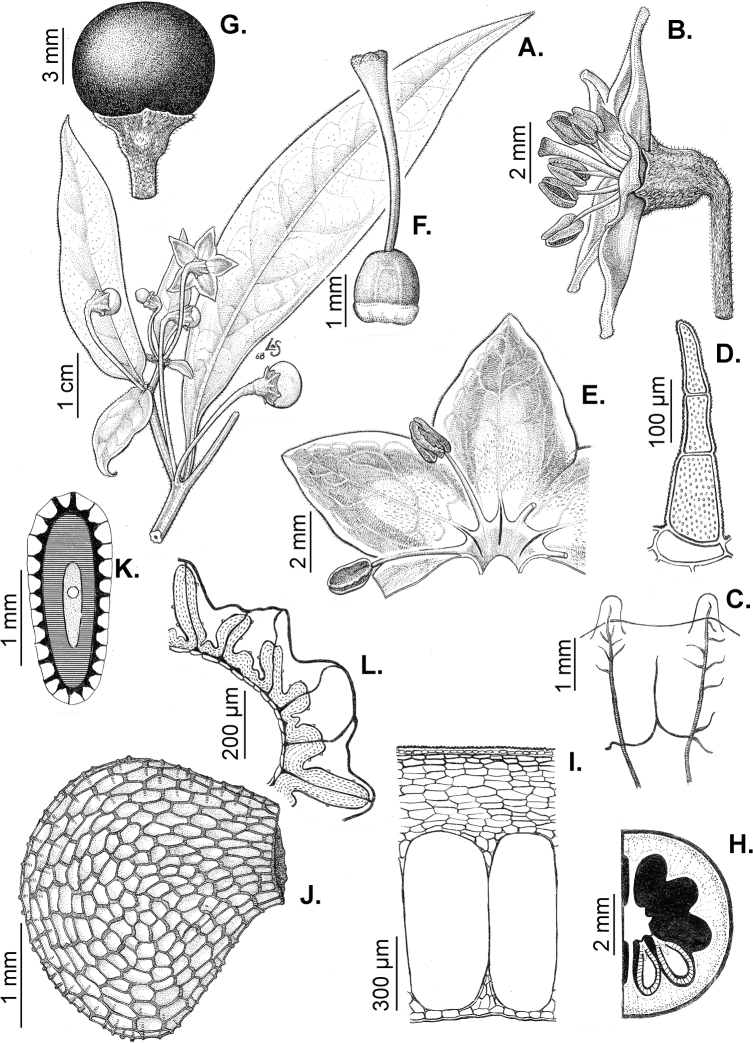
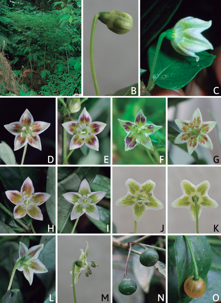
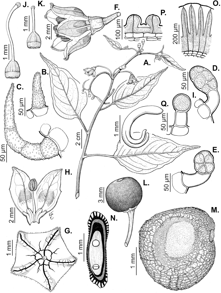
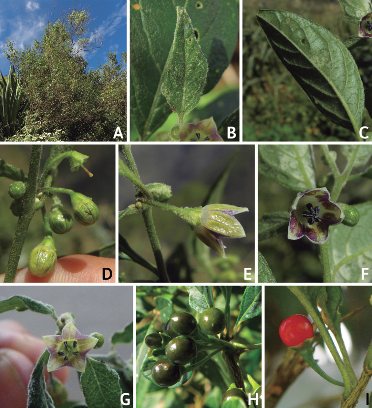
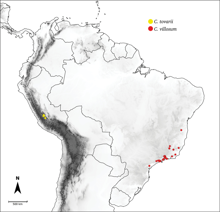
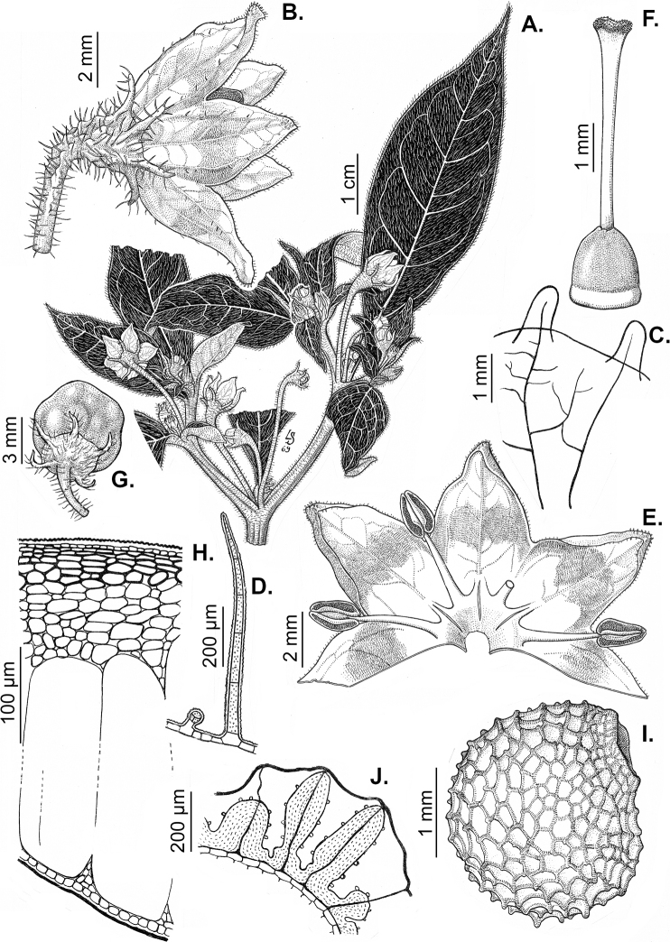
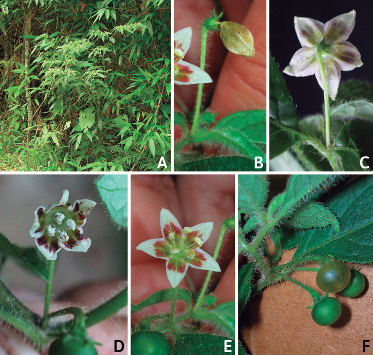
Capsicum L. (tribe Capsiceae, Solanaceae) is an American genus distributed ranging from the southern United States of America to central Argentina and Brazil. The genus includes chili peppers, bell peppers, ajíes, habaneros, jalapeños, ulupicas and pimientos, well known for their economic importance around the globe. Within the Solanaceae, the genus can be recognised by its shrubby habit, actinomorphic flowers, distinctive truncate calyx with or without appendages, anthers opening by longitudinal slits, nectaries at the base of the ovary and the variously coloured and usually pungent fruits. The highest diversity of this genus is located along the northern and central Andes. Although Capsicum has been extensively studied and great advances have been made in the understanding of its taxonomy and the relationships amongst species, there is no monographic treatment of the genus as a whole. Based on morphological and molecular evidence studied from field and herbarium specimens, we present here a comprehensive taxonomic treatment for the genus, including updated information about morphology, anatomy, karyology, phylogeny and distribution. We recognise 43 species and five varieties, including C.mirum Barboza, sp. nov. from São Paulo State, Brazil and a new combination C.muticum (Sendtn.) Barboza, comb. nov.; five of these taxa are cultivated worldwide (C.annuumL.var.annuum, C.baccatumL.var.pendulum (Willd.) Eshbaugh, C.baccatumL.var.umbilicatum (Vell.) Hunz. & Barboza, C.chinense Jacq. and C.frutescens L.). Nomenclatural revision of the 265 names attributed to chili peppers resulted in 89 new lectotypifications and five new neotypifications. Identification keys and detailed descriptions, maps and illustrations for all taxa are provided.

Keywords: America; Capsicum; chili peppers; cytogenetics; morphology; phylogeny; taxonomy.


Figures

Figure 1.
Figure 1.
Capsicum phylogeny. Cladogram summarising findings from Carrizo García et al. (2016) and Barboza et al. (2019, 2020b). Only one sample per species has been included; species names have been updated. Clades distinguished are labelled. Asterisks indicate branches with moderate/low support, letters ‘d’ clades with domesticated taxa.
Figure 2.
Figure 2.
Plant development in C.chacoenseA monopodial vegetative growth B first dichotomy of the main stem and start of sympodial growth C initial branching with three branches. Photos by G.E. Barboza.
Figure 3.
Figure 3.
Eglandular trichomes of Capsicum species A–D, F, H, K, M, O simple trichomes E, I papillae G, J, L, N, P branched trichomes. Scale bar: 50 μm (A, B, E, H–J); 100 μm (C, D, F, G, K–P).
Figure 4.
Figure 4.
Glandular trichomes of Capsicum species. Scale bar: 50 μm (A–G, I); 100 μm (H, J).
Figure 5.
Figure 5.
Flower morphology in Capsicum species AC.rabeniiBC.annuumvar.glabriusculumCC.lanceolatumDC.schottianumEC.frutescensFC.eximiumGC.eshbaughiiHC.cornutumIC.galapagoenseJC.recurvatumKC.cardenasiiLC.lycianthoidesMC.chacoenseNC.baccatumvar.baccatumOC.caballeroi. Abbreviations. im interpetalar membrane sp staminal plaque.
Figure 6.
Figure 6.
A–G Fruit morphology in Capsicum species H, J epicarp structure AC.chinenseBC.baccatumvar.baccatumCC.hookerianumDC.lanceolatumEC.coccineumFC.regaleGC.schottianumH epicarp with regular epidermal cells I epicarp with some sclereids amongst the regular epidermal cells J epicarp exclusively with sclereids. Abbreviation. sc sclereids. Scale bar: 10 μm (H, I, J).
Figure 7.
Figure 7.
Fruit anatomy in Capsicum species A, D–FC.baccatumvar.pendulumBC.baccatumvar.umbilicatumC, G, HC.pubescensA fruit, in cross section (note giant cells in the pericarp) B one locule of a fruit, in cross section (note the absence of giant cells in the pericarp) C, D epicarp and some layers of mesocarp (in D, observe cuticular wedges) E sector of pericarp (the arrow indicates the increase of the cell size ending in the giant cells) F detail of two adjacent giant cells G sector of homogeneous endocarp H sclereids of the endocarp. Abbreviations. c cuticle, cw cuticular wedge, epc epidermal cells, gc giant cells, p pericarp. Scale bars: 1 mm (A, B, E); 10 μm (C, D, H); 100 μm (F, G).
Figure 8.
Figure 8.
Seed morphology and seed coat structure ACapsicumchinenseBC.chacoense, longitudinal section C, DC.schottianum, cross section (D detail of the seed coat structure) EC.annuumvar.annuum, detail of seed coat structure (the rectangle indicates a cell of the seed coat) F, GC.eximium, cross sections at the seed margin and seed body, respectively HC.dimorphum, cross section at the seed body. Abbreviations. aw anticlinal cell wall, bp beak prominence, em embryo, en endosperm, h hilum, im inferior seed margin, ipw inner periclinal wall, opw outer periclinal wall, sb seed body, sc seed coat, sm superior seed margin, w seed width, l seed length. Scale bars: 200 μm (A–C); 100 μm (D, F, H); 20 μm (E, G).
Figure 9.
Figure 9.
Seeds and seed coat morphology in species of the Annuum Clade A–DC.annuumvar.annuumE–HC.annuumvar.glabriusculumI–LC.frutescensM–PC.chinenseQ–TC.galapagoenseA, I seeds with testa partly digested B seed coat with the external periclinal cell wall partly removed C, D cross section of the seed at the seed margin and seed body, respectively; E, M, N, Q untreated seeds showing subterminal hilum (E, N) and medial hilum (Q) F, J, O, P, S, T detail of a non-digested portion of the seed coat G, K testa pattern with the external periclinal cell wall removed H, L detail of testa cells R hilum. Abbreviation. opw outer periclinal cell wall. Scale bars: 200 μm (A, E, I, M, N, Q); 20 μm (B, F–H, K, L, P, S, T); 50 μm (C, J, O, R); 10 μm (D).
Figure 10.
Figure 10.
Seeds and seed coat morphology in species of the Baccatum Clade A–DC.baccatumvar.baccatumEC.baccatumvar.pendulumF–HC.baccatumvar.umbilicatumI–LC.chacoenseM–OC.rabenii.A, I, M seeds with testa partly digested B, G detail of a non-digested portion of the seed coat C, H, J, K, O testa pattern of treated seeds showing anticlinal cell walls with fibrils (C, K), papillae (H) and ridge (O) D detail of a testa cell E, F untreated seeds L cross section of the seed at the seed body N seed showing the subterminal elliptical hilum. Scale bars: 10 μm (H); 20 μm (B–D, G, K, L, O); 100 μm (J); 200 μm (A, E, F, I, M, N).
Figure 11.
Figure 11.
Seeds and seed coat morphology in species of the Atlantic Forest Clade A–DC.campylopodiumE–HC.carassenseI–LC.cornutumM–PC.friburgenseQ–TC.mirabileA untreated seed B, I, Q seeds with testa partly digested C, F, K, O, S marginal testa pattern of treated seeds (note pillar-like outgrowth in K, O, S) D, G, H, L, P, T testa pattern at the seed body of treated seeds showing anticlinal cell walls papillate and punctate E, J, M, R treated seeds (in J showing lateral prominence) N hilar zone with a linear hilum. Abbreviation. lp lateral prominence. Scale bars: 200 μm (A, B, E, I, J, M, Q, R); 100 μm (C, G, K, N, O, S); 20 μm (D, F, H, L, P, T).
Figure 12.
Figure 12.
Seeds and seed coat morphology in species of the Atlantic Forest Clade A–CC.muticumD–GC.pereiraeH–KC.mirumL–PC.schottianumQ–TC.recurvatumA, E, I, N, Q treated seeds B, F, J, O, S marginal testa pattern of treated seeds with pillar-like outgrowths C, G, P, T testa pattern at the seed body of treated seeds showing anticlinal cell walls papillate and punctate D, H, L untreated seeds K detail of papillate anticlinal cell walls M detail of a non-digested portion of the seed coat R hilar zone with a linear hilum. Scale bars: 200 μm (A, D, E, H, I, L, N, Q); 100 μm (B, F, J, O, R); 20 μm (C, G, K, M, P, S, T).
Figure 13.
Figure 13.
Seeds and seed coat morphology in species of the Atlantic Forest Clade A–DC.hunzikerianumE–IC.villosumA, G seeds with testa digested B seed showing medial and linear hilum C, H marginal testa pattern of treated seeds D, I testa pattern at the seed body of treated seeds showing anticlinal cell walls papillate (D) and punctate (I) E untreated seed F detail of a non-digested portion of the seed coat. Scale bars: 200 μm (A, B, E, G); 20 μm (C, D, F, I); 100 μm (H).
Figure 14.
Figure 14.
Seeds and seed coat morphology in species of the Andean Clade A–DC.dimorphumE–HC.geminifoliumI–LC.lanceolatumM–PC.longifoliumQ–TC.lycianthoidesA, Q, S seeds untreated B, E, I, J, M seeds with testa digested C, G, K, O testa pattern at the seed body of treated seeds showing anticlinal cell walls punctate (C, J, O) and with fibrils (G, K, O) D detail of ridge H, L, P detail of testa cells F, N hilar zone R, T detail of a non-digested portion of the seed coat. Scale bars: 200 μm (A, B, E, I, J, M, Q, S); 100 μm (C, K, N, O); 20 μm (D, F, G, H, L, P, R, T).
Figure 15.
Figure 15.
Seeds and seed coat morphology in species of the Andean Clade A–DC.hookerianumE–HC.piuranumI–KC.rhomboideumA untreated seed B, E treated seeds C, D, G, J, K testa pattern of treated seeds showing anticlinal cell walls punctate (C, D, J, K) and papillate (G) F hilar zone H detail of testa cells densely papillate I seed partly digested. Scale bars: 200 μm (A, B, E, I); 20 μm (C, D, F, G, H, J, K).
Figure 16.
Figure 16.
Seeds and seed coat morphology in species of the Bolivian Clade A–DC.caballeroiE–HC.ceratocalyxI–LC.minutiflorumM–PC.neeiQ–TC.coccineumA, Q seeds with testa partly digested B, F, G, J, O, R marginal testa pattern of treated seeds showing anticlinal cell walls punctate and densely papillate (G, O), punctate (J) and with ridge (R) C, H, K, P, S detail of testa cells D, L, T detail of papillae E, I, M treated seeds N linear hilum. Scale bars: 300 μm (A, E, I, M, N, Q); 50 μm (B, F, G, H, O, P); 20 μm (C, J, K, L, R, S); 5 μm (D, T).
Figure 17.
Figure 17.
Seeds and seed coat morphology in species of the Caatinga, Longidentatum, Flexuosum and Tovarii Clades A–DC.caatingaeE–HC.parvifoliumI–LC.longidentatumM–OC.flexuosumP–RC.tovariiA, M, P seeds with testa partly digested (in A hilum in terminal position, P hilum subterminal) B detail of a non-digested portion of the seed coat C, F, J, N, Q marginal testa pattern of treated seeds showing anticlinal cell walls papillate (F), with fibrils (J), punctate (N) and with fringe (Q) D, G, K, O, R detail of testa cells E, I treated seeds H, L papillae on anticlinal cell walls Scale bars: 200 μm (A, E, I, M, P); 20 μm (B, D, G, H, J, K, L, N, O, Q, R); 100 μm (C, F).
Figure 18.
Figure 18.
Seeds and seed coat morphology in species of the Purple corolla and Pubescens Clades A–DC.cardenasiiE–HC.eshbaughiiI–LC.eximiumM–PC.pubescensA, M untreated seeds (in A hilum terminal, M hilum medial) B treated seed C, G, K, O testa pattern of treated seeds showing anticlinal cell walls with fringe (C) and fibrils (G) D, H, L, M detail of testa cells E, I, N seeds with testa partly digested (in I hilum subterminal) F, J detail of a non-digested portion of the seed coat. Scale bars: 200 μm (A, B, E, I, M, N); 20 μm (C, D, G, H, J, K, L, P); 50 μm (F, O).
Figure 19.
Figure 19.
Capsicum geographic distribution. Georeferenced collection points of all wild Capsicum species/varieties. Circles are coloured by clades (Andean: orange; Atlantic Forest: bright green; Flexuosum: bright light blue; Caatinga: lilac; Longidentatum: dark blue; Bolivian: yellow; Purple corolla: red; Tovarii: fuchsia; Baccatum: dark green; Annuum: pale light blue).
Figure 20.
Figure 20.
Capsicumannuumvar.annum. Lectotype (BM). Copyright The Trustees of the Natural History Museum, London. Reproduced with permission.
Figure 21.
Figure 21.
Capsicumannuumvar.annuumA flower bud on pendent pedicel B flower with connivent anthers C flower with hexamerous corolla (note nectar droplets on the limb) and style near the same length as the anthers D flower with heptamerous purple corolla E flower with pentamerous corolla and style exceeding the anthers F, H mature fruits on pendent pedicels G mature fruits on upright pedicels A–H no specimen vouchers, photos by G.E. Barboza and C. Carrizo García taken at different greenhouses.
Figure 22.
Figure 22.
Capsicumannuumvar.glabriusculumA reproductive branch B eglandular trichome of the leaf C flower D section of the calyx showing the venation E opened corolla F gynoecium G fruit H anatomical detail of the pericarp (note the giant cell in the mesocarp) I seed J seed, in cross section K structure of seed coat at the seed margin L structure of seed coat at the seed body M embryo. A–H from Singleton 195I–M from Scolnik 19An329. Drawn by L. Ochoa. Published in Hunziker (2001), reproduced with permission.
Figure 23.
Figure 23.
Capsicumannuumvar.glabriusculumA apex of a reproductive branch B flower bud on geniculate pedicel C–F flowers in anthesis showing variations in corolla, stamens and style colouration G immature fruit H mature and immature fruits A, B, D, F from Barboza et al. 5049, photos by G.E. Barboza C, E, HCarrizo García 102, photos by C. Carrizo García GLeiva González et al. 2105, photo by S. Leiva González.
Figure 24.
Figure 24.
Distribution of C.annuumvar.glabriusculum.
Figure 25.
Figure 25.
Capsicumbaccatumvar.baccatumA flowering branch B calyx C glandular trichome of the calyx D eglandular trichome of the calyx E flower, upper view F sector of opened corolla G gynoecium H fruit I seed J seed, in cross section K embryo A–G from Hunziker 7350H–K from Hunziker 1579. Drawn by N. de Flury. Published in Barboza (2013), courtesy of the Board of the Instituto Darwinion (San Isidro, Buenos Aires, Argentina), reproduced with permission.
Figure 26.
Figure 26.
Capsicumbaccatumvar.baccatumA plant B flower bud C purple flower bud D, E, F flowers at anthesis, different views G young fruiting branch showing ovary with long and short styles H immature fruits I mature fruit A, C from Barboza 4913B, D–F, H from Barboza 5038G from Barboza 2431 bis, I from Barboza et al. 3419. Photos by G.E. Barboza.
Figure 27.
Figure 27.
Distribution of C.baccatumvar.baccatum, C.baccatumvar.pendulum and C.baccatumvar.umbilicatum.
Figure 28.
Figure 28.
Capsicumbaccatumvar.pendulumA plant B flower bud C flower, in pre-anthesis D flower short-styled, longitudinal section E flower long-styled, lateral view F–H flowers hexamerous showing connivent anthers and not connivent anthers I immature fruit J–M mature fruit A, B, E from Barboza 4886C, D, I, K, L, M no specimen vouchers (cult. Pairumani, Cochabamba-Bolivia) F, G, H from Palombo 3J from Barboza et al. 4824. Photos by G.E. Barboza.
Figure 29.
Figure 29.
Capsicumbaccatumvar.umbilicatumA reproductive branch B flower C section of the calyx showing the venation D, E eglandular trichomes of the abaxial surface of the calyx F glandular trichome of the adaxial surface of the calyx G sector of opened corolla H eglandular trichome of the corolla lobes I, J anthers, in dorsal and ventral views, respectively K gynoecium with long style L gynoecium with short style M ovary trilocular, in cross section N seed O seed, in cross section P structure of seed coat at the seed margin Q structure of seed coat at the seed body R embryo. From Rodríguez s.n. (CORD 241). Drawn by N. de Flury. Published in Hunziker (1998), reproduced with permission.
Figure 30.
Figure 30.
Capsicumbaccatumvar.umbilicatumA plant B flower bud C flower, lateral view D flower short-styled E flower long-styled F flower hexamerous G flower, seen from behind H immature fruit I, J mature fruits A–I from Barboza 5163, photos by G.E. Barboza J from Carrizo García 101, photo by C. Carrizo García.
Figure 31.
Figure 31.
CapsicumbenoistiiA flowering branch B flower C calyx D section of the calyx showing the venation E eglandular trichome of the calyx F opened corolla G gynoecium with long style H gynoecium with short style. From Benoist 4204. Drawn by N. de Flury. Published in Barboza et al. (2019), reproduced with permission.
Figure 32.
Figure 32.
Distribution of C.benoistii, C.caatingae, C.caballeroi, C.campylopodium and C.cardenasii
Figure 33.
Figure 33.
CapsicumcaatingaeA flowering branch B branched trichome from leaf C flower D calyx E, F, G glandular trichomes of the calyx and leaf venation H eglandular trichome of the calyx I sector of opened corolla J eglandular trichome of the corolla lobes K gynoecium L ovary, in cross section M node of a fruiting branch N fruit, in cross section O seed P seed, in cross section Q embryo. From Hunziker 25233. Drawn by N. de Flury. Published in Barboza et al. (2011), reproduced with permission.
Figure 34.
Figure 34.
CapsicumcaatingaeA flowering branch B young fruiting branch C immature fruits (note the purple pedicels) D node of a fruiting branch, some fruits already fallen down. No specimen voucher. Photos taken in Federal University of Viçosa (Minas Gerais) by C. dal Zovo (Associazione PepperFriends).
Figure 35.
Figure 35.
CapsicumcaballeroiA fruiting branch B flower C calyx D opened corolla E gynoecium F fruit G seed H seed, in longitudinal section. From Wood 11102. Drawn by P. Peralta.
Figure 36.
Figure 36.
CapsicumcaballeroiA plant B leaf pairs C major leaf D flower bud E flower F flower, in front view G immature fruit H mature fruit I mature fruit, in cross section, showing the seeds A from Barboza et al. 4907B, C, E, F, HBarboza et al. 3655D, G, I from Barboza et al. 4908. Photos by G.E. Barboza and S. Leiva González.
Figure 37.
Figure 37.
CapsicumcampylopodiumA flowering branch B eglandular trichome of the leaf C flower D calyx E section of the calyx showing the venation F flower, upper view G sector of opened corolla H gynoecium I fruit J fruit, upper view K fruit, in cross section L anatomical detail of the pericarp (note the giant cell in the mesocarp) M seed N seed, in cross section O structure of seed coat at the seed margin P structure of seed coat at the seed body Q embryo. From Hunziker 25116. Drawn by L. Sánchez. Published in Hunziker (2001), reproduced with permission.
Figure 38.
Figure 38.
CapsicumcampylopodiumA plant B flower C immature fruit D–F diagrams of different stages of fruit development D ovary, in cross section, showing the locules and the number of ovules E young fruit, in cross section (the lateral arrows indicate the fruit is flattened around the centre) F mature depressed fruit (one locule), in longitudinal section, showing the two seeds occupying the whole locule A from Barboza et al. 2057, photo by G.E. Barboza B, C from Bianchetti et al. 511, photos by L. Bianchetti.
Figure 39.
Figure 39.
CapsicumcarassenseA flowering branch B–D leaf morphology E eglandular trichome of the stem F glandular trichome of the calyx G, H flower buds in different stages of development I flower J opened corolla K fruit L fruiting calyx. From Bianchetti et al. 1364. Drawn by L. Bianchetti. Published in Barboza et al. (2020a), reproduced with permission.
Figure 40.
Figure 40.
CapsicumcarassenseA plant, showing the typical lanceolate leaves B inflorescence with geniculate pedicels C flower, in front view D fruit A–C from Stehmann 6344D from Agra 7268. Photos by J.R. Stehmann. Published in Barboza et al. (2020a), reproduced with permission.
Figure 41.
Figure 41.
Distribution of C.carassense, C.ceratocalyx and C.chacoense
Figure 42.
Figure 42.
CapsicumcardenasiiA flowering branch B eglandular trichome of the calyx C glandular trichome of the corolla D glandular trichome of the adaxial surface of the calyx E flower F section of the calyx showing the venation G sector of opened corolla H gynoecium I fruit J anatomical detail of the pericarp (note the giant cell in the mesocarp) K seed L seed, in cross section M structure of seed coat at the seed margin N structure of seed coat at the seed body O embryo A–H from Eshbaugh 1527I–O from Eshbaugh 2046 J. Drawn by N. de Flury.
Figure 43.
Figure 43.
CapsicumcardenasiiA plant B flower bud C, D flower, in lateral view E, F flower, in front view (note the different colouration inside the corolla) G immature fruits H mature fruit A, B, D, E, G from Barboza 4882, C, F, H from Barboza 4881. Photos by G.E. Barboza.
Figure 44.
Figure 44.
CapsicumceratocalyxA fruiting branch B node with pedicels scars and base of two pedicels C flower D calyx E opened corolla F fruit G seed H seed, in longitudinal section A–E from Beck 28089, F–I from Seidel & Hinojosa 1267. Drawn by P. Peralta.
Figure 45.
Figure 45.
CapsicumchacoenseA root B fruiting branch C eglandular trichome of the leaf D flower E sector of opened corolla F gynoecium G fruit H seed I seed, in cross section J embryo A, C–F from Hunziker 18572B, G–J from Hunziker et al. 25388. Drawn by N. de Flury. Published in Barboza (2013), courtesy of the Board of the Instituto Darwinion (San Isidro, Buenos Aires, Argentina), reproduced with permission.
Figure 46.
Figure 46.
CapsicumchacoenseA plant B flower bud C flower, in pre-anthesis D flower, in front view E immature fruit F mature fruit A–F from Barboza et al. 4910. Photos by G.E. Barboza.
Figure 47.
Figure 47.
Capsicumchinense (A–H from wild plants I–M from domesticated plants) A plant B flower buds C flower on geniculate pedicel D flower, in front view E flower, in full anthesis F immature fruit G fruiting pedicel H mature fruit I flowering branch J flowers on pendent pedicels K, L immature fruits M mature fruit A–H no specimen vouchers (cult. in Banco de Germoplasma de Hortaliças, Embrapa/Hortaliças, Brasília-DF, Brazil), photos taken in situ by L. Bianchetti I–M no specimen vouchers (cult. in Pairumani, Cochabamba-Bolivia), photos taken in situ by G.E. Barboza.
Figure 48.
Figure 48.
Distribution of C.chinense.
Figure 49.
Figure 49.
CapsicumcoccineumA reproductive branch B flower C calyx with appendages D section of the calyx showing the venation E eglandular trichome of the calyx F opened corolla G, H anthers, dorsal and ventral views, respectively I gynoecium J node with fruits K anatomical detail of the pericarp (note the giant cell in the mesocarp) L seed, in cross section M structure of seed coat at the seed margin N structure of seed coat at the seed body O embryo A, B, D–I, K from Woytkowski 1176C, J from Nee & Saldías 35956L–O from Nee 44503. Drawn by J. de Ugarte.
Figure 50.
Figure 50.
CapsicumcoccineumA plant B fruiting node with many pedicels scars C young immature fruit D immature fruit with calyx reflexed E mature fruit with calyx reflexed. From Barboza et al. 4921. Photos by G.E. Barboza.
Figure 51.
Figure 51.
Distribution of C.coccineum and C.cornutum.
Figure 52.
Figure 52.
CapsicumcornutumA leaf B eglandular trichomes of the leaf C flowering branch D sector of opened corolla E gynoecium F fruit G fruit (one carpel), in longitudinal section H anatomical detail of the pericarp (note the giant cell in the mesocarp) I seed J seed, in cross section K structure of seed coat at the seed margin L embryo A–G from Hunziker 19557 H–L from Kuhlmann 4321. Drawn by L. Sánchez. Modified from Hunziker (1971), reproduced with permission.
Figure 53.
Figure 53.
CapsicumcornutumA plant B flower on geniculate pedicel C flower, in lateral view, showing the calyx D flower, in front view E immature fruit. From Barboza & Cosa 2525. Photos by G.E. Barboza.
Figure 54.
Figure 54.
CapsicumdimorphumA flowering branch B flower; C section of the calyx showing the venation; D sector of opened corolla; E gynoecium; F fruit G anatomical detail of the pericarp (note the absence of giant cells in the mesocarp) H seed; I seed, in cross section J structure of seed coat at the seed margin K structure of seed coat at the seed body L embryo A, B, D, E from Jaramillo et al. 2698C from Sneidern 3044F–L from Cuatrecasas 8572. Drawn by L. Ochoa.
Figure 55.
Figure 55.
CapsicumdimorphumA plant B, C flower buds of different colour D, E flowers, in lateral view, with different corolla colour outside F, G flowers, in front view, with different colouration patterns in the corolla within H immature fruit I mature fruit A, H, I from Beltrán 140, photos by G. Beltrán B–G from Orejuela R. et al. 2685, photos by G.E. Barboza.
Figure 56.
Figure 56.
Distribution of C.dimorphum and C.eshbaughii.
Figure 57.
Figure 57.
CapsicumeshbaughiiA flowering branch B eglandular trichome of the leaf C flower D, E furcate glandular trichomes of the pedicel F fruiting calyx G glandular trichome of the abaxial surface of the calyx H, I glandular trichomes of the adaxial surface of the calyx J sector of opened corolla K gynoecium L fruit M seed N embryo A–E, G–K from Eshbaugh 1943 bF, L–N from Nee 36164. Drawn by P. Peralta. Published in Barboza (2011), reproduced with permission.
Figure 58.
Figure 58.
CapsicumeshbaughiiA plant B flower buds C flower, in pre-anthesis D–G stellate corollas with different colouration patterns H flower buds and flower in anthesis, seen from behind I immature fruit J mature fruits A, E, H–J from Carrizo García 67, photos by G.E. Barboza and C. Carrizo García B, C, D, F, G from Palombo 19, photos by N. Palombo.
Figure 59.
Figure 59.
CapsicumeximiumA flowering branch B flower C eglandular trichome of the calyx D opened corolla E, F anthers, dorsal and ventral views, respectively G gynoecium H fruit I seed J seed, in cross section K embryo A from Hunziker 7346B–K from Hunziker 1907. Drawn by N. de Flury. Published in Barboza (2013), courtesy of the Board of the Instituto Darwinion (San Isidro, Buenos Aires, Argentina), reproduced with permission.
Figure 60.
Figure 60.
CapsicumeximiumA plant (wet habitat) B plant (dry habitat) C main fissured stem D young inflorescence E–G flower buds with different colouration H–K flowers, in front view, with different corolla colouration within L flower, in lateral view M, N flowers seen from behind O mature fruits A, G, N from Barboza et al. 4914B, E, M from Barboza et al. 4885C, K from Barboza et al. 4903 D from Wageningen Netherlands University germplasm collection F, I from Barboza et al. 4895H, O from Barboza 1919 (cult.) J from Barboza et al. 4896L from Barboza et al. 3543A–C, E–O photos by G.E. Barboza, D photo by P. Bosland.
Figure 61.
Figure 61.
Distribution of C.eximium and C.flexuosum.
Figure 62.
Figure 62.
CapsicumflexuosumA flowering branch B flower C calyx D glandular trichome of the abaxial surface of the calyx E eglandular trichome of the adaxial surface of the calyx F sector of opened corolla G glandular trichome of the abaxial surface of the corolla H gynoecium I fruit J seed K seed, in cross section L embryo A from Hatschbach 18030B–H from Subils & Moscone 4273I–L from Hunziker et al. 24993. Drawn by N. de Flury. Published in Barboza (2013), courtesy of the Board of the Instituto Darwinion (San Isidro, Buenos Aires, Argentina), reproduced with permission.
Figure 63.
Figure 63.
CapsicumflexuosumA fruiting branch B flowering node C stellate corolla with greenish spots D, H mature fruits E plant F flower bud G rotate-stellate corolla with purple and greenish spots A–D from Barboza et al. 1034EBarboza et al. 3631F–H from Carrizo García 84. Photos by C. Carrizo García.
Figure 64.
Figure 64.
CapsicumfriburgenseA flowering branch B flower C calyx D sector of opened corolla E gynoecium F fruit G seed. From Bianchetti et al. 393. Drawn by L. Sánchez. Published in Barboza and Bianchetti (2005), reproduced with permission.
Figure 65.
Figure 65.
CapsicumfriburgenseA plant B flower buds C flower, in lateral view D flower, seen from behind E immature fruit A, B from Barboza et al. 2048, photo by G.E. Barboza C–E no specimen vouchers, photos taken in situ by C. dal Zovo (Associazione PepperFriends).
Figure 66.
Figure 66.
Distribution of C.friburgense, C.galapagoense and C.geminifolium.
Figure 67.
Figure 67.
CapsicumfrutescensA reproductive branch B eglandular trichome of the leaf C flower D section of the calyx showing the venation E glandular trichome of the abaxial surface of the calyx F opened corolla G glandular trichome of the abaxial surface of the corolla H, I anthers, dorsal and ventral views, respectively J gynoecium K anatomical detail of the pericarp (note the giant cells in the mesocarp) L seed M seed, in cross section N structure of seed coat at the seed margin O structure of seed coat at the seed body P embryo. From Hunziker 25489. Drawn by N. de Flury.
Figure 68.
Figure 68.
CapsicumfrutescensA plant B flower buds C flower showing the calyx D flower, in lateral view E flower, in front view (note the connivent anthers) F flower after anthers dehiscence G immature (green) and mature (red) fruits H fruit, in longitudinal section showing the seeds A, B, E–G from Barboza & Leiva González 4829C, D, H from Barboza et al. 2041. Photos by G.E. Barboza.
Figure 69.
Figure 69.
Distribution of C.frutescens.
Figure 70.
Figure 70.
CapsicumgalapagoenseA flowering branch B eglandular trichome of the leaf C flower D calyx E section of the calyx showing the venation F sector of opened corolla G gynoecium H fruit I anatomical detail of the pericarp (note the giant cell in the mesocarp) J seed K seed, in cross section L structure of seed coat at the seed margin M structure of seed coat at the seed body N embryo A, B from Stewart 3351C–G from Taylor G11H–N from Schimpf 20. Drawn by L. Ochoa.
Figure 71.
Figure 71.
CapsicumgeminifoliumA flowering branch B flower C glandular trichome of the abaxial surface of the calyx D eglandular calyx of the leaf and calyx E branched trichome of the leaf and calyx F section of the calyx showing the venation G sector of opened corolla gynoecium H gynoecium I fruit J anatomical detail of the pericarp (note the absence of giant cells in the mesocarp) K seed, in cross section L structure of seed coat at the seed margin M structure of seed coat at the seed body N embryo. From Scolnik 1389. Drawn by L. Sánchez.
Figure 72.
Figure 72.
CapsicumgeminifoliumA plant B main stem with lenticels C flowering branch D flower bud E flower, in lateral view, with long calyx appendages F flower, with short calyx appendages G flower, in pre-anthesis H, I corollas, in front view, with different colouration patterns within J immature fruit K mature fruit A, B, D, J from Barboza & Leiva González 4845C, G from Deanna & Leiva González 3E, I from Barboza & Leiva González 4852F, H from Deanna & Leiva González 77K from Orejuela R. 2688A, B, D, E, I, J photos by G.E. Barboza C, F–H photos by R. Deanna K photo by A. Orejuela.
Figure 73.
Figure 73.
CapsicumhookerianumA flowering branch B eglandular trichome of the leaf C flower D section of the calyx showing the venation E glandular trichome of the abaxial surface of the calyx F sector of opened corolla G gynoecium H fruiting branch I fruit J anatomical detail of the pericarp (note the absence of giant cells in the mesocarp) K seed L seed, in cross section M structure of seed coat at the seed margin N structure of seed coat at the seed body O embryo A–G from Asplund 15241H–O from Asplund 15363. Drawn by L. Ochoa.
Figure 74.
Figure 74.
CapsicumhookerianumA plant; B internode with lenticels; C fruiting branch; D immature fruit; E mature fruit; F, G fruiting calyx, front and from behind views, respectively. From Barboza and Leiva González 4826. Photos by G.E. Barboza.
Figure 75.
Figure 75.
Distribution of C.hookerianum and C.lanceolatum.
Figure 76.
Figure 76.
CapsicumhunzikerianumA flowering branch B flower C calyx D sector of opened corolla E, F anthers, dorsal and ventral views, respectively G stigma H fruit I seed A–G from Kuhlmann 2785, H, I from Mattos and Mattos 14254. Drawn by L. Ribulgo. Published in Barboza and Bianchetti (2005), reproduced with permission.
Figure 77.
Figure 77.
CapsicumhunzikerianumA plant B flowering branch C flower bud D, E flowers, in front view, with different corolla colour patterns F flower, seen from behind G immature fruit H mature fruit I seed A, E from Bianchetti et al. 1537, photos by L. Bianchetti B, C, D, F, H, I no specimen vouchers, photos taken in situ by C. dal Zovo (Associazione PepperFriends) G from Barboza 5041, photo by G.E. Barboza.
Figure 78.
Figure 78.
Distribution of C.hunzikerianum, C.longidentatum and C.longifolium.
Figure 79.
Figure 79.
CapsicumlanceolatumA flowering branch B flower C calyx (appendages cut off) D eglandular trichome of the adaxial surface of the calyx E glandular trichome of the abaxial surface of the calyx F sector of opened corolla G gynoecium H fruit I anatomical detail of the pericarp (note the absence of giant cells in the mesocarp) J seed K seed, in cross section L structure of seed coat at the seed margin M structure of seed coat at the seed body N embryo A from Ventura A. 798B–G from Skutch 1450H–N from Beaman 6014. Drawn by L. Ochoa. Published in Hunziker (2001), reproduced with permission.
Figure 80.
Figure 80.
CapsicumlanceolatumA pendent flower B flower, lateral view C flower, in front view D flower showing connivent anthers E immature fruit F mature fruit G fruit, longitudinal section, showing the black seeds. From Carrizo García 73. Photos by C. Carrizo García.
Figure 81.
Figure 81.
CapsicumlongidentatumA reproductive branch B detail of a flowering node C flower bud D flower, upper view E opened corolla (one stamen has been removed) F, G anthers, ventral and dorsal views, respectively H gynoecium I fruit J fruiting calyx K seed L embryo A, D, E, F–H from Melo et al. 4344B, C, I–L from Agra & Barboza 7086. Drawn by L. Ribulgo. Published in Barboza et al. (2011), reproduced with permission.
Figure 82.
Figure 82.
CapsicumlongidentatumA habitat (Caatinga) B plant C typical aspect of the plant in the arid caatinga D flowering branch E pendent flower F flower, in front view G immature fruit H fruiting calyx I seed A–C, G–I from Agra & Barboza 7086D–F from Tabosa et al. 55A–C, G–I photos by G.E. Barboza D–F photos by J.R. Stehmann.
Figure 83.
Figure 83.
CapsicumlongifoliumA reproductive branch B flower C opened corolla D, E, F anthers, ventral, lateral and dorsal views, respectively G gynoecium H ovary, in cross section I fruit J seed. From Barboza & Leiva González 4849. Drawn by S. Leiva González. Published in Barboza et al. (2019), reproduced with permission.
Figure 84.
Figure 84.
CapsicumlongifoliumA plant B internode with lenticels C, D flower buds E Flower, in longitudinal section F flowers showing corolla yellow with brownish centre G flower, in lateral view H, I flowers with completely yellow corollas, upper and lateral views, respectively J, K flowers with yellow corollas and red-brown edges, upper and lateral view, respectively L, M immature fruits N mature fruit A, C, H, I from Barboza & Leiva González 4821B, D, E, L, N from Barboza & Leiva González 4846F, G from Barboza & Leiva González 4850J, K, M from Barboza & Leiva González 4851. Photos by S. Leiva González and G.E. Barboza. Published in Barboza et al. (2019), reproduced with permission.
Figure 85.
Figure 85.
CapsicumlycianthoidesA flowering branch B reproductive nodes with pedicel scars C flower, in pre-anthesis D, E flowers, in lateral view, with variations in calyx morphology and corolla colouration F, G flowers, in front view H immature fruit I mature fruit A, I from Beltrán 85, photos by G. Beltrán B, D, H from Deanna et al. 144C, E–G from Deanna et al. 133, photos by R. Deanna.
Figure 86.
Figure 86.
Distribution of C.lycianthoides, C.minutiflorum and C.mirabile.
Figure 87.
Figure 87.
CapsicumminutiflorumA plant B major leaf C flowering branch D flower bud on geniculate pedicel E flower, in front view F flower, in lateral view G fruiting branch H immature fruit I mature fruit. From Barboza et al. 4918. Photos by G.E. Barboza.
Figure 88.
Figure 88.
CapsicummirabileA flowering branch B, C major and minor leaf D flower bud E flower, in lateral view F, G flowers, upper views H sector of opened corolla I gynoecium J fruit K fruit, in cross section L seed. From Hunziker 19562. Drawn by L. Sánchez.
Figure 89.
Figure 89.
CapsicummirabileA plant B flower buds on geniculate pedicels C flower, in pre-anthesis D flower, seen from behind E, H flowers, in front view, showing corollas with different colouration F flower, in full anthesis G flower, seen from behind I immature fruits J mature fruit A, B, D–J from Barboza & Deanna 5024C from Bianchetti & Bustamante 1518A, B, D–H photos by G.E. Barboza C photo by L. Bianchetti I, J photos by R. Deanna.
Figure 90.
Figure 90.
Capsicummirum. Holotype (CORD). From Barboza et al. 1648. Copyright Universidad Nacional de Córdoba, Argentina. Reproduced with permission.
Figure 91.
Figure 91.
CapsicummirumA plant B flowering branch C bud on geniculate pedicel D flower in pre-anthesis E flower, lateral view F flower, in front view G flower, seen from behind H fruiting calyx I immature fruits. No specimen vouchers. Photos taken in situ by J. Lackey (Associazione PepperFriends).
Figure 92.
Figure 92.
Distribution of C.mirum, C.muticum, C.neei and C.parvifolium.
Figure 93.
Figure 93.
CapsicummuticumA habitat (degraded Atlantic Forest) B plant C stem and leaves showing the dense indument D flower bud on geniculate pedicel E flower, in lateral view F flower, in front view G immature fruit H fruit near maturity. From Barboza et al. 5032. Photos by G.E. Barboza.
Figure 94.
Figure 94.
CapsicumneeiA flowering branch B inflorescence C flower bud D flower E glandular trichome of the pedicels F calyx G eglandular trichome of the outer surface of the calyx H glandular trichome of the inner surface of the calyx I opened corolla J glandular trichome of the inner surface of the corolla K eglandular trichome of the inner surface of the corolla lobes L gynoecium M fruit. From Barboza 4927. Drawn by S. Montecchiesi. Published in Barboza et al. (2019), reproduced with permission.
Figure 95.
Figure 95.
CapsicumneeiA plant B leaves, abaxial surface C flower bud D flower, in lateral view E flower, in front view F flower, seen from behind G immature fruit H, I mature fruits J fruit, longitudinal section, showing the seeds A–G from Barboza et al. 4927H–J from Barboza & Reyes 5040. Photos by G.E. Barboza. Modified from Barboza et al. (2019), reproduced with permission.
Figure 96.
Figure 96.
CapsicumparvifoliumA fruiting branch B branched trichome of the leaf C flower D calyx E opened corolla F, G anthers, dorsal and ventral views, respectively H gynoecium I fruits A, C, E–I from Agra & Barboza 7075B, D from Pickersgill 72–366. Drawn by L. Ribulgo. Published in Barboza et al. (2011), reproduced with permission.
Figure 97.
Figure 97.
CapsicumparvifoliumA habitat (Caatinga) B plant C reproductive branch D flower bud and flower on pendent pedicels E flower, in front view F fruiting calyces and immature fruit G mature fruits H fruiting calyx, inside view. From Agra & Barboza 7075. Photos by G.E. Barboza. Modified from Barboza et al. (2011), reproduced with permission.
Figure 98.
Figure 98.
CapsicumpereiraeA flowering branch B leaf C flower bud D flower E calyx F eglandular trichome of the calyx G opened corolla H gynoecium I fruiting branch J fruit, in cross section K anatomical detail of the pericarp (note the giant cells in the mesocarp) L seed M seed, in cross section N embryo A–J, L–N from Pereira 2245K from Hunziker 25248. Drawn by N. de Flury. Published in Barboza and Bianchetti (2005), reproduced with permission.
Figure 99.
Figure 99.
CapsicumpereiraeA flowering branch B young branch showing lanceolate leaves and buds on pendent pedicels C flower showing mutic calyx D, E flowers, in front view, with different patterns of purple colouration F flower without purple spots A, B, E from Bianchetti et al. 1558F from Bianchetti et al. 1567, photos by L. Bianchetti C, D no specimen vouchers, photos taken in situ by C. dal Zovo (Associazione PepperFriends). Modified from Barboza and Bianchetti (2005), reproduced with permission.
Figure 100.
Figure 100.
Distribution of C.pereirae and C.piuranum.
Figure 101.
Figure 101.
CapsicumpiuranumA reproductive branch B flower C calyx D eglandular trichome of the outer surface of the calyx E opened corolla F, G, H anthers, ventral, dorsal and lateral views, respectively I gynoecium J fruit K seed. From G.E. Barboza & S. Leiva González 4841. Drawn by S. Leiva González. Published in Barboza et al. (2019), reproduced with permission.
Figure 102.
Figure 102.
CapsicumpiuranumA plant B leaves, abaxial surface C Fruiting branch with leaf pair dissimilar in shape and size D flower bud E flower and immature fruit on pendent pedicels F mature fruit G fruit, in cross section, showing placenta and seeds H fruit, in cross section, showing a stone cell at the apex (arrow). From Barboza & Leiva González 4841. Photos by S. Leiva González and G.E. Barboza. Published in Barboza et al. (2019), reproduced with permission.
Figure 103.
Figure 103.
CapsicumpubescensA reproductive branch B trichome of the leaf C calyx D section of the calyx showing the venation E flower, upper view F sector of opened corolla G, H anthers, dorsal and ventral views, respectively I, J, K gynoecium with short, medium and long style, respectively L anatomical detail of the pericarp (note the giant cell in the mesocarp) M seed N seed, in cross section O structure of seed coat at the seed margin P structure of seed coat at the seed body Q embryo. From Hunziker 25484. Drawn by J. de Ugarte.
Figure 104.
Figure 104.
CapsicumpubescensA plant B reproductive nodes C young leaves, adaxial surface D flower bud on geniculate pedicel E–H flowers, in front view, showing 4-7-merous corollas I, J flowers, seen from behind K flower, in lateral view L–O mature fruits from different provenance (L, N from Bolivia, M from Peru, O from Mexico) P fruit, in longitudinal section, showing the black seeds A–D, GBarboza et al. 4889E, P from Barboza et al. 4890F from Palombo 21H from Barboza et al. 1847I from Palombo 22J from Palombo 23K, L no specimen vouchers (cult. Córdoba, Argentina) M no specimen voucher (cult. Huancayo-Peru) N from Carrizo García et al. 35O no voucher specimen (bought in Mexico) A–D, G, H, L, M, O, P photos by G.E. Barboza E, F, I–K, N photos by N. Palombo.
Figure 105.
Figure 105.
Distribution of C.pubescens and C.rabenii.
Figure 106.
Figure 106.
CapsicumrabeniiA plant B leaf, abaxial surface showing pubescence on the veins C reproductive nodes D bud on geniculate pedicel E flower, lateral view F rotate corolla G stellate corolla H flower showing the exserted style I immature fruits J globose mature fruits K ellipsoid mature fruit and fruiting calyx A–I, K from Barboza & Deanna 5003J from Barboza et al. 1646. Photos by G.E. Barboza.
Figure 107.
Figure 107.
CapsicumrecurvatumA reproductive branch B flower, seen from behind C eglandular trichome of the outer surface of the calyx D glandular trichome of the inner surface of the calyx E sector of opened corolla F gynoecium G fruit H anatomical detail of the pericarp (note the giant cells in the mesocarp) I seed J seed, cross section K structure of seed coat at the seed margin L structure of seed coat at the seed body M embryo A, C, D, G, H from Hunziker 20775B, E, F from Hunziker 19546I–M from Barboza et al. 2023. Drawn by L. Ochoa.
Figure 108.
Figure 108.
CapsicumrecurvatumA plant B apical branch, showing leaves abaxial surface C flower bud D flower, in pre-anthesis E flower, in front view F flower, seen from behind G flower showing exserted style H immature fruit I mature fruit. From Barboza & Deanna 5004. Photos by G.E. Barboza.
Figure 109.
Figure 109.
Distribution of C.recurvatum and C.regale.
Figure 110.
Figure 110.
CapsicumregaleA fruiting apical branch B unbranched inflorescence C flower D opened corolla E gynoecium F fruit. From Orejuela R. et al. 3034. Drawn by S. Montecchiesi. Published in Barboza et al. (2020b), reproduced with permission.
Figure 111.
Figure 111.
Capsicumregale. A habitat B apical branch, showing leaf pairs dissimilar in shape and size C abaxial surface of leaf with purple main vein D forked inflorescence; note the scars of the deciduous flowers E flower, in lateral view, on an unbranched elongate inflorescence F, G flowers with and without pigmentation, respectively H–K various stages of fruit maturity K mature fruit showing the constriction between the pedicel and the berry (arrow) A–F, H–K from Orejuela R. et al. 3034, photos by A. Orejuela, P. Gonzáles and G.E Barboza G from Hoyos et al. 127, photo by L. Coca. Published in Barboza et al. (2020b), reproduced with permission.
Figure 112.
Figure 112.
CapsicumrhomboideumA reproductive branch B flower C calyx D eglandular trichome of the calyx E, F branched trichomes of the calyx G glandular trichome of the abaxial surface of the calyx H sector of opened corolla I eglandular trichome of the corolla lobes J gynoecium K fruit L fruit, in cross section M anatomical detail of the pericarp (note the absence of giant cells in the mesocarp) N seed O seed, in cross section P structure of seed coat at the seed margin Q structure of seed coat at the seed body R embryo A from Parra V. 089B–D, G–J from Moore 3831E, F from Haught & Svenson 11621K–R from Acosta Solís 14234. Drawn by N. de Flury. Published in Hunziker (2001), reproduced with permission.
Figure 113.
Figure 113.
CapsicumrhomboideumA plant B main stem with lenticels flower buds C branch showing leaf pair dissimilar in shape and size D reproductive branch E flower bud F flower, in lateral view G flower, seen from behind H, J flower, in front view, showing connivent and not connivent anthers, respectively I node with a pendent flower and pedicels scars K mature fruits A, J from Barboza & Leiva González 4854B, D–F from Leiva González 6585C, G from Barboza et al. 5050H, I, K from M. Scaldaferro 73. Photos by G.E. Barboza.
Figure 114.
Figure 114.
Distribution of C.rhomboideum and C.schottianum.
Figure 115.
Figure 115.
CapsicumschottianumA reproductive branch B flower C section of the calyx showing the venation D eglandular trichome of the calyx E sector of opened corolla F gynoecium G fruit H fruit (one carpel), in cross section I anatomical detail of the pericarp (note the giant cells in the mesocarp) J seed K seed, cross section L structure of seed coat at the seed margin A–L from Hunziker 19577L from Macedo 3079. Drawn by L. Sánchez.
Figure 116.
Figure 116.
CapsicumschottianumA plant B flower buds on geniculate pedicel C flower, in pre-anthesis D–J flowers, in front view, showing variations in the purple and greenish pigmentation of the corolla K, L flowers, seen from behind M flower in lateral view, showing not connivent anthers and style slightly exserted N immature fruits O mature fruit A, CBarboza et al. 1638B, J, K, M from Barboza 5043D, G, L from Barboza & Deanna 5019E from Barboza & Deanna 5017F from Barboza & Deanna 5029H from Barboza & Deanna 5018I, O from Barboza & Deanna 5014N from Barboza & Deanna 5006. Photos by G.E. Barboza and R. Deanna.
Figure 117.
Figure 117.
CapsicumtovariiA flowering branch B, C eglandular trichomes of the leaf D, E glandular trichomes of the leaf F flower G calyx showing the venation H sector of opened corolla I glandular trichome of the abaxial surface of the corolla J gynoecium with long style K gynoecium with short style L fruit M seed N seed, cross section O structure of seed coat at the seed margin P structure of seed coat at the seed body Q embryo. From Eshbaugh E 1137. Drawn by N. de Flury.
Figure 118.
Figure 118.
CapsicumtovariiA plant B leaf, adaxial surface C leaf, abaxial surface D node with flower buds on pendent pedicels E flower, in lateral view F, G flower in front view, showing different pattern of purple colouration H immature fruits I mature fruit. From Barboza 5044. Photos by G.E. Barboza.
Figure 119.
Figure 119.
Distribution of C.tovarii and C.villosum.
Figure 120.
Figure 120.
CapsicumvillosumA flowering branch B flower C section of the calyx showing the venation D eglandular and glandular trichomes of the calyx E sector of opened corolla F gynoecium G fruit H anatomical detail of the pericarp (note the giant cells in the mesocarp) I seed J structure of seed coat at the seed margin A, B, D–F from Hunziker 19566C, H, J from Hunziker 25174G, I from Schwacke s.n. Drawn by L. Sánchez. Modified from Hunziker (1971), reproduced with permission.
Figure 121.
Figure 121.
CapsicumvillosumA plant B flower bud on a geniculate pedicel C flower, seen from behind D flower before full anthesis, front view E flower at full anthesis, front view F fruiting branch, with mature and mature fruits A, B, E, F from Barboza et al. 1655, photos by G.E. Barboza C, D from Barboza & Deanna 5026 bis, photos by R. Deanna.

Similar articles

Cited by

References

    1. Acquadro A, Barchi L, Portis E, Nourdine M, Carli C, Monge S, Valentino D, Lanteri S. (2020) Whole genome resequencing of four Italian sweet pepper landraces provides insights on sequence variation in genes of agronomic value. Scientific Reports 10: e9189. 10.1038/s41598-020-66053-2 - DOI - PMC - PubMed
    1. Adedeji O, Akinniyi TA. (2015) Pollen morphology of some species in the Family Solanaceae. Journal of Advanced Laboratory Research in Biology 6(4): 125–129.
    1. Adsersen H. (1989) The rare plants of the Galápagos Islands and their conservation. Biological Conservation 47: 49–77. 10.1016/0006-3207(89)90019-0 - DOI
    1. Aguilar-Meléndez A, Morell PL, Roose ML, Kim S-C. (2009) Genetic diversity and structure in semiwild and domesticated chiles (Capsicumannuum, Solanaceae) from Mexico. American Journal of Botany 96(6): 1190–1202. 10.3732/ajb.0800155 - DOI - PubMed
    1. Aguilera PM, Debat HJ, Sánchez García Y, Martí DA, Grabiele M. (2014) IAPT chromosome data 18. Taxon 63(6): 1387–1393[E1–E3]. 10.12705/636.37 - DOI

LinkOut - more resources