. 2023 Apr 20;35(5):1572-1592.
doi: 10.1093/plcell/koad029.

The Raf-like MAPKKK INTEGRIN-LINKED KINASE 5 regulates purinergic receptor-mediated innate immunity in Arabidopsis

Affiliations

The Raf-like MAPKKK INTEGRIN-LINKED KINASE 5 regulates purinergic receptor-mediated innate immunity in Arabidopsis

Daewon Kim et al. Plant Cell. .

Abstract

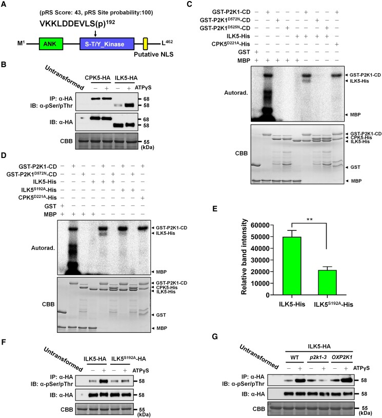
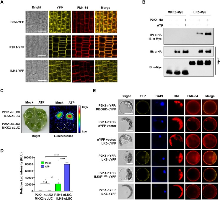
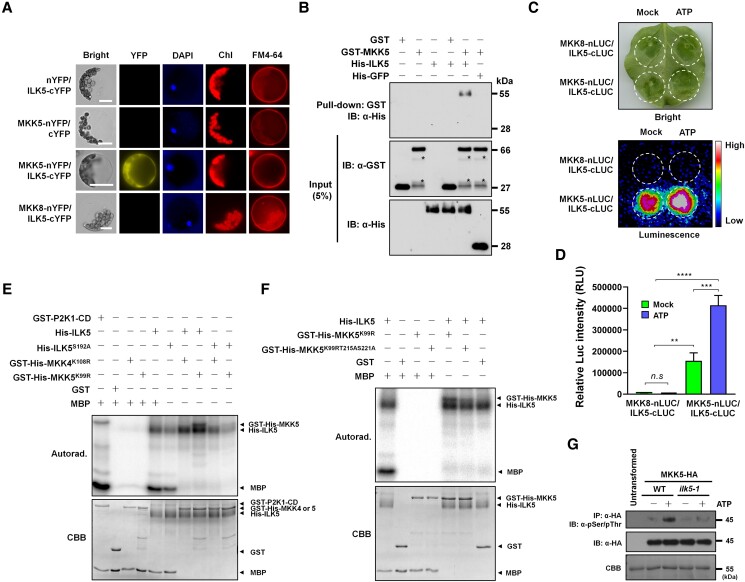
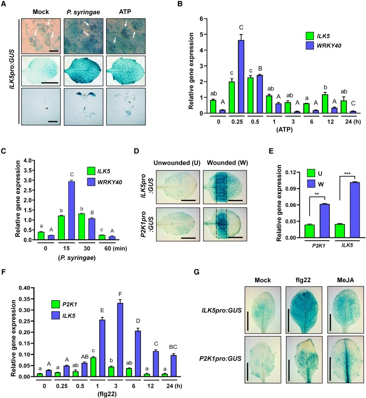
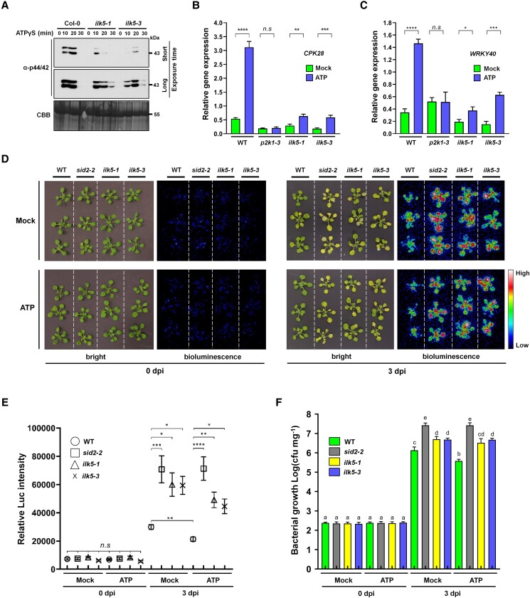
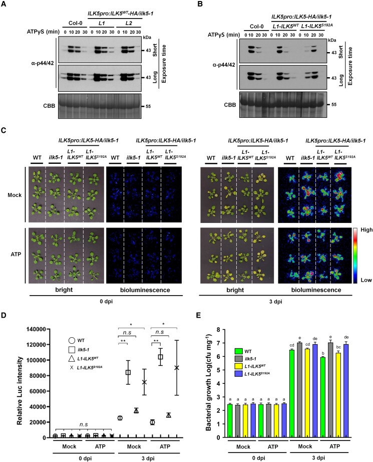
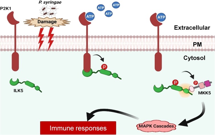
Mitogen-activated protein (MAP) kinase signaling cascades play important roles in eukaryotic defense against various pathogens. Activation of the extracellular ATP (eATP) receptor P2K1 triggers MAP kinase 3 and 6 (MPK3/6) phosphorylation, which leads to an elevated plant defense response. However, the mechanism by which P2K1 activates the MAPK cascade is unclear. In this study, we show that in Arabidopsis thaliana, P2K1 phosphorylates the Raf-like MAP kinase kinase kinase (MAPKKK) INTEGRIN-LINKED KINASE 5 (ILK5) on serine 192 in the presence of eATP. The interaction between P2K1 and ILK5 was confirmed both in vitro and in planta and their interaction was enhanced by ATP treatment. Similar to P2K1 expression, ILK5 expression levels were highly induced by treatment with ATP, flg22, Pseudomonas syringae pv. tomato DC3000, and various abiotic stresses. ILK5 interacts with and phosphorylates the MAP kinase MKK5. Moreover, phosphorylation of MPK3/6 was significantly reduced upon ATP treatment in ilk5 mutant plants, relative to wild-type (WT). The ilk5 mutant plants showed higher susceptibility to P. syringae pathogen infection relative to WT plants. Plants expressing only the mutant ILK5S192A protein, with decreased kinase activity, did not activate the MAPK cascade upon ATP addition. These results suggest that eATP activation of P2K1 results in transphosphorylation of the Raf-like MAPKKK ILK5, which subsequently triggers the MAPK cascade, culminating in activation of MPK3/6 associated with an elevated innate immune response.


Conflict of interest statement

Conflict of interest statement. The authors declare no conflict of interest.

Figures

Figure 1
Figure 1
P2K1 directly phosphorylates ILK5. A) Schematic diagram of ILK5 showing the ankyrin repeat (ANK) and serine-threonine or tyrosine kinase (S-T/Y_kinase) domains. Identified phosphopeptide VKKLDDEVLS(p)192 is located in the kinase domain. B) ILK5 protein is phosphorylated by ATPγS treatment in vivo. ILK5-HA protein is expressed in Arabidopsis protoplasts upon addition of 250 μM ATPγS and subsequently immunoprecipitated (IP) by anti-HA antibody beads and immunoblotting (IB) is carried out with an antiphospho-Ser/Thr antibody. CPK5-HA used as a negative control. C) GST-P2K1-CD directly phosphorylates ILK5-His but not CPK5D221A-HA in vitro. Bacterial recombinant ILK5-His protein was incubated with GST-P2K1 cytosolic domain (GST-P2K1-CD) or two P2K1 kinase dead versions (GST-P2K1D572N-CD and GST-P2K1D525N-CD), or GST in an in vitro kinase assay. D) Mutation of ILK5 on Ser192 residue leads to reduced phosphorylation by P2K1 in vitro. Purified GST or GST-P2K1-CD recombinant protein was incubated with ILK5-His or ILK5S192A-His, followed by an in vitro kinase assay. In panel C and D, auto- and transphosphorylation were detected by incorporation of γ-[32P]-ATP. Myelin basic protein (MBP) and CPK5D221A-His were used as a universal substrate and a negative control, respectively. E) Quantification of phosphorylated ILK5 and ILK5S192A protein. The intensity of the phosphorylation signals of ILK5 and ILK5S192A by P2K1-CD (shown in Supplemental Fig. S1B) were measured and analyzed using the Image J and GraphPad Prism 8 program. Data shown as mean ± SEM, n = 4, **P < 0.01, P-value indicates significance relative to band intensity of ILK5WT-His and was determined by unpaired two-tailed Student's t test. F) Mutation of ILK5 at Ser192 residue results in reduced phosphorylation under ATPγS treatment in vivo. Either WT or S192A of ILK5-hemagglutinin (HA) (ILK5S192A) protein is expressed in protoplasts upon addition of 250 μM ATPγS and subsequently immunoprecipitated with an anti-HA antibody and immunoblotting is carried out with an antiphospho-Ser/Thr antibody. G) ILK5 phosphorylation is dependent on P2K1 protein. ILK5-HA protein was expressed in WT, p2k1-3 and OXP2K1 protoplasts with/without 250 μM ATPγS treatment then subjected to IP and IB by anti-HA and antiphospho-Ser/Thr antibodies. In panel B)-(G), protein loading was visualized by Coomassie Brilliant Blue (CBB) staining. Above experiments were repeated at least two times with similar results.
Figure 2
Figure 2
ILK5 interacts with the P2K1 receptor. A) Subcellular localization of ILK5 in plants expressing a 35S:ILK5-YFP construct. Fluorescent confocal images displaying the subcellular distribution of ILK5-YFP protein were detected from primary root tissue of 7-day-old seedlings. Plasma membrane was counter-stained by incubation for 1 min in a FM4-64 solution (5 μM). P2K1-YFP and free-YFP were used as controls. FM4-64 was used as a plasma membrane marker. Merge indicates overlapped YFP and FM4-64 images. Scale bars = 50 μm. B) Co-immunoprecipitation of P2K1 and ILK5 proteins. The indicated constructs were co-infiltrated and transiently expressed in N. benthamiana leaves after addition of 200 μM ATP (+) for 30 min or MES buffer (pH 5.7) as a mock treatment (−). Total protein was used for Co-IP. Anti-HA and anti-Myc antibodies were used. MKK8-Myc was used as a negative control. C) Split-luciferase experiment showing interaction of P2K1-ILK5 protein with/without ATP treatment. Split-luciferase imaging assay was performed after addition of 200 μM ATP or MES buffer (pH 5.7) as a mock treatment in N. benthamiana leaves co-infiltrated with GV3101 expressing P2K1-nLUC and ILK5-cLUC. 1 mM D-luciferin containing 0.01% silwet-L77 was sprayed onto the N. benthamiana leaves and immediately placed in dark conditions for 10 min to quench the fluorescence. The luminescence was monitored and captured using a low light imaging CCD camera (Photek; Photek, Ltd.). Dotted circles indicate the infiltrated area in N. benthamiana leaves. MKK3-cLUC protein was used as a negative control. D) Quantification of P2K1–ILK5 interaction signal intensities with/without ATP treatment. P2K1–ILK5 interaction was monitored, images were captured, and the luciferase signal intensities were quantified using the C-vision/Im32 and analyzed using the GraphPad Prism 8 program. Data shown as mean ± SEM, n = 4 (biological replicates), ****P < 0.0001, ***P < 0.001, **P < 0.01, *P < 0.05, P-value indicates significance relative to MKK3-cLUC and was determined by unpaired two-tailed Student's t test. E) Biomolecular fluorescence complementation (BiFC) assay in Arabidopsis protoplasts. The indicated constructs were transiently transformed into Arabidopsis protoplasts and then incubated under dark conditions for 24 h. The YFP fluorescence was monitored using a Leica DM 5500B compound microscope with Leica DFC290 color digital camera. DAPI and FM4-64 were used as a nuclear marker and plasma membrane marker, respectively. Chl represents chlorophyl auto-fluorescence signal. Merge indicates overlapped image of YFP and FM4-64. RBOHD-cYFP and ILK6-cYFP were used as positive and negative control, respectively. Scale bars = 10 μm. All above experiments were repeated three times (biological replicates) with similar results.
Figure 3
Figure 3
ILK5 interacts with and phosphorylates MKK5 protein. A) BiFC assay in Arabidopsis protoplasts demonstrating ILK5 interaction with MKK5. The indicated constructs were transiently expressed in Arabidopsis protoplasts and transformed protoplasts were incubated under dark conditions for 24 h before observation YFP signal. The YFP fluorescence was monitored using a Leica DM 5500B compound microscope with Leica DFC290 color digital camera. DAPI was used as a nuclear marker. MKK8-nYFP was used as a negative control. Scale bars = 10 μm. B) ILK5 protein directly interacts with MKK5 protein in vitro. Purified recombinant proteins GST and GST-MKK5 were incubated with His-ILK5 or followed by GST-mediated pull-down. His-GFP protein was used as a negative control. Proteins were detected using a-GST and a-His antibodies. C) ILK5 interaction between MKK5 was demonstrated by split-luciferase imaging with/without ATP treatment. Split-luciferase imaging assay was performed after addition of 200 μM ATP or MES buffer (pH 5.7) as a mock treatment in N. benthamiana leaves co-infiltrated with GV3101 expressing MKK5-nLUC and ILK5-cLUC. Dotted circles indicate the infiltrated area in N. benthamiana leaves. MKK8-nLUC was used as a negative control. D) Quantification of ILK5–MKK5 interaction signal intensities under ATP treatment. ILK5–MKK5 interaction was monitored, images were captured, and luciferase signal intensities were quantified using C-vision/Im32 and analyzed using the GraphPad Prism 8. Data shown as mean ± SEM, n = 7 (biological replicates), ****P < 0.0001, ***P < 0.001, **P < 0.01, P-value indicates significance relative to MKK8-cLUC and was determined by unpaired two-tailed Student's t test. E) MKK5 can be phosphorylated by purified from N. benthamiana recombinant ILK5 and His-ILK5S192A protein displays reduced phosphorylation of MKK5K99R protein in vitro. Purified from N. benthamiana recombinant His-ILK5 or His-ILK5S192A recombinant protein were incubated with GST-His-MKK4K108R or GST-His-MKK5K99R, followed by an in vitro kinase assay. F) ILK5 protein phosphorylates the MKK5 activation loop S/TxxxxxS/T motif. Protein purified from N. benthamiana recombinant His-ILK5 protein was incubated with GST-His-MKK5 kinase dead (GST-His-MKK5K99R) and GST-His-MKK5 triple mutation (GST-His-MKK5K99RT215AS221A) or GST in an in vitro kinase assay. In panel E and F, autophosphorylation and transphosphorylation were detected by incorporation of γ-[32P]-ATP. MBP was used as an universal substrate. Protein loading was visualized by CBB staining. G) MKK5 phosphorylation is significantly reduced in ilk5-1 mutant plants. MKK5-HA protein was expressed in WT and ilk5-1 protoplasts with/without 250 μM ATP treatment, then subjected to IP and IB using anti-HA and antiphospho-Ser/Thr antibodies. Protein loading was visualized by CBB staining. All above experiments were repeated two times with similar results.
Figure 4
Figure 4
ILK5 is highly induced after addition of ATP or P. syringae DC3000 inoculation. A) Histochemical analysis of ILK5 gene expression after ATP treatment or P. syringae DC3000 inoculation. ILK5promoter:GUS transgenic plants were constructed in WT Arabidopsis plants transformed with a chimeric ILK5promoter:GUS including 2 kb of the putative ILK5 promoter (5′ region of the ILK5 gene) fused to the GUS coding sequence. The expression patterns of the ILK5promoter:GUS transgenic plant was detected by histochemical staining in a 10 d-old seedling rosette leaf treated with 200 μM ATP or inoculated with P. syringae DC3000 (OD600 = 0.05) after 1 h. Arrows in upper panel indicate the stained guard cells. Scale bars = upper, 20 μm; middle and bottom, 4 mm. B) Reverse transcription quantitative PCR (RT-qPCR) analysis of ILK5 transcripts after ATP (200 μM) treatment. C) ILK5 gene expression was measured by RT-qPCR analysis in response to P. syringae inoculation. In panel B and C, total RNA was isolated from 10-days old seeding plants at each time point and 2 μg of total RNAs were used in this experiment. The SAND reference gene was used for data normalization. WRKY40 was used as an inducible marker gene for the response to ATP treatment (n = 3) and P. syringae inoculation (n = 3). D) Histochemical analysis of ILK5promoter:GUS and P2K1promoter:GUS expression in response to wounding. The expression patterns of the ILK5promoter:GUS and P2K1promoter:GUS transgenic plants were detected by histochemical staining of 2-wk-old GUS transgenic plants after wounding. Scale bars = 4 mm. E) RT-qPCR analysis of ILK5 and P2K1 gene expression in response to wounding. Data shown as mean ± SEM, n = 2 (biological replicates), ***P < 0.001, ** P < 0.01, P-value was determined and analyzed using the GraphPad Prism 8 by unpaired two-tailed Student's t test. F) RT-qPCR analysis of ILK5 and P2K1 gene expression (n = 3) at different time points in response to flg22 treatment. The SAND reference gene was used for data normalization. G) Histochemical analysis of 10-day-old seedling ILK5promoter:GUS and P2K1promoter:GUS in response to flg22 (100 nM) and MeJA (1 μM). In panel B, C and F, data shown as mean ± SEM, Different letters above the bars indicates significant differences (P < 0.05). P-value was determined and analyzed using the GraphPad Prism 8 by one-way ANOVA followed by Tukey's multiple comparisons. All above experiments were repeated two times (biological replicates) with similar results. Scale bars = 2 mm.
Figure 5
Figure 5
ILK5 is required for plant innate immunity. A) Phosphorylation of MPK3/6 detected in ilk5 mutants by immunoblotting using antiphospho 44/42 antibody. ATPγS (250 μM) was added and incubated for the times shown. Total protein was extracted at each time point and IB was performed with an antiphospho 44/42 MAPK antibody. CBB staining of protein was used as a loading control. These experiments were repeated three times with similar results. B) and C) RT-qPCR analysis of CPK28 and WRKY40 transcripts in p2k1-3, ilk5-1, and ilk5-3 mutant backgrounds after ATP (250 μM) treatment. Total RNA was isolated from 2-wk-old plants and 2 μg of total RNA was used in this experiment. The SAND reference gene was used for data normalization. Data shown as mean ± SEM, WRKY40; (n = 4), CPK28; (n = 6), ****P < 0.0001, ***P < 0.001, **P < 0.01, *P < 0.05, P-value indicates significance relative to mock treatment and was determined and analyzed using the GraphPad Prism 8 by unpaired two-tailed Student's t test. These experiments were repeated three times with similar results. D) Three-wk-old plants were flood inoculated with luxCDABE-tagged P. syringae DC3000 suspension (5 × 10⁶ CFU ml−1) containing 0.025% (v/v) Silwet L-77 with or without the addition of ATP (250 μM). Bioluminescence was detected using a low light capture CCD camera at the time of inoculation (0 d) and 3 d post inoculation. E) Quantification of bioluminescence signal intensities. The signal of luxCDABE-tagged P. syringae DC3000 was monitored, images were captured, and the luciferase signal intensities were quantified using the C-vision/Im32 program (n = 9). F) Bacterial colonization was determined by plate counting (n = 8). In panel E and F, data shown as mean ± SEM, ****P < 0.0001, ***P < 0.001, **P < 0.01, *P < 0.05, P-value and different letters above the bars indicates significance relative to WT plants and was determined and analyzed using the GraphPad Prism 8 by two-way ANOVA followed by Tukey’s multiple comparisons test. These experiments were repeated three times (biological replicates) with similar results.
Figure 6
Figure 6
Expression of the ILK5S192A mutant protein increases plant susceptibility to P. syringae infection. A) Phosphorylation of MPK3/6 detected in complemented lines in which ILK5 expression was driven by the native promoter (L1 and L2) by immunoblotting using antiphospho 44/42 antibody. ATPγS (250 μM) was added and incubated for the times shown. B) Phosphorylation of MPK3/6 was detected in complemented lines where the ILK5WTor ILK5S192A protein were expressed from the ILK5 native promoter in ilk5-1 mutant background (L1-ILK5WT or L1-ILK5S192A). Immunoblotting was performed using antiphospho 44/42 antibody with 250 μM ATP. In A and B, total protein was extracted at each time point after addition of 250 μM ATP. CBB staining of protein was used as a loading control. These experiments were repeated three times with similar results. C) Three-wk-old plants were flood inoculated with luxCDABE-tagged P. syringae DC3000 suspension in the presence of 250 μM ATP. Inoculated plants (Col-0, ilk5-1, L1-ILK5WT-HA, or L1-ILK5S192A-HA complemented lines) were used and bioluminescence was detected using a low light capture CCD camera either at the time of inoculation (0 d) and 3 d post inoculation. D) Quantification of luminescence signal intensities. The signal of luxCDABE-tagged P. syringae DC3000 for each plant was monitored, images were captured, and the luciferase signal intensities were quantified using the C-vision/Im32 program. E) Bacterial colonization was determined by plate counting. In panel C (n = 6) and D (n = 8), data shown as mean ± SEM, ****P < 0.0001, ***P < 0.001, **P < 0.01, *P < 0.05, P-value and different letters above the bars indicates significance relative to WT plants and was determined and analyzed using the GraphPad Prism 8 by two-way ANOVA followed by Tukey’s multiple comparisons test. These experiments were repeated three times (biological replicates) with similar results.
Figure 7.
Figure 7.
Hypothetical model for the role of ILK5 in eATP signaling. Upon addition of the activating ligand eATP, the P2K1 receptor is rapidly auto-phosphorylated and then directly interacts and phosphorylates its downstream target, the ILK5 protein, leading to innate immune response via MAPK cascades.

Similar articles

Cited by

References

    1. Ahsan N, Huang Y, Tovar-Mendez A, Swatek KN, Zhang J, Miernyk JA, Xu D, Thelen JJ. A versatile mass spectrometry-based method to both identify kinase client-relationships and characterize signaling network topology. J Proteome Res. 2013:12(2):937–948. 10.1021/pr3009995 - DOI - PMC - PubMed
    1. Asai T, Tena G, Plotnikova J, Willmann MR, Chiu W-L, Gomez-Gomez L, Boller T, Ausubel FM, Sheen J. MAP Kinase signalling cascade in Arabidopsis innate immunity. Nature. 2002:415(6875):977–983. 10.1038/415977a - DOI - PubMed
    1. Bagchi S, Liao Z, Gonzalez FA, Chorna NE, Seye CI, Weisman GA, Erb L. The P2Y2 nucleotide receptor interacts with αv integrins to activate go and induce cell migration. J Biol Chem. 2005:280(47):39050–39057. 10.1074/jbc.M504819200 - DOI - PubMed
    1. Balagué C, Gouget A, Bouchez O, Souriac C, Haget N, Boutet-Mercey S, Govers F, Roby D, Canut H. The Arabidopsis thaliana lectin receptor kinase LecRK-I.9 is required for full resistance to Pseudomonas syringae and affects jasmonate signalling. Mol Plant Pathol. 2017:18(7):937–948. 10.1111/mpp.12457 - DOI - PMC - PubMed
    1. Besteiro MAG, Ulm R. Phosphorylation and stabilization of Arabidopsis MAP kinase phosphatase 1 in response to UV-B stress*. J Biol Chem. 2013:288(1):480–486. 10.1074/jbc.M112.434654 - DOI - PMC - PubMed

Publication types

MeSH terms

Substances