

Promotion of Joint Degeneration and Chondrocyte Metabolic Dysfunction by Excessive Growth Hormone in Mice
- PMID: 36762426
- PMCID: PMC10313765
- DOI: 10.1002/art.42470
Promotion of Joint Degeneration and Chondrocyte Metabolic Dysfunction by Excessive Growth Hormone in Mice
Abstract
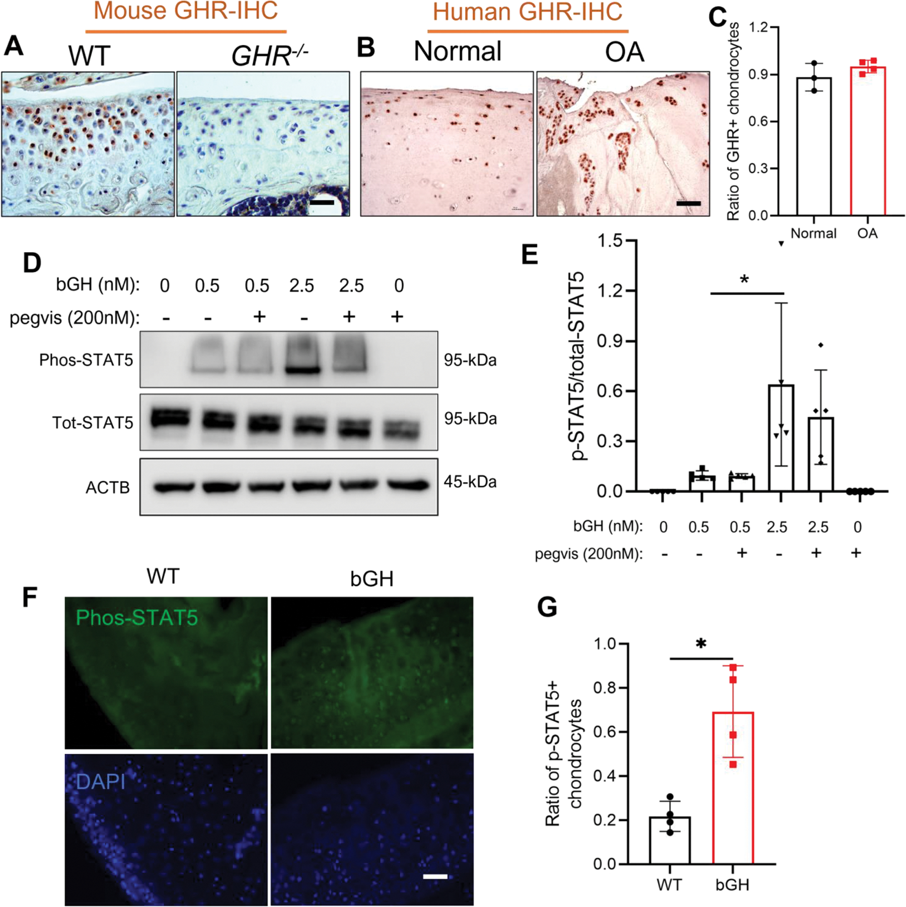
Objective: Many patients with acromegaly, a hormonal disorder with excessive growth hormone (GH) production, report pain in joints. We undertook this study to characterize the joint pathology of mice with overexpression of bovine GH (bGH) or a GH receptor antagonist (GHa) and to investigate the effect of GH on regulation of chondrocyte cellular metabolism.
Methods: Knee joints from mice overexpressing bGH or GHa and wild-type (WT) control mice were examined using histology and micro-computed tomography for osteoarthritic (OA) pathologies. Additionally, cartilage from bGH mice was used for metabolomics analysis. Mouse primary chondrocytes from bGH and WT mice, with or without pegvisomant treatment, were used for quantitative polymerase chain reaction and Seahorse respirometry analyses.
Results: Both male and female bGH mice at ~13 months of age had increased knee joint degeneration, which was characterized by loss of cartilage structure, expansion of hypertrophic chondrocytes, synovitis, and subchondral plate thinning. The joint pathologies were also demonstrated by significantly higher Osteoarthritis Research Society International and Mankin scores in bGH mice compared to WT control mice. Metabolomics analysis revealed changes in a wide range of metabolic pathways in bGH mice, including beta-alanine metabolism, tryptophan metabolism, lysine degradation, and ascorbate and aldarate metabolism. Also, bGH chondrocytes up-regulated fatty acid oxidation and increased expression of Col10a. Joints of GHa mice were remarkably protected from developing age-associated joint degeneration, with smooth articular joint surface.
Conclusion: This study showed that an excessive amount of GH promotes joint degeneration in mice, which was associated with chondrocyte metabolic dysfunction and hypertrophic changes, whereas antagonizing GH action through a GHa protects mice from OA development.
© 2023 The Authors. Arthritis & Rheumatology published by Wiley Periodicals LLC on behalf of American College of Rheumatology.
Conflict of interest statement
Figures


Similar articles
-
Growth hormone-receptor disruption in mice reduces osteoarthritis and chondrocyte hypertrophy.Geroscience. 2024 Oct;46(5):4895-4908. doi: 10.1007/s11357-024-01230-z. Epub 2024 Jun 3. Geroscience. 2024. PMID: 38831184 Free PMC article.
-
Excess Growth Hormone Triggers Inflammation-Associated Arthropathy, Subchondral Bone Loss, and Arthralgia.Am J Pathol. 2023 Jun;193(6):829-842. doi: 10.1016/j.ajpath.2023.02.010. Epub 2023 Mar 3. Am J Pathol. 2023. PMID: 36870529 Free PMC article.
-
The fate of chondrocyte in osteoarthritic cartilage of transgenic mice expressing bovine GH.Osteoarthritis Cartilage. 2004 Jul;12(7):543-51. doi: 10.1016/j.joca.2004.04.002. Osteoarthritis Cartilage. 2004. PMID: 15219569
-
Effects of shear stress on articular chondrocyte metabolism.Biorheology. 2000;37(1-2):95-107. Biorheology. 2000. PMID: 10912182 Review.
-
Molecular regulation of articular chondrocyte function and its significance in osteoarthritis.Histol Histopathol. 2011 Mar;26(3):377-94. doi: 10.14670/HH-26.377. Histol Histopathol. 2011. PMID: 21210351 Review.
Cited by
-
Burn-Induced Gut Microbiota Dysbiosis Aggravates Skeletal Muscle Atrophy by Tryptophan-Kynurenine Mediated AHR Pathway Activation.Adv Sci (Weinh). 2025 Apr;12(14):e2409296. doi: 10.1002/advs.202409296. Epub 2025 Feb 14. Adv Sci (Weinh). 2025. PMID: 39950940 Free PMC article.
-
Growth hormone-receptor disruption in mice reduces osteoarthritis and chondrocyte hypertrophy.Geroscience. 2024 Oct;46(5):4895-4908. doi: 10.1007/s11357-024-01230-z. Epub 2024 Jun 3. Geroscience. 2024. PMID: 38831184 Free PMC article.
-
IGF1 drives Wnt-induced joint damage and is a potential therapeutic target for osteoarthritis.Nat Commun. 2024 Oct 24;15(1):9170. doi: 10.1038/s41467-024-53604-8. Nat Commun. 2024. PMID: 39448593 Free PMC article.
References
-
- Layton MW, Fudman EJ, Barkan A, Braunstein EM, and Fox IH. Acromegalic arthropathy. Characteristics and response to therapy. Arthritis Rheum. 1988;31(8):1022–7. - PubMed
-
- Colao A, Pivonello R, Scarpa R, Vallone G, Ruosi C, and Lombardi G. The acromegalic arthropathy. J Endocrinol Invest. 2005;28(8 Suppl):24–31. - PubMed
-
- Tornero J, Castaneda S, Vidal J, and Herrero-Beaumont G. Differences between radiographic abnormalities of acromegalic arthropathy and those of osteoarthritis. Arthritis Rheum. 1990;33(3):455–6. - PubMed
-
- Kropf LL, Madeira M, Vieira Neto L, Gadelha MR, and de Farias ML. Functional evaluation of the joints in acromegalic patients and associated factors. Clin Rheumatol. 2013;32(7):991–8. - PubMed
-
- Miller A, Doll H, David J, and Wass J. Impact of musculoskeletal disease on quality of life in long-standing acromegaly. Eur J Endocrinol. 2008;158(5):587–93. - PubMed
Publication types
MeSH terms
Substances
Supplementary concepts
Grants and funding
LinkOut - more resources
Full Text Sources
