. 2023 Mar 21;51(5):2464-2484.
doi: 10.1093/nar/gkad056.

A riboswitch separated from its ribosome-binding site still regulates translation

Affiliations

A riboswitch separated from its ribosome-binding site still regulates translation

Griffin M Schroeder et al. Nucleic Acids Res. .

Abstract

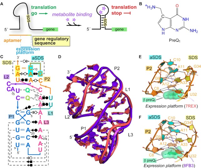
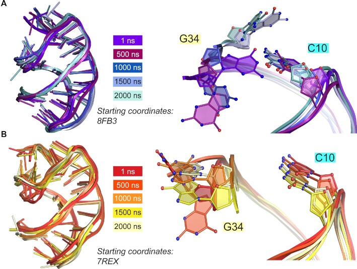
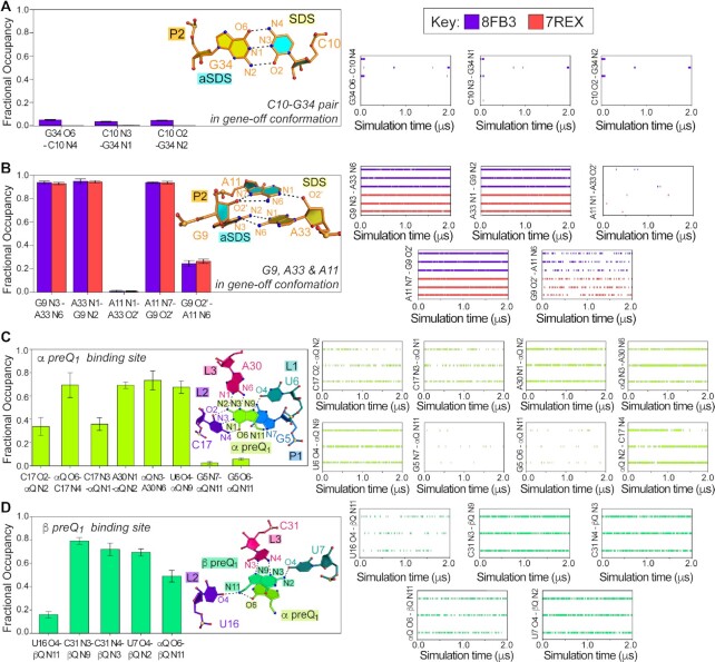
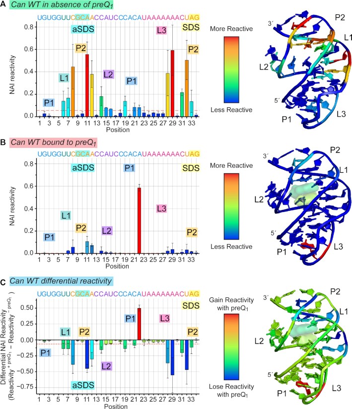
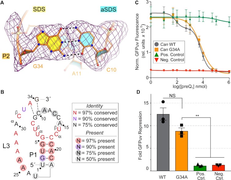
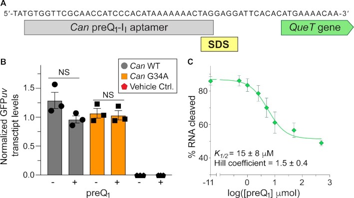
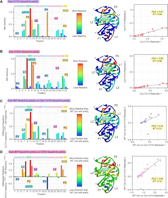
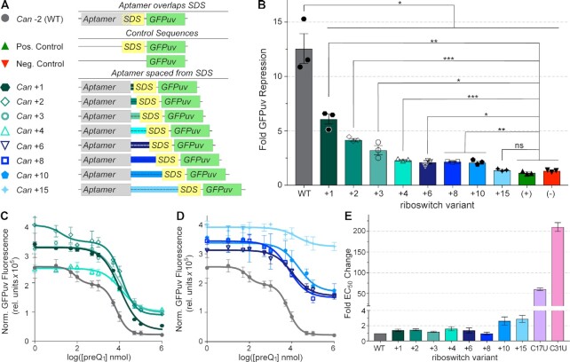
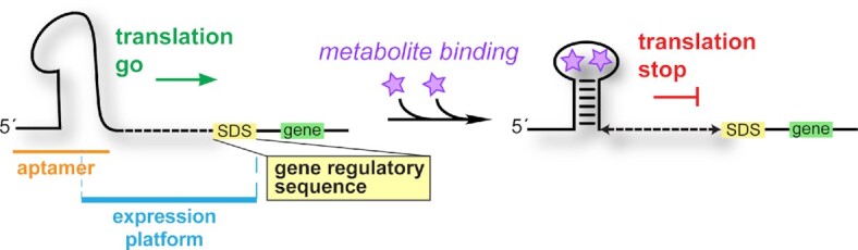
Riboswitches regulate downstream gene expression by binding cellular metabolites. Regulation of translation initiation by riboswitches is posited to occur by metabolite-mediated sequestration of the Shine-Dalgarno sequence (SDS), causing bypass by the ribosome. Recently, we solved a co-crystal structure of a prequeuosine1-sensing riboswitch from Carnobacterium antarcticum that binds two metabolites in a single pocket. The structure revealed that the second nucleotide within the gene-regulatory SDS, G34, engages in a crystal contact, obscuring the molecular basis of gene regulation. Here, we report a co-crystal structure wherein C10 pairs with G34. However, molecular dynamics simulations reveal quick dissolution of the pair, which fails to reform. Functional and chemical probing assays inside live bacterial cells corroborate the dispensability of the C10-G34 pair in gene regulation, leading to the hypothesis that the compact pseudoknot fold is sufficient for translation attenuation. Remarkably, the C. antarcticum aptamer retained significant gene-regulatory activity when uncoupled from the SDS using unstructured spacers up to 10 nucleotides away from the riboswitch-akin to steric-blocking employed by sRNAs. Accordingly, our work reveals that the RNA fold regulates translation without SDS sequestration, expanding known riboswitch-mediated gene-regulatory mechanisms. The results infer that riboswitches exist wherein the SDS is not embedded inside a stable fold.


Figures

Figure 1.
Figure 1.
Gene regulatory scheme for a dual-ligand riboswitch, the preQ1 metabolite and diagrams of Carnobacterium antarcticum (Can) preQ1-II riboswitch co-crystal structures. (A) Mechanism of action of a dual metabolite binding riboswitch in which the Shine-Dalgarno sequence (SDS) is buried upon ligand (star) binding. (B) Chemical diagram of preQ1. (C) Secondary structure of the Can riboswitch based on the co-crystal structure of this investigation. The Leontis-Westhof symbols (88) are shown. (D) All-atom superposition between the new Can riboswitch structure of this investigation and the previous structure (34). (E) Close-up stick model of the expression platform from 7REX. Rings of the SDS are filled yellow; aSDS rings are filled cyan. (F) Expression platform of the new Can riboswitch structure 8FB3 from this study.
Figure 2.
Figure 2.
Position G34 is highly flexible in molecular dynamics (MD) trajectories while C10 remains stationary. (A) Superposition of global Can riboswitch coordinates from 1, 500, 1000, 1500 and 2000 ns timepoints from a representative MD trajectory starting from PDB code 8FB3 (left); close-up view of superimposed C10-G34 interaction at the same timepoints (right). (B) Same as (b) but with a representative trajectory from 7REX starting coordinates.
Figure 3.
Figure 3.
Hydrogen bonds in molecular dynamics simulations show dissolution of the C10–G34 aSDS–SDS pair but preservation of interactions involving A33 and interactions to preQ1 in the binding pocket. (A) Bar plots show average MD trajectory occupancies (n = 6) for the C10–G34 interaction of the Can riboswitch; here and elsewhere the ball-and-stick diagram is from the starting co-crystal structure of this investigation (PDB code 8FB3). Bar plots and occupancy versus simulation time plots show interactions derived the new Can riboswitch structure (PDB code 8FB3, purple) and the previous structure (PDB code 7REX, red). (B) Bar plots showing average MD trajectory occupancies (n = 6) for the G9•A33•A11 interaction. Occupancy versus simulation time plots are as described in panel A. (C) Bar plots showing average MD trajectory occupancies (8FB3 starting coordinates, n = 6) for preQ1 in the α-site of the binding pocket interacting with nearby nucleotides. Occupancy versus simulation time plots correspond to individual trajectories for the new Can riboswitch structure (PDB code 8FB3). (D) Bar plots showing average MD trajectory occupancies (8FB3 starting coordinates, n = 6) for preQ1 in the β-site of the binding pocket interacting with nearby nucleotides and the α-site preQ1. Occupancy versus simulation time plots are as described in panel C.
Figure 4.
Figure 4.
Chemical-modification profiles of the WT Can riboswitch show preQ1-dependent flexibility losses in live cells. (A) ReCo-icSHAPE-seq profile in live bacteria grown in the absence of preQ1 and modified in cells by NAI. The acylation reactivities represent the average of biological replicates (n = 2). Here and elsewhere, nucleotides are colored based on their location within the pseudoknot described in Figure 1C. The average reactivity at each nucleotide was heat-mapped onto the co-crystal structure of this investigation. In this plot and others, the dashed red line shows the standard deviation of reactivity from helical regions, which represents a minimal acylation baseline. (B) ReCo-icSHAPE-seq profile as described in (A), except cells were grown in the presence of 70 μM preQ1. PreQ1 molecules depicted as surface models were not analyzed by chemical modification but are shown to emphasize their presence during RNA modification. (C) Average differential SHAPE-seq reactivity based on profiles from (A) and (B). The Pearson coefficient (PCC) of biological replicates in each condition is shown in Supplementary Figure S9.
Figure 5.
Figure 5.
The C10–G34 base pair in the expression platform is not conserved and is dispensable for gene-regulatory function. (A) Close-up view of the WT Can riboswitch co-crystal structure (PDB code 8FB3) showing the C10–G34 canonical pair between aSDS and SDS sequences. The mFo – DFc polder electron density map (89) at 3.0 Å resolution for chain A is contoured at 3.0σ; the nucleotides from the final model (shown) were omitted from the phase calculation. (B) Consensus model for the preQ1-II (class I type I) riboswitch mapped onto the Can riboswitch sequence used in this investigation. The model is adapted from (36). (C) Dose–response analysis of the WT Can preQ1 riboswitch and the G34A mutant. Normalized GFPuv fluorescence emission measured from live cells is shown as a function of preQ1 added into the growth medium. The positive control is missing the Can riboswitch before an intact SDS; the negative control contains an inverted aSDS sequence in place of the SDS. (D) Fold repression of the GFPuv reporter gene based on assays in (C). EC50 and fold repression values for each construct are provided in Supplementary Table S3.
Figure 6.
Figure 6.
The Can riboswitch blocks ribosome assembly but does not affect transcription levels to achieve gene regulation. (A) Genomic sequence of Can (GenBank: CP010796.1) from nucleotide position 2093555 to 2093612. The sequences corresponding to the Can riboswitch aptamer, Shine-Dalgarno sequence (SDS) and start codon of the downstream preQ1 transport gene, queT (36) are shown. (B) qPCR analysis of GFPuv transcripts controlled by the Can riboswitch. QueF knockout E. coli cells containing Can WT, G34A or no plasmid (vehicle) were grown in the presence and absence of preQ1 with whole cell RNA extracted and transcript levels quantified. All transcripts were quantified relative to transcript levels of the cysG reference gene and then normalized to levels of ampR derived from the same low-copy-number plasmid used to express GFPuv (see Materials and Methods and Supplementary Figure S2). Transcript analysis is the average of biological replicates and error bars correspond to S.E.M. (n = 3 biological replicates). (C) RelE cleavage assay showing preQ1-dependent ribosome assembly on a transcript containing the WT Can riboswitch joined to the downstream queT ORF. The plot of cleavage versus preQ1 was fit to an isotherm that resulted in a Hill coefficient of 1.5 ± 0.4. Error bars correspond to S.E.M (n = 3 biological replicates).
Figure 7.
Figure 7.
Chemical-modification profiles of mutant Can riboswitches show higher reactivity than WT in the presence of preQ1. (A) Acylation reactivities of bound-state C17U mutants derived from ReCo-icSHAPE-seq. In-cell reactivity values were heat-mapped onto the Can riboswitch co-crystal structure of this investigation (PDB code 8FB3). In this plot and others, the dashed red line shows the standard deviation of reactivity from helical regions, which represents a minimal acylation baseline. The Pearson Correlation Coefficient (PCC) of biological replicates (n = 2) is shown (right) along with the fit to a line. (B) Acylation reactivities of bound-state C31U as described in panel A. (C) Differential SHAPE-seq reactivity based on the C17U reactivity profile from (A). Positive values indicate higher reactivity for the mutant compared to WT, suggesting greater flexibility. The corresponding PCC is shown. (D) Differential SHAPE-seq reactivity based on the C31U profile from (B) calculated as in (C); the corresponding PCC is shown. A22 in the tight P1–L3 turn loses reactivity in the mutant. Additional reactivity profiles for each mutant in the presence of preQ1 and differential reactivity plots compared to WT Can are shown in Supplementary Figure S6.
Figure 8.
Figure 8.
WT Can riboswitches separated from the SDS by unstructured spacers support preQ1-dependent regulation of GFPuv gene expression. (A) Organization of the WT riboswitch showing overlap between the aptamer and SDS. Unstructured spacers (dashed lines) were inserted between the 3′-end of the WT riboswitch and the naturally occurring downstream SDS. The GFPuv reporter gene was described previously (24,57). (B) Fold repression of the WT riboswitch relative to variants with increasing spacer lengths. Student t-tests show significance of repression for various confidence intervals; ns means not significant. Controls are described in Figure 5C. (C, D) Normalized GFPuv fluorescence versus preQ1 dose response curves for spacer variants in panel A. (E) Fold change in preQ1 EC50 from the dose response curves in panels C and D; the C17U and C31U mutant data were adapted from (34) (https://creativecommons.org/licenses/by/4.0/). EC50 and fold repression values for each construct are provided in Supplementary Table S3.
Figure 9.
Figure 9.
Proposed translation regulation by a metabolite-sensing riboswitch that attenuates translation without SDS sequestration. The mechanism of action recognizes that the ligand-dependent aptamer structure interferes with translation in a manner that does not require the SDS to engage in a mutually exclusive conformational change. The efficacy of gene regulation depends upon the proximity of the SDS to the ligand-dependent aptamer fold. The expression platform includes a spacer sequence (dashed line) and the SDS.

Similar articles

Cited by

References

    1. McCown P.J., Corbino K.A., Stav S., Sherlock M.E., Breaker R.R.. Riboswitch diversity and distribution. RNA (New York, N.Y.). 2017; 23:995–1011. - PMC - PubMed
    1. Breaker R.R. Riboswitches and the RNA world. Cold Spring Harb. Perspect. Biol. 2012; 4:a003566. - PMC - PubMed
    1. Nahvi A., Sudarsan N., Ebert M.S., Zou X., Brown K.L., Breaker R.R.. Genetic control by a metabolite binding mRNA. Chem. Biol. 2002; 9:1043. - PubMed
    1. Breaker R.R. The biochemical landscape of riboswitch ligands. Biochemistry. 2022; 61:12. - PMC - PubMed
    1. Motika S.E., Ulrich R.J., Geddes E.J., Lee H.Y., Lau G.W., Hergenrother P.J.. A Gram-negative antibiotic active through inhibition of an essential riboswitch. J. Am. Chem. Soc. 2020; 142:10856–10862. - PMC - PubMed

Publication types