

Brain monitoring after cardiac arrest
- PMID: 36762679
- PMCID: PMC9994800
- DOI: 10.1097/MCC.0000000000001023
Brain monitoring after cardiac arrest
Abstract
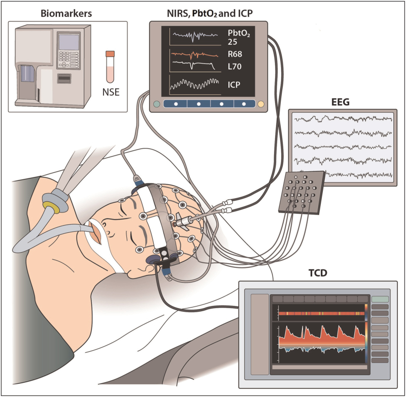
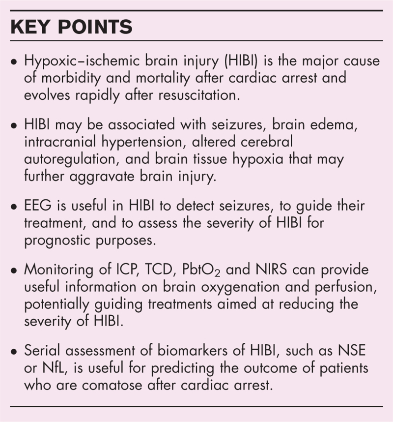
Purpose of review: To describe the available neuromonitoring tools in patients who are comatose after resuscitation from cardiac arrest because of hypoxic-ischemic brain injury (HIBI).
Recent findings: Electroencephalogram (EEG) is useful for detecting seizures and guiding antiepileptic treatment. Moreover, specific EEG patterns accurately identify patients with irreversible HIBI. Cerebral blood flow (CBF) decreases in HIBI, and a greater decrease with no CBF recovery indicates poor outcome. The CBF autoregulation curve is narrowed and right-shifted in some HIBI patients, most of whom have poor outcome. Parameters derived from near-infrared spectroscopy (NIRS), intracranial pressure (ICP) and transcranial Doppler (TCD), together with brain tissue oxygenation, are under investigation as tools to optimize CBF in patients with HIBI and altered autoregulation. Blood levels of brain biomarkers and their trend over time are used to assess the severity of HIBI in both the research and clinical setting, and to predict the outcome of postcardiac arrest coma. Neuron-specific enolase (NSE) is recommended as a prognostic tool for HIBI in the current postresuscitation guidelines, but other potentially more accurate biomarkers, such as neurofilament light chain (NfL) are under investigation.
Summary: Neuromonitoring provides essential information to detect complications, individualize treatment and predict prognosis in patients with HIBI.
Copyright © 2023 The Author(s). Published by Wolters Kluwer Health, Inc.
Conflict of interest statement
Figures
References
-
- Nolan JP, Sandroni C, Bottiger BW, et al. . European Resuscitation Council and European Society of Intensive Care Medicine guidelines 2021: postresuscitation care. Intensive Care Med 2021; 47:369–421. - PMC - PubMed
-
The 2021 Guidelines on Post-Resuscitation Care resulted from a wide expert consensus and a collaboration between the European Resuscitation Council and the European Society of Intensive Care Medicine. The section on prognostication after cardiac arrest is based on a systematic review of 94 studies (2013-2020).
-
- Alkhachroum A, Appavu B, Egawa S, et al. . Electroencephalogram in the intensive care unit: a focused look at acute brain injury. Intensive Care Med 2022; 48:1443–1462. - PMC - PubMed
-
This extensive narrative review written by a group of major experts in the field describes the clinical applications of the EEG in intensive care.
-
- Hirsch LJ, Fong MWK, Leitinger M, et al. . American Clinical Neurophysiology Society's Standardized Critical Care EEG terminology: 2021 version. J Clin Neurophysiol 2021; 38:1–29. - PMC - PubMed
-
This updated edition of the ACNS terminology, originally published in 2013, represents a standard for the definition and the nomenclature of the EEG patterns in critical care.
Publication types
MeSH terms
Substances
LinkOut - more resources
Full Text Sources
Medical
Research Materials
