Review
. 2023 Jun 8;72(3):164-177.
doi: 10.1093/jmicro/dfad014.

HIV-1 transmission: modelling and direct visualization in the third dimension

Affiliations
Review

HIV-1 transmission: modelling and direct visualization in the third dimension

Charles A Coomer et al. Microscopy (Oxf). .

Abstract

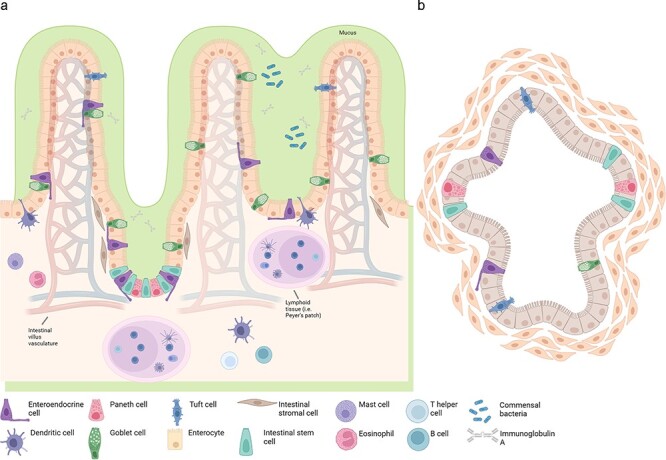
Identifying initial events of mucosal entry of human immunodeficiency virus type-1 (HIV-1) in laboratory-based, physiologically relevant and high-throughput contexts may aid in designing effective strategies to block local transmission and spread of HIV-1. Several paradigms have been posited for how HIV-1 crosses mucosal barriers to establish infection based on two dimensional (2D) culture-based or animal-based models. Nevertheless, despite these models stemming from 2D culture and animal studies, monolayers of cells poorly replicate the complex niche that influences viral entry at mucosal surfaces, whereas animal models often inadequately reproduce human disease pathophysiology and are prohibitively expensive. Organoids, having never been directly utilized in HIV-1 transmission investigations, may offer a compromise between 2D culture and animal models as they provide a platform that mimics the biophysical and biochemical niche of mucosal tissues. Importantly, observation of events downstream of viral inoculation is potentially accessible to researchers via an array of microscopy techniques. Because of the potential insights organoids may provide in this context, we offer this review to highlight key physiological factors of HIV-1 transmission at common mucosal sites and a discussion to highlight how many of these factors can be recapitulated in organoids, their current limitations and what questions can initially be addressed, particularly using a selective inclusion of quantitative light microscopy techniques. Harnessing organoids for direct observation of HIV-1 entry at mucosal sites may uncover potential therapeutic targets which prevent the establishment of HIV-1 infection.

Keywords: HIV-1 entry; imaging HIV-1 in tissue; mucosal immunity; two-photon FLIM metabolism; two-photon microscopy.


Figures

Fig. 1.
Fig. 1.
Current biological experimental systems and their relative level of increasing complexity in reproducing human infectious pathology. Schematic representation of the current culturing techniques available and their ability to reproduce the complexity of human–HIV interactions: from 2D monolayer cancer cell lines to human studies. Human cancer cell lines have played a vital fundamental role in determining underpinning pathways involved in HIV transmission and have been critical in screening and characterizing therapeutic candidates to prevent or treat acute HIV infection. Additionally, these cells have been utilized to investigate how HIV-1 may interact with epithelial cells at the mucosal barrier. Similarly, primary cells isolated from human tissue or organoid cultures have also been paramount in generating more physiologically relevant data to answer similar questions. Nevertheless, these two models fail to recapitulate the in vivo 3D microenvironment, induce cell differentiation, possess natural tissue-specific cell heterogeneity and perform tissue-specific functions. Oftentimes, many of these limitations are bypassed using other models such as fixed tissue explants or animal models in order to get closer to mimicking human HIV-1 pathophysiology. Nevertheless, fixed tissue models often only offer indirect evidence to support certain investigations and the fixing and explanting process may alter variables (e.g. cell migration) critical for the particular scientific question. Investigations using NHP models are dependent on the species, viral strains used for infection, genetic pedigree of the individual monkeys and humanized murine models are met with challenges regarding poor lymph node development, humoral responses, engraftment and species-specific cytokines. To balance this spectrum of experimental platforms, their benefits and drawbacks, organoids enable the mimicry of the intricate 3D organization and architecture of tissue, tissue-specific functions, natural host microbiota and the cell heterogeneity found in natural tissue in addition to being reproducible and subject to live-cell imaging. Therefore, organoids offer a compromise on the tissue culture platform spectrum between 2D monotypic cancer cell lines and living organisms.
Fig. 2.
Fig. 2.
Key routes of HIV-1 penetration in the FGT. The human vagina and ectocervix are insulated by a multi-layered stratified, non-keratinized squamous epithelium. Nevertheless, HIV-1 can penetrate the FGT via multitudinous mechanisms, the following of which that are mentioned form a non-exhaustive list. (a) Free or cell-associated HIV virions may become trapped in cervicovaginal mucous. These mucous-trapped cells and virions may then subsequently have sufficient exposure time to the epithelial surface, such that free virions or virions produced by attached HIV-infected donor cells may navigate through gaps between epithelial cells, be captured and transcytosed by epithelial cells or be captured and trafficked to endocytic compartments by epithelial-resident Langerhans cells. (b) Free virions or virions from infected donor cells may fuse with intraepithelial CD4+ T cells. (c) Cell-associated virions may be transmitted to epithelial cells which may then be transcytosed to later target and infect underlying stromal macrophages. Cell-associated virions may also be transmitted to CD4+ T cells via the virological synapse. (d) During sexual intercourse, physical abrasions of the epithelium commonly occur, often in regions where stromal papillae projections, which are enriched in immune cells, nearly reach the luminal surface. Importantly, potentially free virions or cell-associated virions from infected donor cells may pass through these abrasions to directly contact the plethora of target cells within the mucosa or stroma. Because resident HIV-1 target cells cluster in these regions, numerous infection foci are created. (e) Stromal projections also are docking points for lymphatic and arteriovenous vasculature and can therefore transport cell-free or cell-associated virions to the systemic circulation or local lymph nodes. (f) Conflicting reports indicate that it is possible that free HIV-1 virions may transcytose through or productively basal epithelial cells throughout the FGT, including uterine epithelial cells (not pictured). (g) Viruses within the stroma are capable of being taken up by or (conflictingly) productively infect stromal DCs in order to subsequently transfer virions to susceptible CD4+ T cells via the viral synapse, followed by an explosive increase in productive infection, whereby generated virions can continue to infect other susceptible cells (e.g. macrophages). (h) Cytokines produced by stromal leucocytes and epithelial cells in response to commensal bacteria or sexually transmitted infections may act to recruit HIV-1 susceptible target cells to increase the target population of susceptible leucocytes. (i) The junction between the stratified squamous epithelial barrier that characterizes the ectocervix and the high-turnover, single-layer columnar epithelial layer of the endocervix, is known as the transformation zone. Because of its delicate monolayer, the endocervix may be the preferential site of HIV-1 entry in the FGT.
Fig. 3.
Fig. 3.
Comparison of intestinal tissue explants vs. their organoid counterparts. (a) Structure and cell type specification of explanted intestinal epithelium. The intestinal epithelium consists of stem cells, Paneth cells (small intestine), goblet cells, tuft cells, enteroendocrine cells, enterocytes (small intestine)/colonocytes (colon), stromal cells and microfold cells (not pictured) overlying lymphoid tissue. Importantly, explanted tissue often temporarily preserves lymphatic tissue and microvasculature (pictured), however immune cells often emigrate out of the explants early. (b) Pluripotent stem-cell-derived intestinal organoids, although containing intestinal epithelium consists of stem cells and their derivatives, in addition to mesenchymal cells, mucous and any co-cultured cells (which varies and is thus, not pictured). Created in BioRender.
Fig. 4.
Fig. 4.
Principle of two-photon microscopy. (a) Two-photon microscopy (right panel) offers a number of advantages as compared with conventional sinlge-photon excitation approaches (left panel). In two-photon microscopy, the transition between the highest occupied molecular orbital toward the lowest unoccupied molecular orbital is excited by two photons simultaneously. The total energy of these two photons is the same as the one needed for one-photon excitation events (which depends on the particular fluorophore to be excited/studied). Longer wavelengths imply lower energy and therefore less phototoxicity. Also, less scatter is obtained with two- or three-photon microscopy enabling deeper tissue imaging (b).
Fig. 5.
Fig. 5.
Principle of two-photon microscopy coupled to TCSPC FLIM. Coupling of two-photon excitation with TCSPC FLIM allows measurement of endogenous-free and -bound NAD(P)H relative concentrations in live cells and tissue. The optical path of a typical two-photon FLIM microscope is shown. A Ti:Sa pulsed femtosecond laser is directed toward the dichroic and is also coupled to a photo-diode and TCSPC electronics. The emitted photons arrive to the photon counting detector with a particular delay relative to the two-photon femtosecond pulsed (normally tuned at 80 MHz, i.e. one pulse every 12.5 ns). The computer provides the fluorescence decay or the right coordinates for the phasor plot (right panels). For non-fitting approaches, only around 100 photons per pixel are enough to recover the average lifetime and the minimal fraction of interacting donor if interested in detecting FRET between two fluorophores (donor and acceptor); for instance, FRET-based biosensors.
Fig. 6.
Fig. 6.
Real-time single virus tracking in live cells. (a) HIV-1 pseudoviruses can be labelled with lipophilic dyes such as DiD in the envelope and with Gag-eGFP that is cleaved in mature viruses. When hemifusion occurs in the plasma membrane, DiD signal dilutes and vanishes and the tracked HIV-1 particle changes colour from yellow to green. If full fusion occurs, the cleaved eGFP monomers are released to the cytosol and the green signal disappears. If the HIV-1 particle fuses within endosomal compartments, the DiD signal remains in the endosomal membrane upon hemifusion. Full fusion is detected when the eGFP signal disappears, and therefore the particle changes from yellow to red. (b) Example of TZM-bl cells exposed to double-labelled HIV-1 viruses. Scale bar = 10 μm. (c) Examples of double-labelled HIV-1 particles either exposed to viruses at 4°C and subject to spinoculation (2100 G, left panels) where endocytosis occurs for both HIV-1 particles pseudotyped with JRFL Env and VSG-G Env and only exposed to double-labelled HIV-1 virions exposed at 4°C (right panels). TZM-bl cells were expressing Rab5-mCherry to highlight early endosomal compartments. The micrographs were 40 × 40 μm.

Similar articles

References

    1. UNAIDS (2016) Global AIDS Update 2016. World Health Organization, 422.
    1. Amico P et al. (2010) HIV spending as a share of total health expenditure: an analysis of regional variation in a multi-country study. PLoS One 5: 1–8. - PMC - PubMed
    1. Shattock R J et al. (2011) AIDS. Turning the tide against HIV. Science (1979) 333: 42–43. - PubMed
    1. Xu H et al. (2013) Mucosal immunology of HIV infection. Immunol. Rev. 254: 10–33. - PMC - PubMed
    1. Fackler O T et al. (2014) Adding new dimensions: towards an integrative understanding of HIV-1 spread. Nat. Rev. Microbiol. 12: 563–571. - PMC - PubMed