

2023 update of template tables for reporting biomolecular structural modelling of small-angle scattering data
- PMID: 36762858
- PMCID: PMC9912924
- DOI: 10.1107/S2059798322012141
2023 update of template tables for reporting biomolecular structural modelling of small-angle scattering data
Abstract
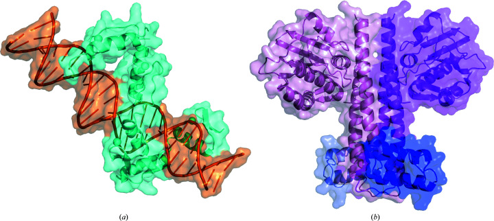
In 2017, guidelines were published for reporting structural modelling of small-angle scattering (SAS) data from biomolecules in solution that exemplified best-practice documentation of experiments and analysis. Since then, there has been significant progress in SAS data and model archiving, and the IUCr journal editors announced that the IUCr biology journals will require the deposition of SAS data used in biomolecular structure solution into a public archive, as well as adherence to the 2017 reporting guidelines. In this context, the reporting template tables accompanying the 2017 publication guidelines have been reviewed with a focus on making them both easier to use and more general. With input from the SAS community via the IUCr Commission on SAS and attendees of the triennial 2022 SAS meeting (SAS2022, Campinas, Brazil), an updated reporting template table has been developed that includes standard descriptions for proteins, glycosylated proteins, DNA and RNA, with some reorganization of the data to improve readability and interpretation. In addition, a specialized template has been developed for reporting SAS contrast-variation (SAS-cv) data and models that incorporates the additional reporting requirements from the 2017 guidelines for these more complicated experiments. To demonstrate their utility, examples of reporting with these new templates are provided for a SAS study of a DNA-protein complex and a SAS-cv experiment on a protein complex. The examples demonstrate how the tabulated information promotes transparent reporting that, in combination with the recommended figures and additional information best presented in the main text, enables the reader of the work to readily draw their own conclusions regarding the quality of the data and the validity of the models presented.
Keywords: SANS; SAXS; biomolecular structural modelling; contrast variation; small-angle scattering; template tables.
open access.
Figures
Similar articles
-
2017 publication guidelines for structural modelling of small-angle scattering data from biomolecules in solution: an update.Acta Crystallogr D Struct Biol. 2017 Sep 1;73(Pt 9):710-728. doi: 10.1107/S2059798317011597. Epub 2017 Aug 18. Acta Crystallogr D Struct Biol. 2017. PMID: 28876235 Free PMC article.
-
Small Angle Scattering and Structural Biology: Data Quality and Model Validation.Adv Exp Med Biol. 2018;1105:77-100. doi: 10.1007/978-981-13-2200-6_7. Adv Exp Med Biol. 2018. PMID: 30617825 Review.
-
Publication guidelines for structural modelling of small-angle scattering data from biomolecules in solution.Acta Crystallogr D Biol Crystallogr. 2012 Jun;68(Pt 6):620-6. doi: 10.1107/S0907444912012073. Epub 2012 May 17. Acta Crystallogr D Biol Crystallogr. 2012. PMID: 22683784
-
Recent advances in small-angle scattering and its expanding impact in structural biology.Structure. 2022 Jan 6;30(1):15-23. doi: 10.1016/j.str.2021.09.008. Epub 2021 Dec 13. Structure. 2022. PMID: 34995477 Review.
-
Report of the wwPDB Small-Angle Scattering Task Force: data requirements for biomolecular modeling and the PDB.Structure. 2013 Jun 4;21(6):875-81. doi: 10.1016/j.str.2013.04.020. Structure. 2013. PMID: 23747111 Review.
Cited by
-
Design of intrinsically disordered protein variants with diverse structural properties.Sci Adv. 2024 Aug 30;10(35):eadm9926. doi: 10.1126/sciadv.adm9926. Epub 2024 Aug 28. Sci Adv. 2024. PMID: 39196930 Free PMC article.
-
Selected advances in small-angle scattering and applications they serve in manufacturing, energy and climate change.J Appl Crystallogr. 2023 May 29;56(Pt 3):787-800. doi: 10.1107/S1600576723003898. eCollection 2023 Jun 1. J Appl Crystallogr. 2023. PMID: 37284276 Free PMC article.
-
Structural dynamics of IDR interactions in human SFPQ and implications for liquid-liquid phase separation.Acta Crystallogr D Struct Biol. 2025 Jul 1;81(Pt 7):357-379. doi: 10.1107/S2059798325005303. Epub 2025 Jun 27. Acta Crystallogr D Struct Biol. 2025. PMID: 40574713 Free PMC article.
-
Iron-sensing and redox properties of the hemerythrin-like domains of Arabidopsis BRUTUS and BRUTUS-LIKE2 proteins.Nat Commun. 2025 Apr 24;16(1):3865. doi: 10.1038/s41467-025-58853-9. Nat Commun. 2025. PMID: 40274781 Free PMC article.
-
Structural basis for the recognition and ubiquitylation of type-2 N-degron substrate by PRT1 plant N-recognin.Nat Commun. 2025 Aug 21;16(1):7817. doi: 10.1038/s41467-025-63282-9. Nat Commun. 2025. PMID: 40841552 Free PMC article.
References
-
- Aalst, W. M. P. van der, Bichler, M. & Heinzl, A. (2017). Bus. Inf. Syst. Eng. 59, 311–313.
-
- Arnold, O., Bilheux, J. C., Borreguero, J. M., Buts, A., Campbell, S. I., Chapon, L., Doucet, M., Draper, N., Ferraz Leal, R., Gigg, M. A., Lynch, V. E., Markvardsen, A., Mikkelson, D. J., Mikkelson, R. L., Miller, R., Palmen, K., Parker, P., Passos, G., Perring, T. G., Peterson, P. F., Ren, S., Reuter, M. A., Savici, A. T., Taylor, J. W., Taylor, R. J., Tolchenov, R., Zhou, W. & Zikovsky, J. (2014). Nucl. Instrum. Methods Phys. Res. A, 764, 156–166.
-
- Bergmann, A., Orthaber, D., Scherf, G. & Glatter, O. (2000). J. Appl. Cryst. 33, 869–875.
MeSH terms
Substances
LinkOut - more resources
Full Text Sources
