. 2023 Feb 23;66(4):2773-2788.
doi: 10.1021/acs.jmedchem.2c01837. Epub 2023 Feb 10.

Lessons for Oral Bioavailability: How Conformationally Flexible Cyclic Peptides Enter and Cross Lipid Membranes

Affiliations

Lessons for Oral Bioavailability: How Conformationally Flexible Cyclic Peptides Enter and Cross Lipid Membranes

Stephanie M Linker et al. J Med Chem. .

Abstract

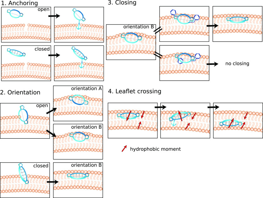
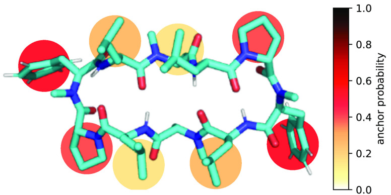
Cyclic peptides extend the druggable target space due to their size, flexibility, and hydrogen-bonding capacity. However, these properties impact also their passive membrane permeability. As the "journey" through membranes cannot be monitored experimentally, little is known about the underlying process, which hinders rational design. Here, we use molecular simulations to uncover how cyclic peptides permeate a membrane. We show that side chains can act as "molecular anchors", establishing the first contact with the membrane and enabling insertion. Once inside, the peptides are positioned between headgroups and lipid tails─a unique polar/apolar interface. Only one of two distinct orientations at this interface allows for the formation of the permeable "closed" conformation. In the closed conformation, the peptide crosses to the lower leaflet via another "anchoring" and flipping mechanism. Our findings provide atomistic insights into the permeation process of flexible cyclic peptides and reveal design considerations for each step of the process.


Conflict of interest statement

The authors declare no competing financial interest.

Figures

Figure 1
Figure 1
Summary of the four steps for the passive membrane permeation of conformationally flexible cyclic peptides. The peptide is shown in blue and the membrane in orange. Peptides in the closed conformation are indicated by their intramolecular hydrogen bonds (dashed lines). Side-chain residues that anchor the peptide in the membrane are depicted with an anchor symbol. The hydrophobic moment is pointing from the polar to the apolar part of a molecule.
Figure 2
Figure 2
(A) Backbone scaffold and amino acid composition of the cyclic decapeptide (CDP) series used in this work. The colored residues were systematically replaced according to Table 1. The backbone scaffold was reported by Fouché et al., and is kept constant. (B) Schematic workflow showing the different conditions, total simulation time, and number of observed events. Unbiased MD simulations were used to elucidate the membrane permeation pathway of CDPs. Biasing along steps of this pathway was used to enrich sampling and to obtain starting structures for new unbiased simulations.
Figure 3
Figure 3
(A) Snapshot of CDP 1 directly before anchoring to the membrane. The atoms of the POPC headgroups are colored in blue; the atoms of the tails are colored in orange. Thermal fluctuations of the lipids can lead to temporary headgroup gaps and the apolar tails underneath become exposed (in this snapshot the gaps manifest as orange patches). If the CDP is close to such a transient gap, its apolar side chains can “anchor” to the lipid membrane. (B) Trajectory of CDP 1 entering a membrane. The z-position of the CDP is indicated with black dots. The position of the headgroup and tail region are indicated with dark gray and light gray lines, respectively. The angle between the normal vectors of the peptide and the membrane is shown in blue. The RMSD with respect to the closed conformation of the CDP is shown in orange. (C) Snapshot of the CDP while anchoring to the membrane. A hydrogen bond between the CDP backbone and the polar headgroup atoms can stabilize the anchoring. (D) During the anchoring process, three consecutive hydrogen bonds are formed. The distance between the hydrogen-bond pairs are shown in red. The z-positions of the CDP and the membrane are shown as in panel B.
Figure 4
Figure 4
“Anchor quality” of the different side chains for the CDPs in open (left) and closed (right) conformations. The anchor probability was determined as the fraction of successful anchoring events after pulling the CDPs on that respective side chain toward the membrane. The probabilities are averaged over the eight CDPs.
Figure 5
Figure 5
(A) Representation of how cyclic peptides insert into lipid membranes using the example of CDP 3. The coordinates of the peptide are projected onto its distance from the bilayer center and its orientation with respect to the membrane plane. The heatmap shows the distribution of the simulation time spent in this phase space with darker color corresponding to more simulation time. Regions of interest are highlighted with simulation snapshots. The regions corresponding to orientation A and orientation B are marked with a dotted box. (B) Visualization of Table 2. The amino acid composition of the CDPs determines their preference for orientation A or B; proline and phenylalanine residues favor orientation B, and leucine residues favor orientation A.
Figure 6
Figure 6
Representative snapshots of orientation A and B using the example of CDP 3. The peptide is shown at the polar/apolar interface of the membrane and in a top view. To reach orientation B from orientation A, the peptide rotates by roughly 180° around its major axis. In addition, the ϕ-angles of leucine residues 3 and 8 show a ∼160° shift such that the leucine side chains are approximately aligned with the lipid tails. A cartoon was added for visual guidance.
Figure 7
Figure 7
(A) Representative closing simulation using the example of CDP 6. The peptide starts in an open conformation and closes inside the membrane via a half-closed structure. The closing of the peptide is traced by its RMSD with respect to the closed reference conformation (orange line). Inlays show selected simulation snapshots. Shaded areas correspond to the time point of the inlays. The dotted line indicates the z-position of the peptide. The membrane position is shown for reference (gray). The peptide stays in orientation B for the whole simulation (blue line). (B) Heatmap comparison of the orientation/position of all simulation frames in open conformations (here for CDP 6) versus the orientation/position of the 12 closing trajectories. Whereas open peptides prefer orientation A, all open peptides that close during our simulations originate from orientation B.
Figure 8
Figure 8
Phenylalanine can act as a lock that prevents closing in the membrane. (left) Phenylalanine can adopt two distinct positions in orientation B. In the unlocked position, both phenylalanine residues point toward the aqueous phase. In the “locked” position, at least one phenylalanine residue is rotated and points toward the membrane center. All closing events originate from the unlocked position. (right) Simulation of CDP 8 that shows an unlocking and a closing event. The closing of the peptide is traced by the RMSD with respect to the closed reference (orange line). The red line indicates the relative position of phenylalanine at position 5 with respect to the ring plane of the peptide. Inlays show selected simulation snapshots. Shaded areas correspond to the time point of the inlays. The dotted line indicates the z-position of the peptide. The membrane position is shown for reference (gray). The peptide first adopts orientation B in the locked position after entering the membrane. After a rotation of phenylalanine residue 5 to the unlocked position, the peptide starts closing.
Figure 9
Figure 9
(A) CDP 3 crossing from the upper to the lower leaflet. Inlays show representative simulation snapshots. Shaded areas correspond to the time point of the inlays. Upon passing the membrane center, the peptide undergoes a flip along its major axis (blue line). (B) Zoom-in on the peptide anchoring in the lower leaflet. Lipid tails from the upper and lower leaflet are colored differently to help distinguishing the two leaflets.
Figure 10
Figure 10
Ability of the different side chains of the CDPs to anchor in the lower membrane leaflet when the peptide is inserted in the upper leaflet. Membrane crossings were observed only for peptides in the closed conformation. The anchor probability was determined as the fraction of successful anchoring events after pulling the CDPs on that respective side chain toward the lower leaflet.
Figure 11
Figure 11
Schematic free-energy surfaces and the corresponding rate-limiting barriers for the impermeable CDPs 1 and 8 (conformational closure for CDP 1 and anchoring for CDP 8). The free energies are projected to the conformation of the peptide and its position with respect to the membrane. Design advice for lowering barriers and thus improving the passive permeability are indicated.

References

    1. Yudin A. K. Macrocycles: Lessons from the Distant Past, Recent Developments, and Future Directions. Chem. Sci. 2015, 6, 30–49. 10.1039/C4SC03089C. - DOI - PMC - PubMed
    1. Mallinson J.; Collins I. Macrocycles in New Drug Discovery. Future Med. Chem. 2012, 4, 1409–1438. 10.4155/fmc.12.93. - DOI - PubMed
    1. Giordanetto F.; Kihlberg J. Macrocyclic Drugs and Clinical Candidates: What Can Medicinal Chemists Learn from Their Properties?. J. Med. Chem. 2014, 57, 278–295. 10.1021/jm400887j. - DOI - PubMed
    1. Villar E. A.; Beglov D.; Chennamadhavuni S.; Porco J. A.; Kozakov D.; Vajda S.; Whitty A. How Proteins Bind Macrocycles. Nature Chem. Biol. 2014, 10, 723–731. 10.1038/nchembio.1584. - DOI - PMC - PubMed
    1. Ermert P. Design, Properties and Recent Application of Macrocycles in Medicinal Chemistry. CHIMIA 2017, 71, 678–702. 10.2533/chimia.2017.678. - DOI - PubMed

Publication types

LinkOut - more resources