. 2023 May;165(4):536-549.
doi: 10.1111/jnc.15781. Epub 2023 Mar 1.

Apolipoprotein E ε4 modulates astrocyte neuronal support functions in the presence of amyloid-β

Affiliations

Apolipoprotein E ε4 modulates astrocyte neuronal support functions in the presence of amyloid-β

Rebecca M Fleeman et al. J Neurochem. 2023 May.

Abstract

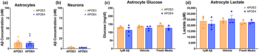
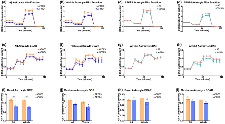
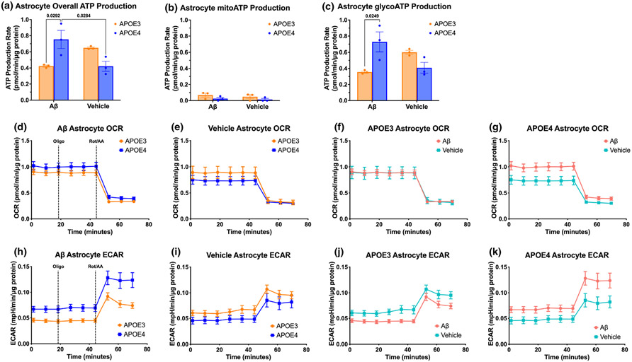
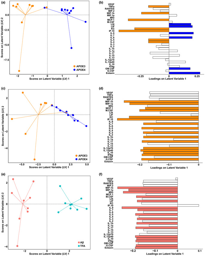
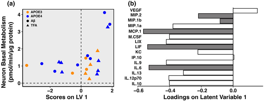
Apolipoprotein E (APOE) is a lipid transporter produced predominantly by astrocytes in the brain. The ε4 variant of APOE (APOE4) is the strongest and most common genetic risk factor for Alzheimer's disease (AD). Although the molecular mechanisms of this increased risk are unclear, APOE4 is known to alter immune signaling and lipid and glucose metabolism. Astrocytes provide various forms of support to neurons, including regulating neuronal metabolism and immune responses through cytokine signaling. Changes in astrocyte function because of APOE4 may therefore decrease neuronal support, leaving neurons more vulnerable to stress and disease insults. To determine whether APOE4 alters astrocyte neuronal support functions, we measured glycolytic and oxidative metabolism of neurons treated with conditioned media from APOE4 or APOE3 (the common, risk-neutral variant) primary astrocyte cultures. We found that APOE4 neurons treated with conditioned media from resting APOE4 astrocytes had similar metabolism to APOE3 neurons treated with media from resting APOE3 astrocytes, but treatment with astrocytic conditioned media from astrocytes challenged with amyloid-β (Aβ), a key pathological protein in AD, caused APOE4 neurons to increase their basal mitochondrial and glycolytic metabolic rates more than APOE3 neurons. These changes were not because of differences in astrocytic lactate production or glucose utilization, but instead correlated with increased glycolytic ATP production and a lack of cytokine secretion in response to Aβ. Additionally, we identified that astrocytic cytokine signatures could predict basal metabolism of neurons treated with the astrocytic conditioned media. Together, these findings suggest that in the presence of Aβ, APOE4 astrocytes alter immune and metabolic functions that result in a compensatory increase in neuronal metabolic stress.

Keywords: ATP; amyloid-β; astrocytes; cytokines; glucose; glycolysis; immunometabolism.


Conflict of interest statement

CONFLICT OF INTEREST STATEMENT

The authors declare that they have no conflict of interests with the content of this article.

Figures

FIGURE 1
FIGURE 1
APOE4 neurons increase oxidative and glycolytic output in response to ACM from Aβ-treated astrocytes. Mitochondrial stress test oxygen consumption rates (OCRs) of APOE3 and APOE4 neurons treated for 72 hours with (a) fresh neuronal media, (b) vehicle-treated astrocyte conditioned media (ACM), (c) 1 μM Aβ-treated ACM, or (d) glial media that had not been on cells. Neurons were treated with sequential injections of mitochondrial inhibitors oligomycin A (Oligo), FCCP, and rotenone/antimycin A (Rot/AA). Corresponding extracellular acidification rates (ECARs) of mitochondrial stress tests for APOE3 and APOE4 neurons treated for 72 hours with (e) fresh neuronal media, (f) vehicle-treated ACM, (g) 1 μM Aβ-treated ACM, or (h) glial media that had not been on cells. Quantifications of basal OCR (i) and ECAR (j) of neuronal mitochondrial stress tests. N of 7 to 8 individual neonates from 3 or more separate litters per group for all experiments. (i, j) Unpaired t-test, two-tailed, df = 13, (i) t = 1.452, p = 0.170; (j) t = 1.479, p = 0.163.
FIGURE 2
FIGURE 2
Aβ internalization, glucose consumption, and lactate production are similar in APOE3 and APOE4 astrocytes. (a) Concentration of Aβ remaining after 72-h incubation of 1 μM Aβ on astrocytes. (b) Concentration of Aβ remaining after 72-h incubation of Aβ-treated ACM on neurons. Levels of glucose (c) and lactate (d) in astrocyte media after 72-h treatment with 1 μM Aβ, vehicle, or fresh glial media. N of 8 to 10 individual neonates from 3 or more separate litters per group (a, b), N of 3 to 4 (c, d). (a, b) Unpaired t-test, two-tailed, (a) df = 16, t = 0.170, p = 0.867; (b) df = 5, t = 0.522, p = 0.624. (c, d) Two-way ANOVA; (c) df = 14, ptreatment = 0.358, pgenotype = 0.068, pinteraction = 0.885; (d) df = 12, ptreatment = 0.544, pgenotype = 0.949, pinteraction = 0.679.
FIGURE 3
FIGURE 3
APOE4 astrocytes have lower metabolic function than APOE3 astrocytes but increased glycolytic response to Aβ. Mitochondrial stress test OCR (a, b) and ECAR (e, f) of APOE3 and APOE4 astrocytes treated for 72 h with 1 μM Aβ or vehicle. APOE3 mitochondrial stress test OCR (c) and ECAR (g) of astrocytes treated for 72 h with 1 μM Aβ or Vehicle. APOE4 mitochondrial stress test OCR (d) and ECAR (h) of astrocytes treated for 72 h with 1 μM Aβ or Vehicle. Quantifications of APOE3 and APOE4 basal and maximal OCR (i, j) and ECAR (k, l). N of 3 individual neonates per group. (i–k) Two-way ANOVA, df = 8; (i) ptreatment = 0.449, pgenotype <0.001, pinteraction = 0.602, followed by Tukey's test; (j) ptreatment = 0.047, pgenotype = 0.003, pinteraction = 0.975, followed by Tukey's test; (k) ptreatment = 0.904, pgenotype = 0.408, pinteraction = 0.314; (l) ptreatment = 0.261, pgenotype = 0.084, pinteraction = 0.491.
FIGURE 4
FIGURE 4
APOE4 astrocytes increase glycolytic ATP production more than APOE3 astrocytes. Total (a), mitochondrial (b), and glycolytic (c) ATP production rate comparisons between APOE3 and APOE4 astrocytes treated with 1 μM Aβ or Vehicle. OCR (d–g) and ECAR (h–k) of Seahorse ATP rate tests on APOE3 and APOE4 astrocytes treated for 72 h with 1 μM Aβ, with serial injections of mitochondrial inhibitors oligo and Rot/AA. N of three individual neonates per group. (a–c) Two-way ANOVA, df = 8; (a) ptreatment = 0.436, pgenotype = 0.450, pinteraction = 0.003, followed by Tukey's test; (b) ptreatment = 0.453, pgenotype = 0.087, pinteraction = 0.841; (c) ptreatment = 0.613, pgenotype = 0.238, pinteraction = 0.004, followed by Tukey's test.
FIGURE 5
FIGURE 5
Cytokine signatures of APOE4 astrocytes demonstrate a lack of response to pathological Aβ. Partial least squares discriminant analysis (PLSDA) of vehicle-treated APOE3 and APOE4 ACM, separated by genotype (accuracy: 70.17% (5LV); confidence: 95.06%), scores plot (a) and loadings (b). Cytokines with an above average contribution to the predictive accuracy of our model, calculated by variable importance in projection scores (VIPs), are highlighted in orange. PLSDA scores (c) and loadings (d) of 1 μM Aβ-treated APOE3 and APOE4 ACM, separated by genotype (accuracy: 72.13% (2LV); confidence: 98.80%), with VIPs highlighted in orange. PLSDA scores (e) and loadings (f) of APOE3 ACM, separated by treatment with 1 μM Aβ or vehicle (accuracy: 67.22% (5LV); confidence: 96.39%), VIPs highlighted in salmon. N of 9 to 10 individual neonates per group. Accuracy determined by cross-validation (CV) with one-third of the data; confidence determined by comparing predictive CV accuracy of our model to the distribution of CV accuracies of 100 random models.
FIGURE 6
FIGURE 6
Astrocytic cytokine signatures predict basal metabolism of neurons treated with ACM. (a) Scores plot and (b) loadings for partial least squares regression (PLSR) of APOE3 and APOE4 ACM cytokine protein levels against basal neuron metabolism (as measured by oxygen consumption rate) of corresponding ACM-treated neurons (root mean squared error of cross-validation (RMSECV) of 0.765 (1LV); Confidence: 100%). Cytokines with an above average contribution to our model, calculated by variable importance in projection score, highlighted in gray; N = 22 biological replicates. Accuracy determined by performing cross-validation (CV) with one-third of the data; confidence determined by comparing predictive CV accuracy of our model to the distribution of CV accuracies of 100 random models.

Similar articles

Cited by

References

    1. Allaman I, Gavillet M, Belanger M, Laroche T, Viertl D, Lashuel HA, & Magistretti PJ (2010). Amyloid-β aggregates cause alterations of astrocytic metabolic phenotype: Impact on neuronal viability. Journal of Neuroscience, 30, 3326–3338. - PMC - PubMed
    1. Arnaud L, Benech P, Greetham L, Stephan D, Jimenez A, Jullien N, García-González L, Tsvetkov PO, Devred F, Sancho-Martinez I, Izpisua Belmonte JC, Baranger K, Rivera S, & Nivet E (2022). APOE4 drives inflammation in human astrocytes via TAGLN3 repression and NF-κB activation. Cell Reports, 40, 111200. - PubMed
    1. Batiuk MY, Martirosyan A, Wahis J, de Vin F, Marneffe C, Kusserow C, Koeppen J, Viana JF, Oliveira JF, Voet T, Ponting CP, Belgard TG, & Holt MG (2020). Identification of region-specific astrocyte subtypes at single cell resolution. Nature Communications, 11(1), 1–15. - PMC - PubMed
    1. Bayraktar OA, Bartels T, Holmqvist S, Kleshchevnikov V, Martirosyan A, Polioudakis D, Ben Haim L, AMH Y, Batiuk MY, Prakash K, Brown A, Roberts K, Paredes MF, Kawaguchi R, Stockley JH, Sabeur K, Chang SM, Huang E, Hutchinson P, … Rowitch DH (2020). Astrocyte layers in the mammalian cerebral cortex revealed by a single-cell in situ transcriptomic map. Nature Neuroscience, 23(4), 500–509. - PMC - PubMed
    1. Beattie EC, Stellwagen D, Morishita W, Bresnahan JC, Ha BK, Von Zastrow M, Beattie MS, & Malenka RC (2002). Control of synaptic strength by glial TNFα. Science, 1979(295), 2282–2285. - PubMed

Publication types