. 2023 Feb 10;18(2):e0277478.
doi: 10.1371/journal.pone.0277478. eCollection 2023.

Expression, purification, and characterisation of the p53 binding domain of Retinoblastoma binding protein 6 (RBBP6)

Affiliations

Expression, purification, and characterisation of the p53 binding domain of Retinoblastoma binding protein 6 (RBBP6)

Bonnie L Russell et al. PLoS One. .

Abstract

RBBP6 is a 250 kDa eukaryotic protein known to be a negative regulator of p53 and essential for embryonic development. Furthermore, RBBP6 is a critical element in carcinogenesis and has been identified as a potential biomarker for certain cancers. RBBP6's ability to interact with p53 and cause its degradation makes it a potential drug target in cancer therapy. Therefore, a better understating of the p53 binding domain of RBBP6 is needed. This study presents a three-part purification protocol for the polyhistidine-tagged p53 binding domain of RBBP6, expressed in Escherichia coli bacterial cells. The purified recombinant domain was shown to have structure and is functional as it could bind endogenous p53. We characterized it using clear native PAGE and far-UV CD and found that it exists in a single form, most likely monomer. We predict that its secondary structure is predominantly random coil with 19% alpha-helices, 9% beta-strand and 14% turns. When we exposed the recombinant domain to increasing temperature or known denaturants, our investigation suggested that the domain undergoes relatively small structural changes, especially with increased temperature. Moreover, we notice a high percentage recovery after returning the domain close to starting conditions. The outcome of this study is a pure, stable, and functional recombinant RBBP6-p53BD that is primarily intrinsically disordered.


Conflict of interest statement

The authors have declared that no competing interests exist.

Figures

Fig 1
Fig 1. Recombinant expression of RBBP6 p53BD.
SDS-PAGE analysis of expression of the RBBP6-p53BD showing soluble "sup" and insoluble "Pe" fractions for (A): BL21 (DE3) cells over post-induction time of 2 to 16 hours, (B): BL21 (DE3) cells inducer concentrations from 0.1 mM to 1.0 mM, (C): NiCo21 (DE3) cells over post-induction time of 2 to 16 hours, (D): NiCo21 (DE3) cells inducer concentrations from 0.1mM to 1.0mM. (E): Shuffle® T7 express cells over post-induction time of 2 to 16 hours, (F): Shuffle® T7 express inducer concentrations from 0.1 mM to 1.0 mM. The position of overexpressed protein bands is marked with pink arrows. The protein molecular weight marker (Thermo Scientific, 26610) has sizes in kDa, marked on the gel.
Fig 2
Fig 2. Protein conformation of RBBP6-p53BD.
(A): Shows western blot analysis of supernatant fractions of cell lysate from transformed Shuffle® T7 express, BL21 (DE3), and NiCo21 (DE3) E.coli cell lines using an anti-polyhistidine antibody. The RBBP6-p53BD is shown with a pink box. (B): After SDS-PAGE the two protein bands indicated (red and green boxes) were isolated for trypsin digested LC-MS analysis after partial purification of the supernatant from NiCo21 (DE3) cells. The protein molecular weight marker (Thermo Scientific, 26610) has sizes in kDa, marked on the gel.
Fig 3
Fig 3. Three-part purification of the RBBP6-p53BD.
Chromatograph with A280 effluent (blue), elution gradient (green), conductivity (red) and Flow-through (FT), elution and fractions collected (FX) depicted for (A): Part 1, hydrophobic interaction chromatography with 1 M ammonium sulphate in binding buffer (B): Part 2 hydrophobic interaction chromatography with 1.4 M ammonium sulphate in binding buffer and (C): Part 3, nickel IMAC chromatography. SDS-PAGE analysis of protein sample loaded onto the column, the Flow-through (FT), and certain fractions collected "FX" where X represents the fraction number for (D): Part 1, (E): Part 2, and (F): Part 3. The RBBP6-p53BD is indicated in flow-through (FT) or fractions (FX) with a pink box. The protein molecular weight marker (Thermo Scientific, 26610) has sizes in kDa, marked on the gel.
Fig 4
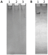
Fig 4. Clear native PAGE analysis of the RBBP6-p53BD.
Clear native PAGE analysis of purified RBBP6-p53BD. (A): Tris-HCl buffer, with pH 8.3. Lane 1 is the RBBP6-p53BD in 50 mM sodium phosphate, pH 7. Lane 2 is the RBBP6-p53BD in 50 mM sodium phosphate, pH 7 after being heated to and cooled from 90°C to 20°C, as described in section 2.8. Lane 3 is the RBBP6-p53BD in 50 mM sodium phosphate, pH 7 with 2 M urea. (B): Imidazole-HEPES buffer, pH7.4. Lane 1 is the RBBP6-p53BD in 50 mM sodium phosphate, pH 7, and Lane 2 is the RBBP6-p53BD in 50 mM sodium phosphate, pH 7 after being heated to and cooled from 90°C to 20°C, as described in section 2.8. Protein samples were not equalised before loading onto the gels.
Fig 5
Fig 5. Far-UV CD spectrum of the RBBP6-p53BD.
Far UV CD spectra of the RBBP6-p53BD, showing 250 nm to 195 nm for 2.6 μM RBBP6-p53BD in 20 mM sodium phosphate, pH 7.
Fig 6
Fig 6. I-TASSER software prediction of secondary and tertiary structure.
Top three 3D conformation models predicted by I–TASSER, with predicted c-Score shown on each model. Alpha-helices are shown in red, beta-strands in yellow, and loops shown in green. The structure was obtained from the protein data bank (PDB) files produced by I-TASSER and viewed with PyMOL Molecular Graphics System (2.5.2 Edu), Schrödinger, LLC.
Fig 7
Fig 7. Far-UV circular dichroism monitoring of thermal unfolding of RBBP6 p53BD.
(A): Far UV CD spectrum for the RBBP6-p53BD from 20°C and 90°C (dark blue to dark red). (B): Spectrum for the RBBP6-p53BD in the native state (blue), when heated to 90°C (red) and cooled back to 20°C (black). The heat unfolding curves for the RBBP6-p53BD at (C): 222nm, (D): 215nm, and (E): 208nm between 20°C and 90°C.
Fig 8
Fig 8. RBBP6 p53BD in the presence of denaturants.
(A): Far-UV CD Spectra between 250 nm and 195 nm for the RBBP6-p53BD from 0 to 8 M urea (B): Far UV-CD curves at 215 nm and 222 nm for 0 to 8 M urea. (C): Far-UV CD Spectra between 250 nm and 195 nm for the RBBP6-p53BD from 0 to 6M guanidinium chloride. (D): Far UV-CD curves at 215 nm and 22 2nm for 0 to 6 M guanidinium chloride.
Fig 9
Fig 9. Western blot analysis of Co-IP assays performed using anti-p53 and anti-polyhistidine on HEK293 T cells.
Two separate Western blots were performed to investigate the presence of endogenous p53 and recombinant polyhistidine-tagged RBBP6 p53BD in Co-IP assays performed using protein A agarose with anti-p53 (lanes 2 and 3) and anti-polyhistidine (lanes 4 and 5) as probes. Lane 1 contains the HEK293 T cells cell lysate with purified RBBP6-p53BD which was used in Co-IP assays. Lanes 2 contains the flow-through from the anti-p53 Co-IP assay which consists of the proteins in the cell lysate that did not bind with the p53 antibody. Lanes 3 contains the immuno-complexes isolated during the anti-p53 Co-IP assays. Lanes 4 contains the flow-through from the anti-polyhistidine Co-IP assay which consists of the proteins in the cell lysate that did not bind with the polyhistidine antibody. Lane 3 contains the immuno-complexes isolated during the anti-polyhistidine Co-IP assay. Lane 6 contains the PBS collected from washing the agarose beads before elution for both Co-IP assays combined. Lane 7 contains purified RBBP6-p53BD as a control. Lane 3 in both blots was overexposed, and therefore a shorter exposure time was used for this lane and overlaid on top.

Similar articles

Cited by

References

    1. Pardee AB. G1 events and regulation of cell proliferation. Science. 1989;246(4930):603–8. doi: 10.1126/science.2683075 - DOI - PubMed
    1. Hartwell LH, Weinert TA. Checkpoints: controls that ensure the order of cell cycle events. Science. 1989;246(4930):629–34. doi: 10.1126/science.2683079 - DOI - PubMed
    1. Haupt Y, Maya R, Kazaz A, Oren M. Mdm2 promotes the rapid degradation of p53. Nature. 1997;387(6630):296–9. doi: 10.1038/387296a0 - DOI - PubMed
    1. Poyurovsky MV, Katz C, Laptenko O, Beckerman R, Lokshin M, Ahn J, et al.. The C terminus of p53 binds the N-terminal domain of MDM2. Nat Struct Mol Biol. 2010;17(8):982–9. doi: 10.1038/nsmb.1872 - DOI - PMC - PubMed
    1. Honda R, Tanaka H, Yasuda H. Oncoprotein MDM2 is a ubiquitin ligase E3 for tumor suppressor p53. FEBS Lett. 1997;420(1):25–7. doi: 10.1016/s0014-5793(97)01480-4 - DOI - PubMed

Publication types

MeSH terms