. 2023 Feb 10;18(2):e0281069.
doi: 10.1371/journal.pone.0281069. eCollection 2023.

An in vitro carcinogenesis model for cervical cancer harboring episomal form of HPV16

Affiliations

An in vitro carcinogenesis model for cervical cancer harboring episomal form of HPV16

Weerayut Wongjampa et al. PLoS One. .

Abstract

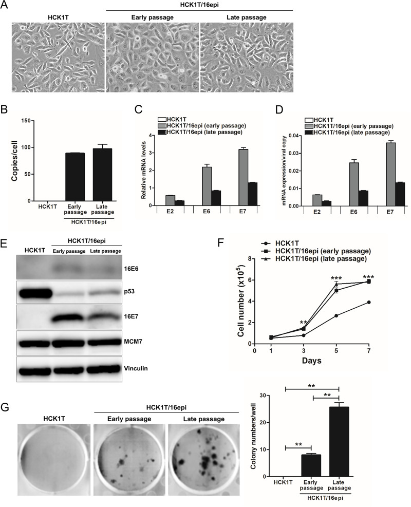
Deregulated expression of viral E6 and E7 genes often caused by viral genome integration of high-risk human papillomaviruses (HR-HPVs) into host DNA and additional host genetic alterations are thought to be required for the development of cervical cancer. However, approximately 15% of invasive cervical cancer specimens contain only episomal HPV genomes. In this study, we investigated the tumorigenic potential of human cervical keratinocytes harboring only the episomal form of HPV16 (HCK1T/16epi). We found that the HPV16 episomal form is sufficient for promoting cell proliferation and colony formation of parental HCK1T cells. Ectopic expression of host oncogenes, MYC and PIK3CAE545K, enhanced clonogenic growth of both early- and late-passage HCK1T/16epi cells, but conferred tumor-initiating ability only to late-passage HCK1T/16epi cells. Interestingly, the expression levels of E6 and E7 were rather lower in late-passage than in early-passage cells. Moreover, additional introduction of a constitutively active MEK1 (MEK1DD) and/or KRASG12V into HCK1T/16epi cells resulted in generation of highly potent tumor-initiating cells. Thus an in vitro model for progression of cervical neoplasia with episomal HPV16 was established. In the model, constitutively active mutation of PIK3CA, PIK3CAE545K, and overexpression of MYC, in the cells with episomal HPV16 genome were not sufficient, but an additional event such as activation of the RAS-MEK pathway was required for progression to tumorigenicity.


Conflict of interest statement

The authors have declared that no competing interests exist.

Figures

Fig 1
Fig 1. Characterization of the HCK1T cells harboring episomal form of HPV16 genome.
(A) Cell morphology of HCK1T/HPV16epi at early and late passage was compared to parental HCK1T cells. Scale bars represent 50 μm. (B) The copy number of HPV16 was measured by quantitative PCR using specific primers to the E6 and L2 ORFs of HPV16. Relative expression levels of HPV16 E6 and E7 mRNA in HCK1T/16epi, showing levels overall (C) and per viral copy, calculated using the following formula: Mean relative mRNA expression/mean relative HPV16 copy number (D). (E) Expression of HPV16 E6 and E7 oncoproteins was detected by western blotting using specific anti-HPV16 E6 and E7 antibodies and the levels of E6 and E7 were compared between early- and late-passage HCK1T/16epi cells. Parental HCK1T cells were included as a negative control. Levels of p53 and MCM7 proteins were also determined to show that E6 and E7 from episomal HPV16 genomes are able to reduce p53 or increase MCM7, a robust indicator of E7 activity [31], respectively. Vinculin was included as a loading control. The ability of the episomal form of HPV16 to promote cell transformation in HCK1T/16epi cells relative to the parental HCK1T cell line was demonstrated by examining growth curves and clonogenic potential. (F) Growth curves of early- as well as late-passage HCK1T/16epi cells and parental HCK1T cells. (G) Clonogenic potential of HCK1T/16epi and parental HCK1T cells. Each bar represents the mean of triplicate values ± SEM. **P ≤ 0.01, ***P ≤ 0.001.
Fig 2
Fig 2. Establishment of an in vitro model for cervical cancer with cells harboring episomal form of HPV16 genome and expression of defined oncogenes.
HCK1T/16epi cells transduced with indicated oncogenes were incubated with or without 1 μg/ml doxycycline (DOX) for 4 days. (A) Cell morphology of HCK1T/16epi cells did not change after expression of the indicated oncogenes. Scale bars represent 50 μm. (B) The expression of individual transgenes including MYC and PIK3CAp110 and their downstream target proteins including AKT and p-AKT were detected by western blotting. The expression levels of HPV16 E6 and E7 proteins in HCK1T/16epi cells were also examined. Vinculin was included as a loading control. (C) Tumor-promoting potential of the MYC and PIK3CAE545K oncogenes were compared between early and late passage of HCK1T/16epi cells by mouse xenografts. 1x10⁶ cells mixed with Matrigel were subcutaneously injected into nude mice. Mice were given 1 mg/ml DOX. The size of each tumor was measured at the indicated time points. (D) Mice were sacrificed after tumor formation then tumor mass was measured. H&E staining of representative tumors isolated from nude mice injected with HCK1T/16epi expressing MYC and PIK3CAE545K. Scale bars represent 50 μm.
Fig 3
Fig 3. Characterization of the HCK1T/16epi cells expressing human oncogenes.
HCK1T/16epi cells transduced with the indicated oncogenes were incubated with or without 1 μg/ml DOX and/or 100 nM 4-OHT for 4 days. Cell morphology of HCK1T/16epi cells from early passage (A) as well as late passage (B) did not change after expression of the indicated oncogenes. Scale bars represent 50 μm. The copy number of HPV16 genomes was determined by quantitative PCR using primers specific to the E6 and L2 ORFs of HPV16, at early passage (C) and at late passage (D). Each bar represents the mean of triplicate values ± SEM. The expression of individual transgenes including MYC, PIK3CAp110, MEK and KRAS, and their downstream target proteins including AKT; p-AKT, mTOR; p-mTOR and ERK; p-ERK were detected by western blotting, in the cells from early passage (E) and late passage (F). The expression levels of HPV16 E6 and E7 proteins in HCK1T/16epi cells were also examined. Vinculin was included as a loading control.
Fig 4
Fig 4. The tumorigenicity of HCK1T/16epi cells expressing oncogenes in nude mice.
Tumor-promoting potentials of the MYC, PIK3CAE545K, MEK1DD and ER-KRASG12V oncogenes were investigated by mouse xenografts. 1x10⁶ cells mixed with Matrigel were subcutaneously injected into nude mice. Mice were given 1 mg/ml DOX and/or 0.2 mg/ml 4-OHT, or vehicle. The size of each tumor was measured at the indicated time points. The results from early-passage (A) and late-passage cells (B) are shown. Transgenic mice were sacrificed after tumor formation then tumor mass was compared for early-passage (C) and late-passage (D) HCK1T/16epi cells. DOX was removed from drinking water for mice injected with late-passage HCK1T/HPV16epi cells with MYC and PIK3CAE545K after tumor size reached more than 400 mm3.
Fig 5
Fig 5. H&E staining of tumor tissues.
H&E staining of representative tumors isolated from nude mice injected with HCK1T/16epi expressing MYC/PIK3CAE545K alone or with MEK1DD and/or ER-KRASG12V, at early passage (A) and at late passage (B). Scale bars represent 50 μm.

Similar articles

Cited by

References

    1. de Sanjose S, Quint WG, Alemany L, Geraets DT, Klaustermeier JE, Lloveras B, et al.. Human papillomavirus genotype attribution in invasive cervical cancer: a retrospective cross-sectional worldwide study. Lancet Oncol. 2010;11(11):1048–56. doi: 10.1016/S1470-2045(10)70230-8 . - DOI - PubMed
    1. Torre LA, Bray F, Siegel RL, Ferlay J, Lortet‐Tieulent J, Jemal A. Global cancer statistics, 2012. CA Cancer J Clin. 2015;65(2):87–108. doi: 10.3322/caac.21262 . - DOI - PubMed
    1. Biryukov J, Meyers C. Papillomavirus infectious pathways: a comparison of systems. Viruses. 2015;7(8):4303–25. doi: 10.3390/v7082823 . - DOI - PMC - PubMed
    1. Groves IJ, Coleman N. Pathogenesis of human papillomavirus‐associated mucosal disease. J Pathol. 2015;235(4):527–38. doi: 10.1002/path.4496 . - DOI - PubMed
    1. Network CGAR. Integrated genomic and molecular characterization of cervical cancer. Nature. 2017;543(7645):378. doi: 10.1038/nature21386 . - DOI - PMC - PubMed

Publication types

MeSH terms

Substances