. 2023 Feb 10;9(6):eade2727.
doi: 10.1126/sciadv.ade2727. Epub 2023 Feb 10.

Subnanometer structure of an enveloped virus fusion complex on viral surface reveals new entry mechanisms

Affiliations

Subnanometer structure of an enveloped virus fusion complex on viral surface reveals new entry mechanisms

Tara C Marcink et al. Sci Adv. .

Abstract

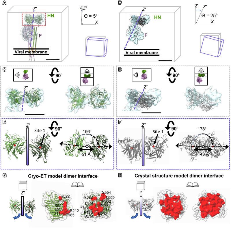
Paramyxoviruses-including important pathogens like parainfluenza, measles, and Nipah viruses-use a receptor binding protein [hemagglutinin-neuraminidase (HN) for parainfluenza] and a fusion protein (F), acting in a complex, to enter cells. We use cryo-electron tomography to visualize the fusion complex of human parainfluenza virus 3 (HN/F) on the surface of authentic clinical viruses at a subnanometer resolution sufficient to answer mechanistic questions. An HN loop inserts in a pocket on F, showing how the fusion complex remains in a ready but quiescent state until activation. The globular HN heads are rotated with respect to each other: one downward to contact F, and the other upward to grapple cellular receptors, demonstrating how HN/F performs distinct steps before F activation. This depiction of viral fusion illuminates potentially druggable targets for paramyxoviruses and sheds light on fusion processes that underpin wide-ranging biological processes but have not been visualized in situ or at the present resolution.


Figures

Fig. 1.
Fig. 1.. Subnanometer resolution of the HN/F viral fusion complex on the virus surface.
(A) Representative central slice from a tomogram of an HPIV3 viral particle. (B) X and Y projections from final subtomogram complex reconstruction using a tight mask around HN and F. (C) Fourier shell correlation (FSC) plots of the HN and F complex with and without a mask and the individual HN- and F-focused refinements. Side views and top view of the final HN (green) and F (pink) reconstruction without (D and E) and with (F and G) prefusion F [Protein Data Bank (PDB) ID: 6MJZ] and HN (PDB ID: 4MZA) models fit into the density. Side view of the prefusion F apex region with clear α-helical densities is shown in the inset. (G) HN and F each separately in top-down view with the models fit into the density. (H) Side view of the HN/F complex reconstruction filtered to 20 Å and with prefusion F (PDB ID: 6MJZ) and HN (PDB ID: 4MZA) models fit into the density. The density threshold level was decreased to reveal the viral membrane. (I) Full-length HN (green model) and F (pink model) derived from a combination of the soluble F structure (PDB ID: 6MJZ) and the AlphaFold model of the F trimer stalk region were fit into our final subvolumes. (J) Subtomogram-averaged volume of the glycoprotein organization on the viral surface (one of the five classes from a particle classification is shown, revealing separated dimers; see also fig. S1 for additional classes). Scale bars, (A) 50 nm, (D) 5 nm, and (J) 10 nm.
Fig. 2.
Fig. 2.. Interactions between HN protomer heads observed in cryo-ET reconstructions compared with the crystal structure of soluble HN dimer.
(A) Full-length cryo-ET AlphaFold model of HN + F. Dashed box: HN protomers individually fit into cryo-ET density. (B) Crystal structure model of dimeric HN (PDB ID: 4MZA) fit into cryo-ET density. Axis of rotation around HN (Z′; purple dashed lines) plotted in relation to viral membrane. When compared to the z axis of viral membrane (gray cube), axis of rotation around HN is tilted 5° for the cryo-ET model (A) and 25° for the crystal structure model (B) (purple cubes). (C) Zoomed-in side, top views of individual HN protomer heads from crystal structure (green) fit into cryo-ET density. (D) Zoomed-in side, top views of HN dimer heads from crystal structure (gray) placed into cryo-ET density by fitting only proximal HN protomer head. (E) Cryo-ET model has 156° between HN protomer heads with respect to the Z′ axis (purple bar) and 51 Å between the HN protomer centers (red spheres). (F) Crystal structure model has 178° between the two HN protomers’ heads with respect to the Z′ axis (purple bar) and 43 Å between the centers. (G and H) Peeling opens the HN dimer for each model so that the interfaces face out. Surfaces that contact the opposing protomer in the cryo-ET model are red (G). Buried surface area in the cryo-ET structure (red) is less than the buried surface area in the crystal structure (H). Scale bars (A to D), 5 nm.
Fig. 3.
Fig. 3.. Loop motif of the HN protomeric head interacting with the apex of F.
(A) Full-length cryo-ET AlphaFold-derived model of HN + F fit into the cryo-ET density. The dashed box indicates the HN/F interaction region under discussion. (B) Zoomed-in view of the final reconstruction focused on the HN/F interaction. (C) Models of HN (green) and F (pink) fit into the density of (A) with the HN loop interacting with the apex of F. (D) Electrostatic potential of the HN/F interaction region under discussion. (E) Individual residues of HN and F (spheres) that are in close interaction with each other at the F apex. (F) Density corresponding to an N-linked glycosylation site that is involved with the HN/F interaction region. The model has one hydrogen bond between the apex of F and the sugar molecule on HN . (G) Schematic side views of the thumb-finger motif that appears to position the HN protomer above F. (H to J) Overlay of measles H (yellow) and Nipah G (red) on the HPIV3 HN protomer (green) with comparison showing the similarities in receptor binding site 1 (H) along with the thumb finger motif (I and J).
Fig. 4.
Fig. 4.. Anti–F-neutralizing antibody and HN both bind at the apex of prefusion F.
(A) Cryo-EM structure of soluble prefusion F (pink) bound to anti–F-neutralizing PIA174 Fab (blue) (PDB ID: 6MJZ). (B and C) Overlay of PIA174 Fab surface (blue) with (B) and without (C) the surface from the cryo-ET model with Fab (blue), HN (green), and F (pink). (D and E) Disruption of viral surface by PIA174 Fab showing regions of viral surface with no density (orange arrows) or F only without HN (pink arrows). (F and G) Subtomogram average without (F) and with (G) the fit of the PIA174 Fab cryo-EM structure. (H) X and Y projection slices from subtomogram reconstruction of selected particles on the viral surface containing PIA174 bound to prefusion F. (I) Plots of FSCs from the final subvolume of the PIA174 Fab bound to F complex with masked and unmasked resolution at the 0.143-Å cutoff value. (J and K) Schematic (J) of assay for PIA174 Fab stabilization of prefusion F (heat temperature–mediated activation of F at 55°C) in the absence of HN. (K) Percent cells with prefusion F after varying incubation durations at 55°C with prefusion-specific PIA174 Fab, normalized to F alone and incubated with Fab at 4°C. ***P ≤ 0.001 by two-way analysis of variance (ANOVA) and Sidak’s post hoc test. (L) Schematic of competition between HN and PIA174. (M) Proportion of PIA174 Fab binding to cells expressing F alone (pink bar) or F + HN (green bar) in presence versus absence of DTSSP. Data are means ± SE from three separate experiments. **P ≤ 0.01 by unpaired two-tailed t test. Scale bars, (D and E) 50 and (F) 10 nm.
Fig. 5.
Fig. 5.. Alterations at key HN/F interface probed with neutralizing anti-F antibody.
(A) The virus that arose under selective pressure of PIA174 Fab is resistant to entry inhibition by PIA174 Fab. Inhibition was quantitated by plaque counting normalized to no treatment. (B) F-A194T is not recognized by PIA174 Ab on cells expressing F-parental or F-A194T and treated with PIA174 Ab. (C to E) Top-down view of the soluble cryo-EM prefusion F structure (PDB ID: 6MJZ) for (C) F (pink surface), (D) F with A194T mutation (orange surface), and (E) F-A194T mutation and PIA174 Fab loop (blue spheres). (F and G) Schematic of glycoprotein combinations used in the next set of experiments: F + HN or F + influenza HA, which tethers via sialic acid receptor engagement but does not complex with, or activate, F, with or without PIA174 Ab. (H) Inhibitory activity of PIA174 Ab in cell-cell fusion assay based on α complementation of β-galactosidase (β-Gal) where cell fusion leads to alpha-omega complementation. Receptor-bearing cells coexpressing HN + F (green), HA + F (red), HN + F-A194T (orange), and HA + F-A194T (purple) were treated with PIA174 Ab (x axis). y axis, % inhibition of fusion by of PIA174 Ab. (I and J) Activation of F by heat assessed at a range of temperatures without (I) or with (J) F-A194T mutation. Readout for F activation is fusion, binding, or release of red blood cells (RBCs) interacted with the F-expressing cells. (K and L) Activation of F by HN was assessed at a range of temperatures without (K) or with (L) F-A194T. Data are means ± SE from at least three separate experiments for (A), (B), and (H) to (L).

References

    1. T. C. Marcink, M. Porotto, A. Moscona, Parainfluenza virus entry at the onset of infection. Adv. Virus Res. 111, 1–29 (2021). - PMC - PubMed
    1. A. Moscona, R. W. Peluso, Fusion properties of cells persistently infected with human parainfluenza virus type 3: Participation of hemagglutinin-neuraminidase in membrane fusion. J. Virol. 65, 2773–2777 (1991). - PMC - PubMed
    1. M. Porotto, M. Murrell, O. Greengard, A. Moscona, Triggering of human parainfluenza virus 3 fusion protein (F) by the hemagglutinin-neuraminidase (HN) protein: An HN mutation diminishes the rate of F activation and fusion. J. Virol. 77, 3647–3654 (2003). - PMC - PubMed
    1. M. Porotto, I. DeVito, S. G. Palmer, E. M. Jurgens, J. L. Yee, C. C. Yokoyama, A. Pessi, A. Moscona, Spring-loaded model revisited: Paramyxovirus fusion requires engagement of a receptor binding protein beyond initial triggering of the fusion protein. J. Virol. 85, 12867–12880 (2011). - PMC - PubMed
    1. M. Porotto, Z. W. Salah, L. Gui, I. DeVito, E. M. Jurgens, H. Lu, C. C. Yokoyama, L. M. Palermo, K. K. Lee, A. Moscona, Regulation of paramyxovirus fusion activation: The hemagglutinin-neuraminidase protein stabilizes the fusion protein in a pretriggered state. J. Virol. 86, 12838–12848 (2012). - PMC - PubMed