

doi: 10.1093/ced/llac065.
Ancillary techniques to improve dermoscopy specificity for skin cancer detection
Affiliations
- PMID: 36763719
- PMCID: PMC10935604
- DOI: 10.1093/ced/llac065
Item in Clipboard
Ancillary techniques to improve dermoscopy specificity for skin cancer detection
Clin Exp Dermatol.
.
Abstract
Although the use of dermoscopy has markedly improved both the sensitivity and specificity for skin cancer detection, there is still opportunity for improvement. Ancillary techniques provide additional ways to assess a lesion with the aim of improving our diagnostic ability with little extra cost. Usage of these techniques can strengthen diagnosis and help reduce unnecessary biopsies of benign lesions.
© The Author(s) 2022. Published by Oxford University Press on behalf of British Association of Dermatologists. All rights reserved. For permissions, please e-mail: journals.permissions@oup.com.
Conflict of interest statement
Conflict of interest: The authors declare that they have no conflict of interest.
Figures
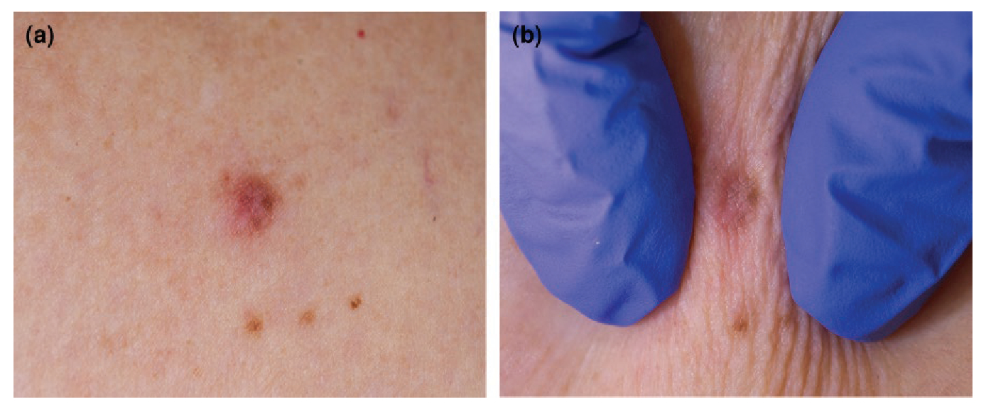
(a) Lesion suspected to be a dermatofibroma (DF); (b) the pinch test illustrates a positive dimple sign, supporting a diagnosis of DF.
(a) Dermoscopic view of a lesion suspected to be an intradermal naevus (IDN); (b) application of horizontal pressure causes the lesion to wobble, supporting the diagnosis of IDN.
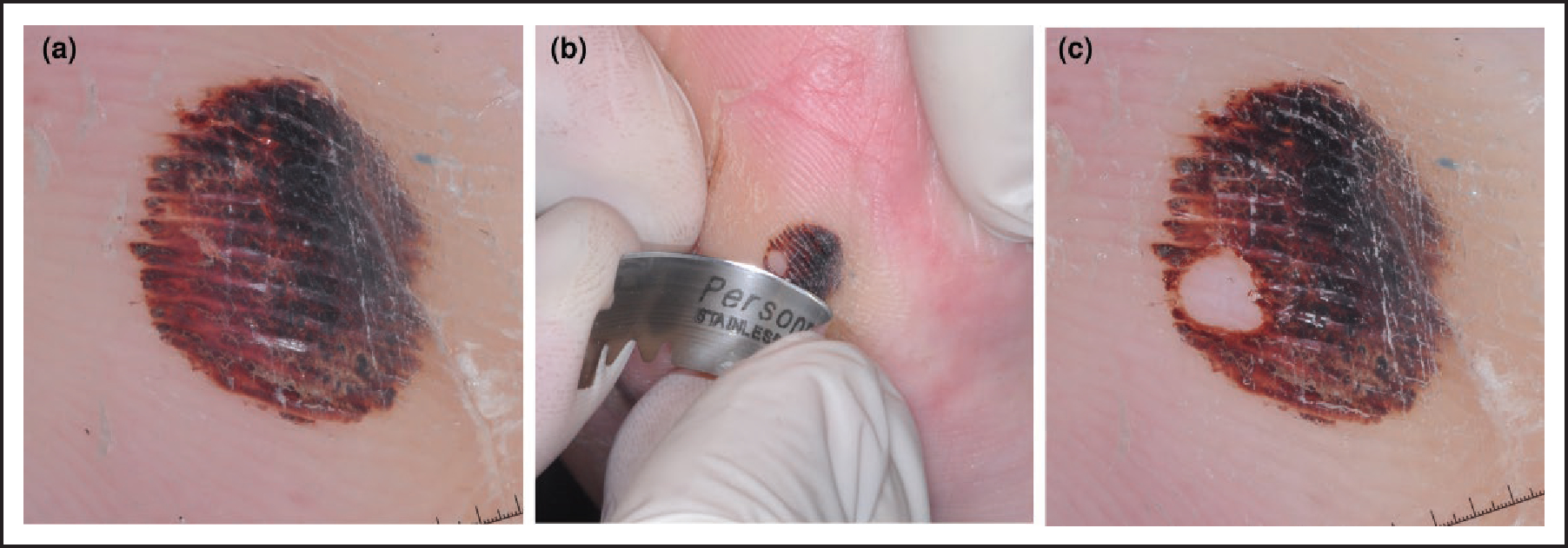
(a) Dermoscopic image of a red/black lesion on the sole with a parallel ridge pattern; (b) scrape test performed using a blade to remove the stratum corneum from part of the lesion; (c) part of the lesion has been removed, confirming a diagnosis of subcorneal haematoma.
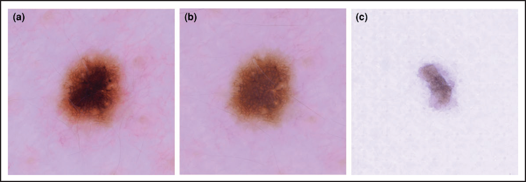
(a) Dermoscopic view of a darkly pigmented lesion with a central blotch; (b) appearance of the lesion following the adhesive tape test, revealing the reticular network of a benign naevus; (c) the pigmented stratum corneum has been removed and is visible on the tape.
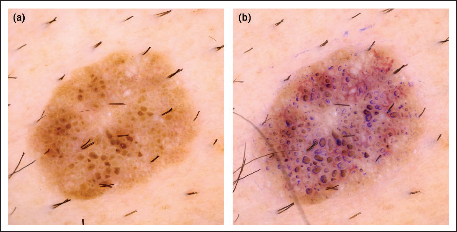
(a) Dermoscopic image of a lesion composed of nonspecific brown clods; (b) following the ink test, the comedo-like openings become easily apparent, aiding diagnosis of a seborrhoeic keratosis.
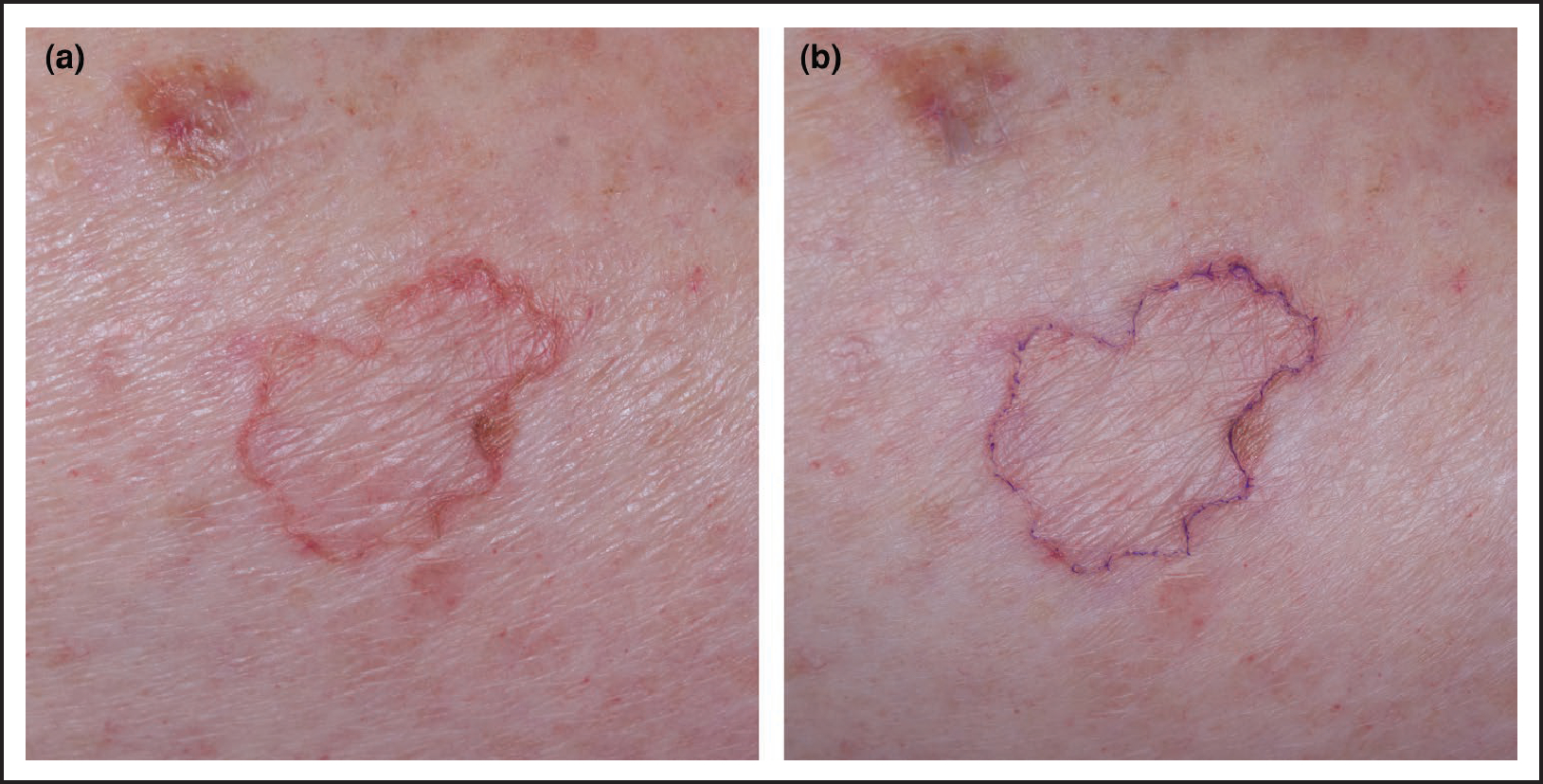
(a) An annular skin lesion with unclear diagnosis; (b) following the ink test, deposition of ink within the furrow of the cornoid lamella confirms the diagnosis of porokeratosis.
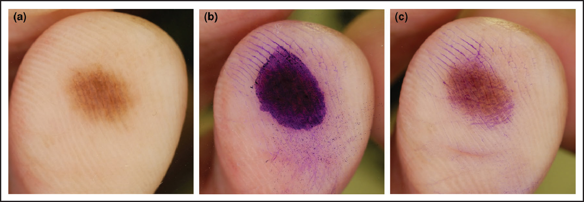
(a) Dermoscopic view of a pigmented acral lesion; it is unclear whether the pigment predominates in the furrows or the ridges. (b) Ink is applied to the lesion as part of the furrow ink test; (c) when the ink is wiped away a parallel furrow pattern becomes evident, indicating a benign naevus.
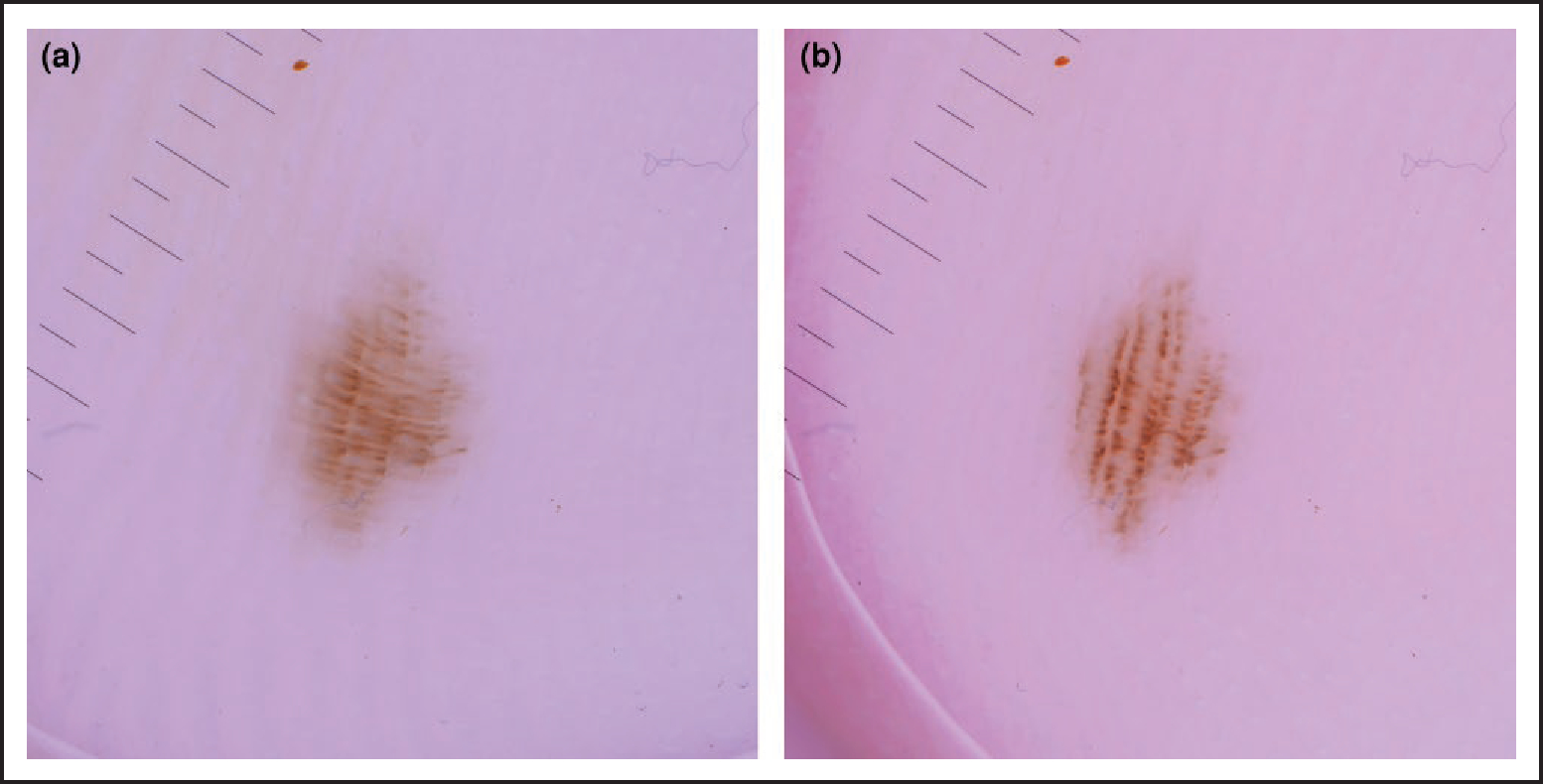
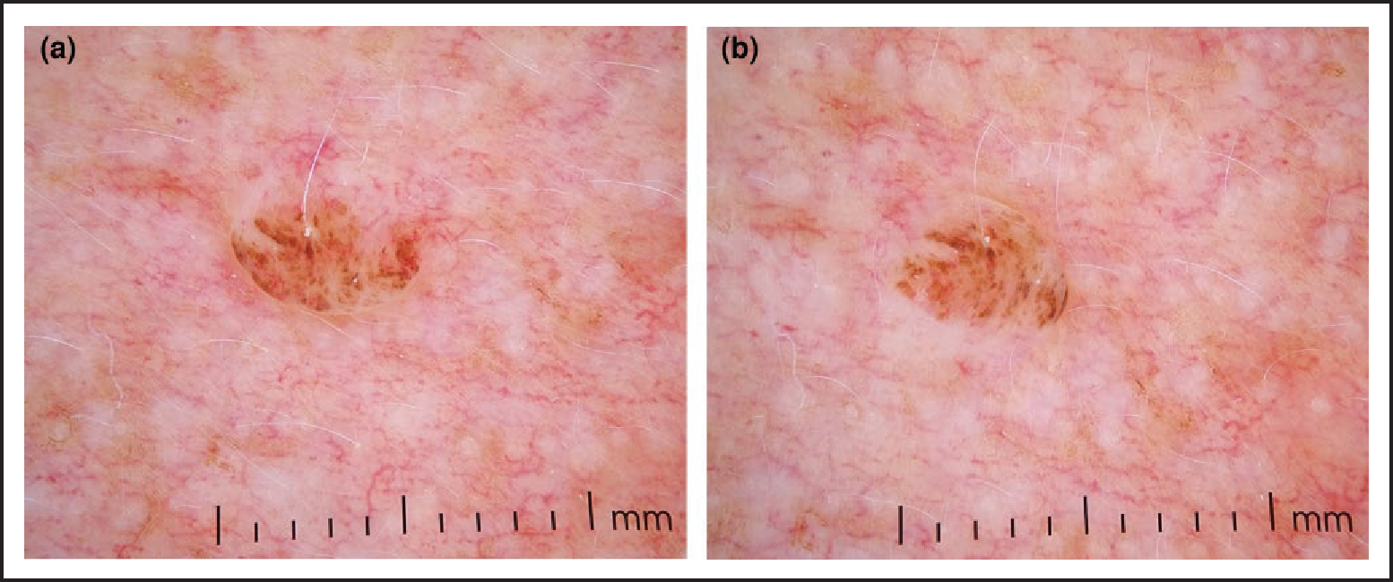
(a) Dermoscopic image of a fibrillar-patterned pigmented lesion on the sole; (b) lateral pressure with the dermatoscope aligns the pigment back to a parallel furrow pattern, indicating a benign naevus.
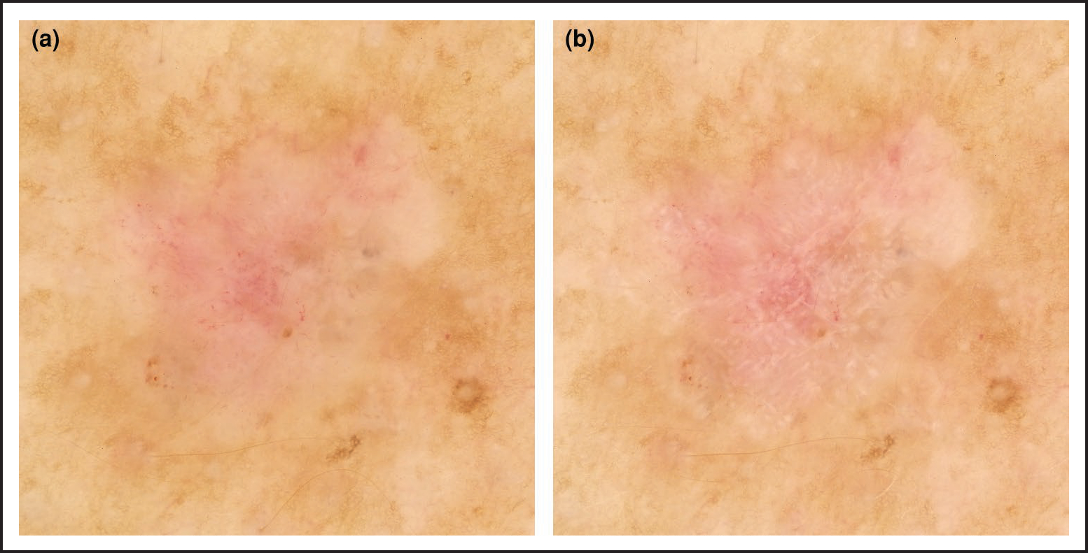
(a) Nonpolarized dermoscopy revealed a largely featureless skin lesion; (b) toggling to polarized dermoscopy allowed identification of shiny white structures, aiding with the diagnosis of skin cancer, in this case a basal cell carcinoma.
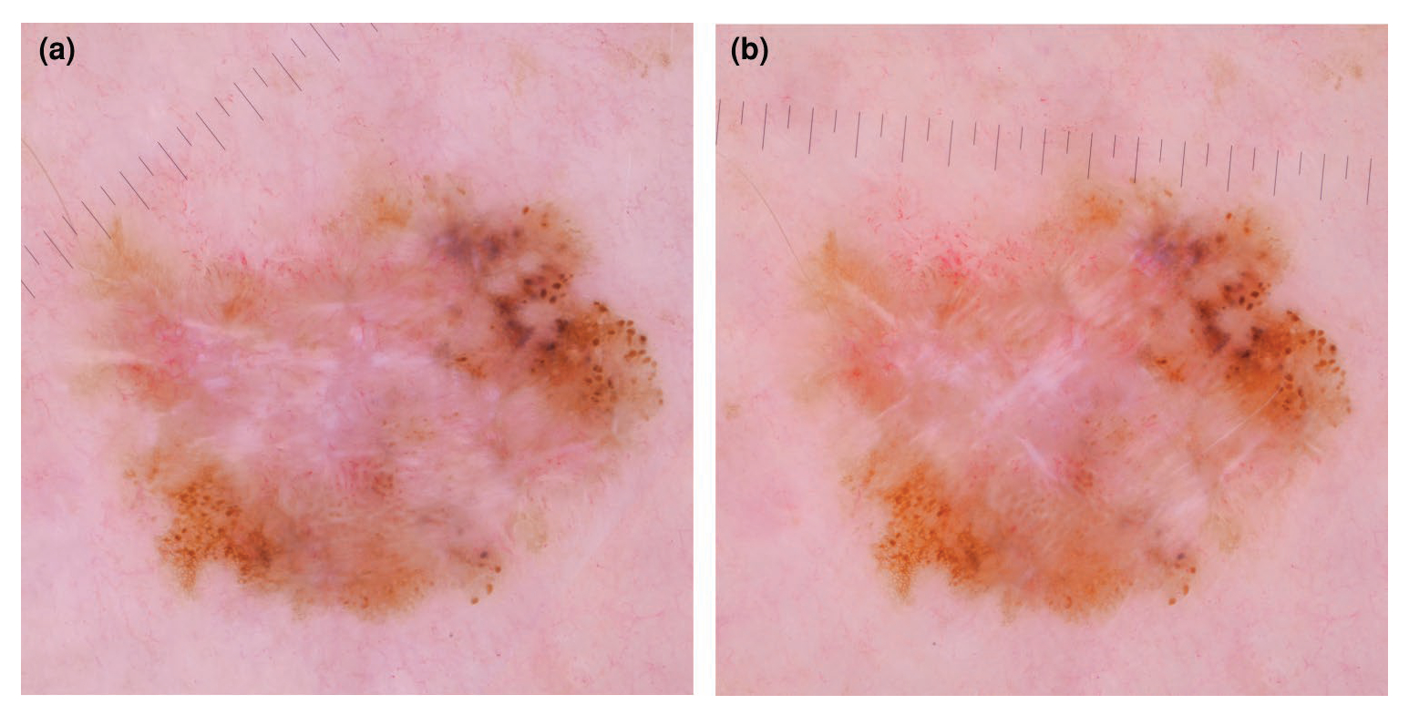
(a) Shiny white structures (SWS) are visible with polarized dermoscopy (PD) but less prominent due to the orientation of the dermatoscope; (b) slow rotation of the dermatoscope up to 180 degrees while using PD can enhance identification of the SWS, in this case supporting a diagnosis of melanoma. This phenomenon is due to the angular dependence of polarized light.
Similar articles
-
Role of In Vivo Reflectance Confocal Microscopy in the Analysis of Melanocytic Lesions.Acta Dermatovenerol Croat. 2018 Apr;26(1):64-67. Acta Dermatovenerol Croat. 2018. PMID: 29782304 Review.
-
Performance of the First Step of the 2-Step Dermoscopy Algorithm.JAMA Dermatol. 2015 Jul;151(7):715-21. doi: 10.1001/jamadermatol.2014.4642. JAMA Dermatol. 2015. PMID: 25714607
-
Dermoscopy patterns of nevi associated with melanoma.G Ital Dermatol Venereol. 2010 Feb;145(1):99-110. G Ital Dermatol Venereol. 2010. PMID: 20197749 Review.
-
Dark pigmented lesions: Diagnostic accuracy of dermoscopy and reflectance confocal microscopy in a tertiary referral center for skin cancer diagnosis.J Am Acad Dermatol. 2021 Jun;84(6):1568-1574. doi: 10.1016/j.jaad.2020.07.084. Epub 2020 Jul 27. J Am Acad Dermatol. 2021. PMID: 32730850
-
Discrimination of cancerous from benign pigmented skin lesions based on multispectral autofluorescence lifetime imaging dermoscopy and machine learning.J Biomed Opt. 2022 Jun;27(6):066002. doi: 10.1117/1.JBO.27.6.066002. J Biomed Opt. 2022. PMID: 35701871 Free PMC article.
References
-
- Vestergaard ME, Macaskill P, Holt PE, Menzies SW. Dermoscopy compared with naked eye examination for the diagnosis of primary melanoma: a meta-analysis of studies performed in a clinical setting. Br J Dermatol 2008; 159:669–76. - PubMed
-
- Welch HG, Mazer BL, Adamson AS. The rapid rise in cutaneous melanoma diagnoses. N Engl J Med 2021; 384:72–9. - PubMed
-
- Argenziano G, Cerroni L, Zalaudek I et al. Accuracy in melanoma detection: a 10-year multicenter survey. J Am Acad Dermatol 2012; 67:54–9. - PubMed
-
- Carli P, De Giorgi V, Crocetti E et al. Improvement of malignant/benign ratio in excised melanocytic lesions in the ‘dermoscopy era’: a retrospective study 1997–2001. Br J Dermatol 2004; 150:687–92. - PubMed
MeSH terms
Grants and funding
LinkOut - more resources
Full Text Sources
Medical
