Review
. 2023 Feb 10:85:165-189.
doi: 10.1146/annurev-physiol-031522-021457.

Flipping Off and On the Redox Switch in the Microcirculation

Affiliations
Review

Flipping Off and On the Redox Switch in the Microcirculation

Máté Katona et al. Annu Rev Physiol. .

Abstract

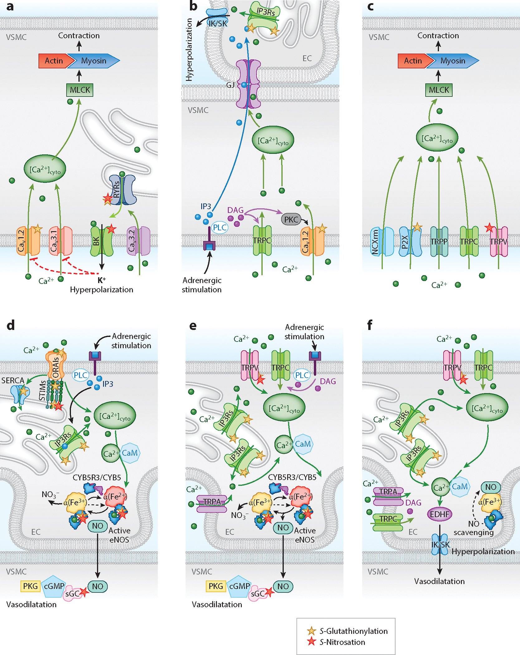
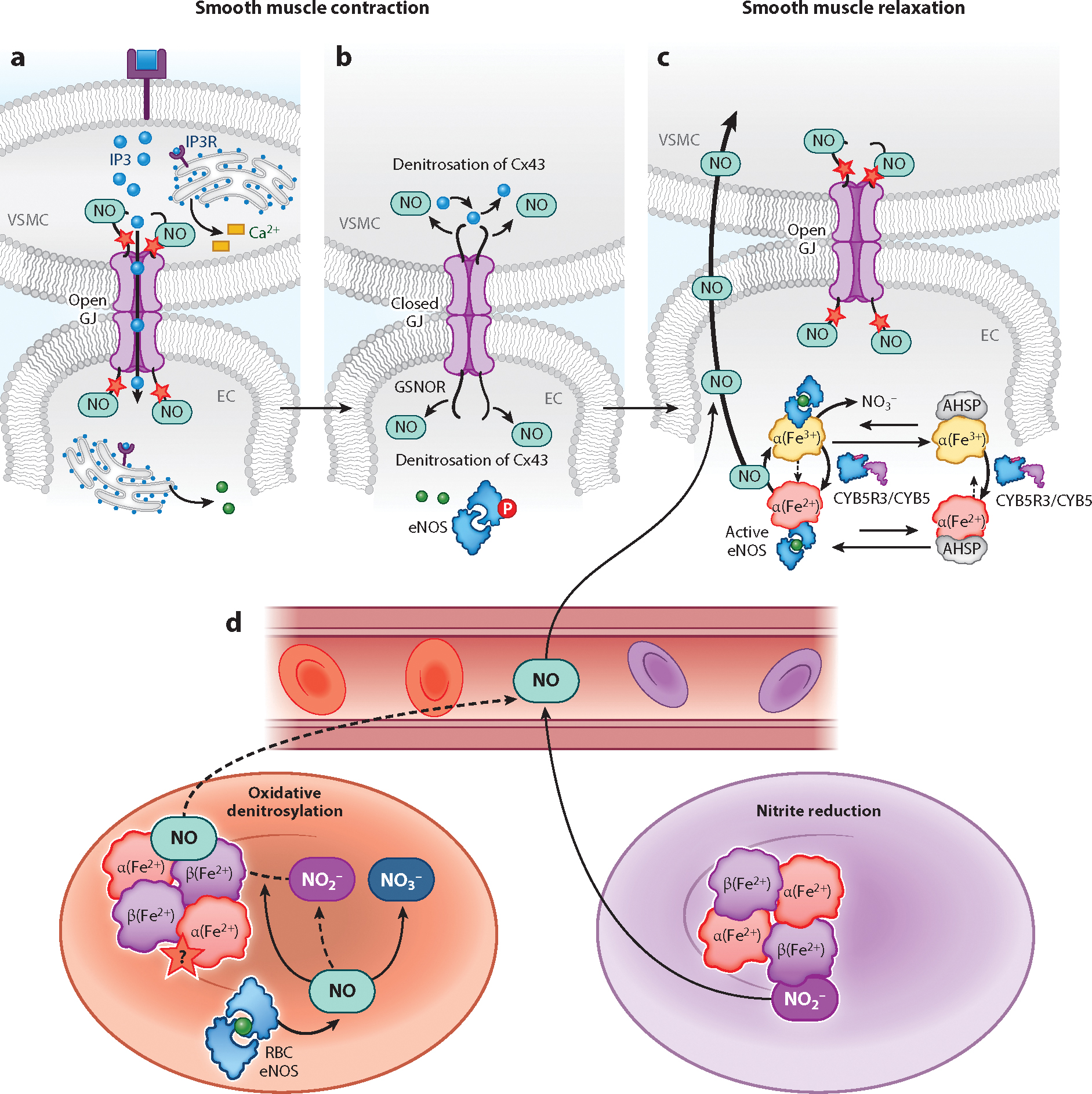
Resistance arteries and arterioles evolved as specialized blood vessels serving two important functions: (a) regulating peripheral vascular resistance and blood pressure and (b) matching oxygen and nutrient delivery to metabolic demands of organs. These functions require control of vessel lumen cross-sectional area (vascular tone) via coordinated vascular cell responses governed by precise spatial-temporal communication between intracellular signaling pathways. Herein, we provide a contemporary overview of the significant roles that redox switches play in calcium signaling for orchestrated endothelial, smooth muscle, and red blood cell control of arterial vascular tone. Three interrelated themes are the focus: (a) smooth muscle to endothelial communication for vasoconstriction, (b) endothelial to smooth muscle cell cross talk for vasodilation, and (c) oxygen and red blood cell interregulation of vascular tone and blood flow. We intend for this thematic framework to highlight gaps in our current knowledge and potentially spark interest for cross-disciplinary studies moving forward.

Keywords: calcium; endothelial; microcirculation; red blood cell; redox; smooth muscle.


Figures

Figure 1
Figure 1
Ca2+ entry through voltage-gated Ca2+ channels (CaV1.2 and CaV3.1) triggers the VSMC contractile machine through the activation of MLCK to promote VSMC contraction. (a) Activation of CaV3.2 channels stimulates RyRs through CICR to trigger Ca2+ release from the SR/ER, potentiating BK channel–mediated hyperpolarization and vasodilatation. The red dotted line indicates inhibition of CaV1.2 and CaV3.1 channel function by hyperpolarization. (b) Adrenergic stimulation induces IP3 and DAG formation by PLC. DAG activates TRPC channels and PKC, which phosphorylates the CaV1.2 channel to increase [Ca2+]cyto and induce vasoconstriction in VSMCs. IP3 and Ca2+ can diffuse from VSMCs to the EC through the GJs of the myoendothelial junctions to increase EC [Ca2+]cyto and limit vasoconstriction by activating IK/SK channels. (c) Activation of NCXrm and P2X and TRPP, TRPC, and TRPV channels increases VSMC [Ca2+]cyto to induce vasoconstriction through the activation of MLCK/actin-myosin. (d) IP3R-mediated SR/ER Ca2+ release is induced by CICR or adrenergic stimulation through the generation of IP3 by PLC. The subsequent decrease in [Ca2+]SR/ER induces SOCE (mediated by STIM and ORAI) and ER store refilling (facilitated by the activity of SERCA) to increase [Ca2+]cyto for Ca2+–CaM-dependent activation of eNOS and generation of NO. NO diffuses to the VSMCs and causes vasodilatation through the activation of the NO-sGC-cGMP-PKG pathway. (e) Activation of TRPV, TRPC, and TRPA channels through mechanical stimuli and/or adrenergic activation of PLC and generation of DAG and subsequent activation of SR/ER Ca2+ release (IP3R) increase [Ca2+]cyto to activate eNOS and cause VSMC relaxation through the production of NO. (f) Ca2+ influx via TRPC, TRPV, and TRPA channels and/or Ca2+ release from the SR/ER through IP3Rs activates IK/SK channels via Ca2+–CaM. The activation of the IK/SK channels (EDHF) causes EC hyperpolarization and VSMC relaxation. eNOS-derived NO is scavenged by α-globin in the microcirculation. Abbreviations: Ca2+–CaM, calcium-calmodulin; [Ca2+]cyto, cytosolic Ca2+ concentration; [Ca2+]SR/ER, sarcoplasmic/endoplasmic reticulum Ca2+ concentration; CaV1.2, voltage-gated Ca2+ channel 1.2; CaV3.1, voltage-gated Ca2+ channel 3.1; cGMP, cyclic guanosine 3′,5′-cyclic monophosphate; CICR, Ca2+-induced Ca2+ release; CYB5, cytochrome B5; CYB5R3, cytochrome B5; reductase 3; DAG, diacylglycerol; EC, endothelial cell; EDHF, endothelial-derived hyperpolarizing factor; eNOS, endothelial nitric oxide synthase; GJ, gap junction; IK/SK, intermediate and small conductance Ca2+-activated K+ channels; IP3, inositol 1,4,5-trisphosphate; IP3R, IP3 receptor; MLCK, myosin light chain kinase; NCXrm, Na+/Ca2+ exchangers in reverse mode; NO, nitric oxide; ORAI, calcium release–activated calcium channel protein; P2X, purinergic P2X; PKG, protein kinase G; PLC, phospholipase C; RyR, ryanodine receptor; SERCA, SR/ER Ca2+ ATPase; sGC, soluble guanylyl cyclase; SOCE, store-operated Ca2+ entry; STIM, stromal interaction molecule; TRP, transient receptor potential channel; TRPA, ankyrin-rich protein TRP subfamily; TRPC, canonical TRP subfamily; TRPP, polycystic TRP subfamily; TRPV, vanilloid TRP subfamily; VMSC, vascular smooth muscle cell.
Figure 2
Figure 2
(a) S-Nitrosation of Cx43 facilitates the open state of GJs at the myoendothelial junctions to allow the intercellular exchange of signaling molecules. (b) IP3-induced Ca2+ release from the ER activates eNOS through phosphorylation (P circled in red). Denitrosation of Cx43 by GSNOR closes GJs and limits the diffusion of IP3. (c) NO generated by the activity of eNOS, which is mediated by the changes in the α-globin heme redox state. The NO generated during eNOS activity can diffuse from the EC to VSMC to cause vasodilatation and is important in maintaining the open probability of GJs through the S-nitrosation of Cx43. α-Globin is stabilized by AHSP before and complexes with eNOS to facilitate NO production. The redox state of the α-globin heme is regulated by CYB5R3 or eNOS. S-Nitrosation is marked with a red star. (d) In oxygenated RBCs (red), eNOS generates NO that can ultimately participate in vasorelaxation to regulate blood flow and blood pressure. Redox reactions of NO with oxygen, oxyhemoglobin, and deoxyhemoglobin result in nitrite NO2, NO3, and Fe2+-nitrosyl-hemoglobin. During gas exchange, the RBC partially deoxygenates (purple), and diffusible molecules that can signal for vessel relaxation are generated. NO2 undergoes a reductive reaction with deoxyhemoglobin, releasing diffusible NO as the oxygen tension approaches the point where 50% of the RBC hemoglobin has become deoxygenated. Reactive intermediates (NO2 and H2O2) formed in the process also facilitate the release of NO from Fe2+-nitrosyl-hemoglobin (oxidative denitrosylation) and participate in radical–radical interactions that generate the diffusible N2O3 for possible RBC export. The red star marks suspected S-nitrosation (S-nitrosothiol modification of hemoglobin), which is distinct from Fe2+-nitrosyl-hemoglobin formation. Abbreviations: AHSP, α-globin stabilizing protein; Cx43, gap junction–forming vascular connexin; CYB5, cytochrome B5; CYB5R3, cytochrome B5 reductase 3; EC, endothelial cell; eNOS, endothelial nitric oxide synthase; GJ, gap junction; GSNOR, S-nitrosoglutathione reductase; H2O2, hydrogen peroxide; IP3, inositol 1,4,5-trisphosphate; IP3R, IP3 receptor; N2O3, dinitrogen trioxide; NO, nitric oxide; NO2, nitrite; NO3, nitrate; RBC, red blood cell; VMSC, vascular smooth muscle cell.

Similar articles

Cited by

References

    1. Nehls V, Drenckhahn D. 1991. Heterogeneity of microvascular pericytes for smooth muscle type alphaactin. J. Cell Biol. 113:147–54 - PMC - PubMed
    1. Straub AC, Zeigler AC, Isakson BE. 2014. The myoendothelial junction: connections that deliver the message. Physiology 29:242–49 - PMC - PubMed
    1. Rhodin JA. 1967. The ultrastructure of mammalian arterioles and precapillary sphincters. J. Ultrastruct. Res. 18:181–223 - PubMed
    1. Meng J, Lv Z, Zhang Y, Wang Y, Qiao X, et al. 2021. Precision redox: the key for antioxidant pharmacology. Antioxid. Redox Signal. 34:1069–82 - PMC - PubMed
    1. Förstermann U, Sessa WC. 2012. Nitric oxide synthases: regulation and function. Eur. Heart J. 33:829–37 - PMC - PubMed

Publication types

LinkOut - more resources