Review
. 2023 Feb 10:85:137-164.
doi: 10.1146/annurev-physiol-031522-034807.

Pericytes and the Control of Blood Flow in Brain and Heart

Affiliations
Review

Pericytes and the Control of Blood Flow in Brain and Heart

Thomas A Longden et al. Annu Rev Physiol. .

Abstract

Pericytes, attached to the surface of capillaries, play an important role in regulating local blood flow. Using optogenetic tools and genetically encoded reporters in conjunction with confocal and multiphoton imaging techniques, the 3D structure, anatomical organization, and physiology of pericytes have recently been the subject of detailed examination. This work has revealed novel functions of pericytes and morphological features such as tunneling nanotubes in brain and tunneling microtubes in heart. Here, we discuss the state of our current understanding of the roles of pericytes in blood flow control in brain and heart, where functions may differ due to the distinct spatiotemporal metabolic requirements of these tissues. We also outline the novel concept of electro-metabolic signaling, a universal mechanistic framework that links tissue metabolic state with blood flow regulation by pericytes and vascular smooth muscle cells, with capillary KATP and Kir2.1 channels as primary sensors. Finally, we present major unresolved questions and outline how they can be addressed.

Keywords: brain; calcium; electro-metabolic signaling; heart; ion channels; metabolism; neurovascular coupling; pericytes; potassium.


Figures
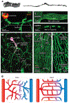
Figure 1
Figure 1
Microcirculation in heart and brain. (a) Original illustrations of pericytes by Zimmermann depicting two pericytes of a precapillary arteriole in cat kidney (left) and a thin-strand pericyte observed on a human capillary (right). Panel adapted with permission from Reference ; copyright 2022 Springer Nature. (b) An arteriole-capillary transition in brain in vivo showing elastin staining (red) of the penetrating arteriole, which abruptly ends at the point of transition into the capillaries. (c, top) In arterioles, RBCs (dark objects silhouetted against green, fluorescent plasma) tumble past one another side by side due to the wider diameter of the vessel. (c, bottom) In capillaries, RBCs squeeze to enter to narrow confines of the vessel and pass in single file. (d) A PA in a mouse brain giving way to highly branching capillaries. In brain, the capillaries (green) can be numbered by branch order, with the assigned number increasing by one with each successive branch point. Pink fluorescence reflects the presence of the Ca2+ indicator GCaMP5, which is expressed under the control of the Acta2 promoter. This highlights both SMCs of the PA and the contractile pericytes of the initial branches of the capillary bed. (e) Confocal image showing the high density of capillaries in heart. DsRed, under the NG2 promoter (pink), is highly expressed in arteriole smooth muscle cells but not in veins, which makes the feeding arteriole and vein readily distinguishable. (f) Zoomed-in image showing the capillary hierarchy in heart bookended by a precapillary arteriole and postcapillary venule. In heart, the precapillary arteriole and capillaries are readily distinguished by their different sizes and different orientations with myocytes. Note the high degree of looping anastomoses compared to brain. (g) Simplified illustrations highlighting the major differences between heart and brain circulatory organization. In brain (left), the capillary branching pattern appears to be somewhat random, branches give way to capillaries that head into the parenchyma in all directions, and anastomoses are much less frequent in this context. In heart (right), the capillaries form regular anastomoses, giving rise to loops that pass through and around adjacent cardiomyocytes. Abbreviations: FITC, fluorescein isothiocyanate; NG2, neuronal-glial antigen 2; PA, penetrating arteriole; PV, penetrating venule; RBC, red blood cell; SMC, smooth muscle cell; TRITC, tetramethylrhodamine isothiocyanate; WGA, wheat germ agglutinin.
Figure 2
Figure 2
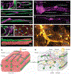
Pericytes in heart and brain microcirculation. (a) Confocal images showing the morphological diversity of capillary pericytes in heart. (i) A pericyte with a TMT leaping from one capillary to another, (ii) an intercapillary pericyte bridging one capillary to a capillary loop with its processes, (iii) an intercapillary pericyte bridging parallel capillaries with its cell body, and (iv) a capillary pericyte in heart. Magenta denotes NG2-DsRed and green FITC-WGA. (b) A contractile pericyte in heart. This confocal image shows an α-actin (pink)-positive pericyte wrapping around arteriole-capillary junctions (green; WGA) with thick processes. (c) Immunostaining image showing that each myocyte is surrounded and supplied by 4–6 capillaries. Panel adapted with permission from Reference ; copyright 2014 Springer Nature. (d) Confocal image showing close contact between a myocyte (pink; actinin) and a capillary (green; WGA) that follows a furrow along the cardiac cell. (e) Diagram illustrating the mixed cell populations and the organization of capillaries (green), myocytes (light red), and pericytes (pink and purple) in heart. Each myocyte is surrounded by 4–6 interconnected, pericyte-embroidered capillaries. Capillaries in heart are easily recognizable by their parallel orientation with myocytes. Contractile pericytes wrap around arteriole-capillary junctions, a vantage from which they may exert profound influence over blood flow. (f) Detailed images showing morphological differences between (i) SMCs, the specialized precapillary sphincter at the border between arterioles and capillaries, contractile pericytes, and (ii) thin-strand pericytes of the brain. Pink denotes DsRed expressed under the NG2 promoter. (g) Expression of eGFP (gold) in astrocytes reveals the extensive endfoot coverage that these cells establish around both arterioles and capillaries. (h) Diagram detailing the mixed population of cells that surround the tortuous vascular network of the brain. Pericytes are nestled at the center of the neurovascular unit and are in close proximity with astrocytic endfeet, neurons, and microglial processes and directly contact the endothelium. Abbreviations: DAPI, 4′,6-diamidino-2-phenylindole; eGFP, enhanced green fluorescent protein; FITC, fluorescein isothiocyanate; IB4, isolectin B4; NG2, neuronal-glial antigen 2; SMC, smooth muscle cell; TMT, tunneling microtube; WGA, wheat germ agglutinin.
Figure 3
Figure 3
Blood flow control by electrical, Ca2+ and electro-metabolic signaling in brain and heart. (Central image) Top-down view of an arteriole enwrapped by SMCs and lined by ECs diving into the brain. On the proximal capillaries close to the arteriole are a population of pericytes (contractile) that express high α-SMA and rapidly regulate underlying vessel diameter. Deeper capillaries are covered by the cell bodies and processes of thin-strand pericytes that express low α-actin and regulate diameter over longer time frames and more modestly. Capillary blood flow is regulated by electrical (a, b), Ca2+ (c), and metabolic factors (a, d, e), all of which involve pericytes. (a) Neuronal activity elevates parenchymal K+ in the brain, which activates Kir channels to hyperpolarize the membrane, initiating electrical signals that propagate to upstream arterioles and contractile pericyte-covered branches, causing dilations. (b) Diagram illustrating the effect of elevated extracellular K+ on the current produced by Kir2.1 channels at −40 mV. Point A illustrates the relatively small current produced by Kir2.1 channels at −40 mV in cECs in the presence of extracellular [K+] of 3 mM which is typical for the brain parenchyma. Resting [K+] in the interstitial spaces of the heart and in blood plasma is higher. When extracellular [K+] around the cECs is elevated to 15 mM from 3 mM, there is an increase in outward current at −40 mV, as shown by point B. The increase in outward current hyperpolarizes the cECs to a potential negative to −40 mV. This counterintuitive effect occurs because of the upward and rightward shift in the IV relationship produced by the increase of [K+]o. Note that the Nernst potential for K+ (EK) moves to a more positive potential, but this is still negative to the resting potential of the cell. (c) Relaxation of contractile pericytes by capillary Ca2+ signals. Neuronal activity drives Ca2+ elevations in ECs of the proximal capillary bed, which couple to the generation of NO.As a gas, NO diffuses into pericytes and promotes relaxation of their contractile machinery, likely through a PKG–dependent mechanism that promotes membrane hyperpolarization and a fall in pericyte Ca2+. (d) Thin-strand pericytes act as metabolic sentinels in the brain that modulate EC electrical activity through KATP channel activity. In situations where the local availability of the key energy substrate glucose is reduced, the fall in intracellular ATP:ADP ratio increases KATP channel open probability, which promotes pericyte hyperpolarization. This hyperpolarization in turn is transmitted via pericyte PSJs into the underlying endothelium, where it modulates the electrical signaling mechanism described in panel a. (e) Local blood flow control by electro-metabolic signaling in heart. Local blood flow is controlled or regulated by electrically connected elements that include myocytes, capillaries, pericytes, and arterial SMCs, with myocytes as master controllers. As cardiac myocytes carry out work, ATP is consumed and ADP is produced and elevated. This leads to the opening of KATP channels in the cardiac myocytes and drives electro-metabolic signaling. KATP opening produces a time-averaged hyperpolarization of the cardiac action potential and the accumulation of K+ in the subspace between myocytes and capillaries. This leads to the hyperpolarization of the other elements in the network through gap junctions. In contrast, the elevation of extracellular K+ (through KATP and other K+ channels opening during repolarization) activates Kir2.1 in the cECs, resulting in cEC hyperpolarization and upstream contractile pericyte and smooth muscle relaxation. Abbreviations: ADP, adenosine diphosphate; α-SMA, alpha-smooth muscle actin; ATP, adenosine triphosphate; Ca2+, calcium; cEC, capillary endothelial cell; EC, endothelial cell; EK, potassium equilibrium potential; eNOS, endothelial nitric oxide synthase; GLUT1, glucose transporter 1; IV, current-voltage; K+, potassium; [K+]o, external potassium concentration; KATP, ATP-sensitive potassium channel; Kir, inward rectifier potassium channel; NO, nitric oxide; PKG, protein kinase G; PSJ, peg-socket junction; SMC, smooth muscle cell.

Similar articles

Cited by

References

    1. Eberth C. 1871. Handbuch der Lehre von der Gewegen des Menschen und der Tiere. Vol. 1. Leipzig, Ger.: Engelmann
    1. Rouget C. 1873. Mémoire sur le développement, la structure et les proprietés physiologiques des capillaires sanguins et lymphatiques. Arch. Physiol. Norm. Pathol 5:603–63
    1. Zimmermann KW. 1923. Der feinere Bau der Blutcapillaren. Z. Anat. Entwicklungsgesch 68(1):29–109
    1. Krogh A. 1920. A contribution to the physiology of the capillaries. Nobel Lecture, Dec. 11. https://www.nobelprize.org/prizes/medicine/1920/krogh/lecture/
    1. Feigl EO. 1983. Coronary physiology. Physiol. Rev 63(1):1–205 - PubMed

Publication types

Substances

LinkOut - more resources