Review
. 2023 Feb 10:85:425-448.
doi: 10.1146/annurev-physiol-031522-084334.

Polycystin Channel Complexes

Affiliations
Review

Polycystin Channel Complexes

Orhi Esarte Palomero et al. Annu Rev Physiol. .

Abstract

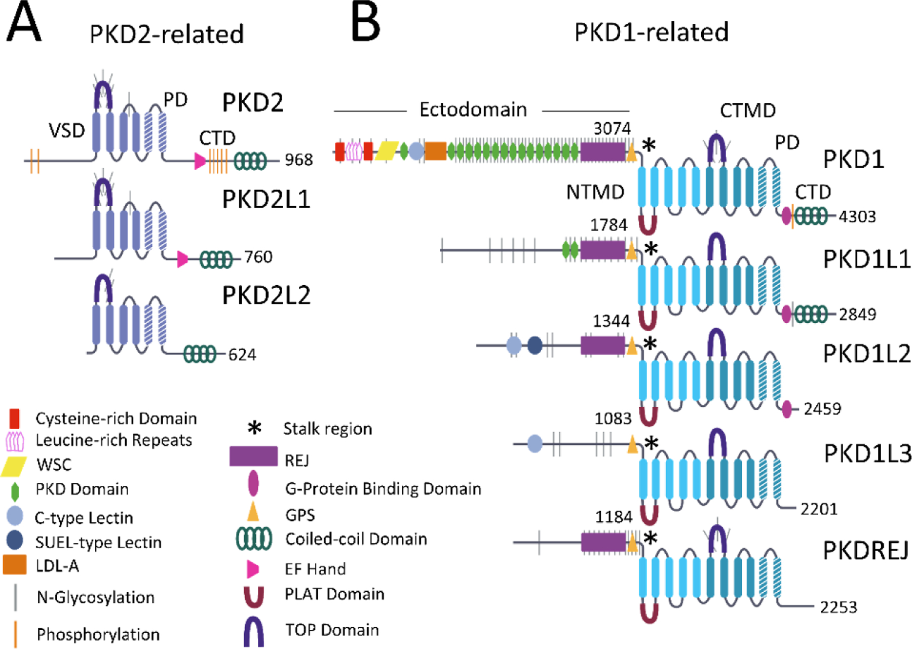
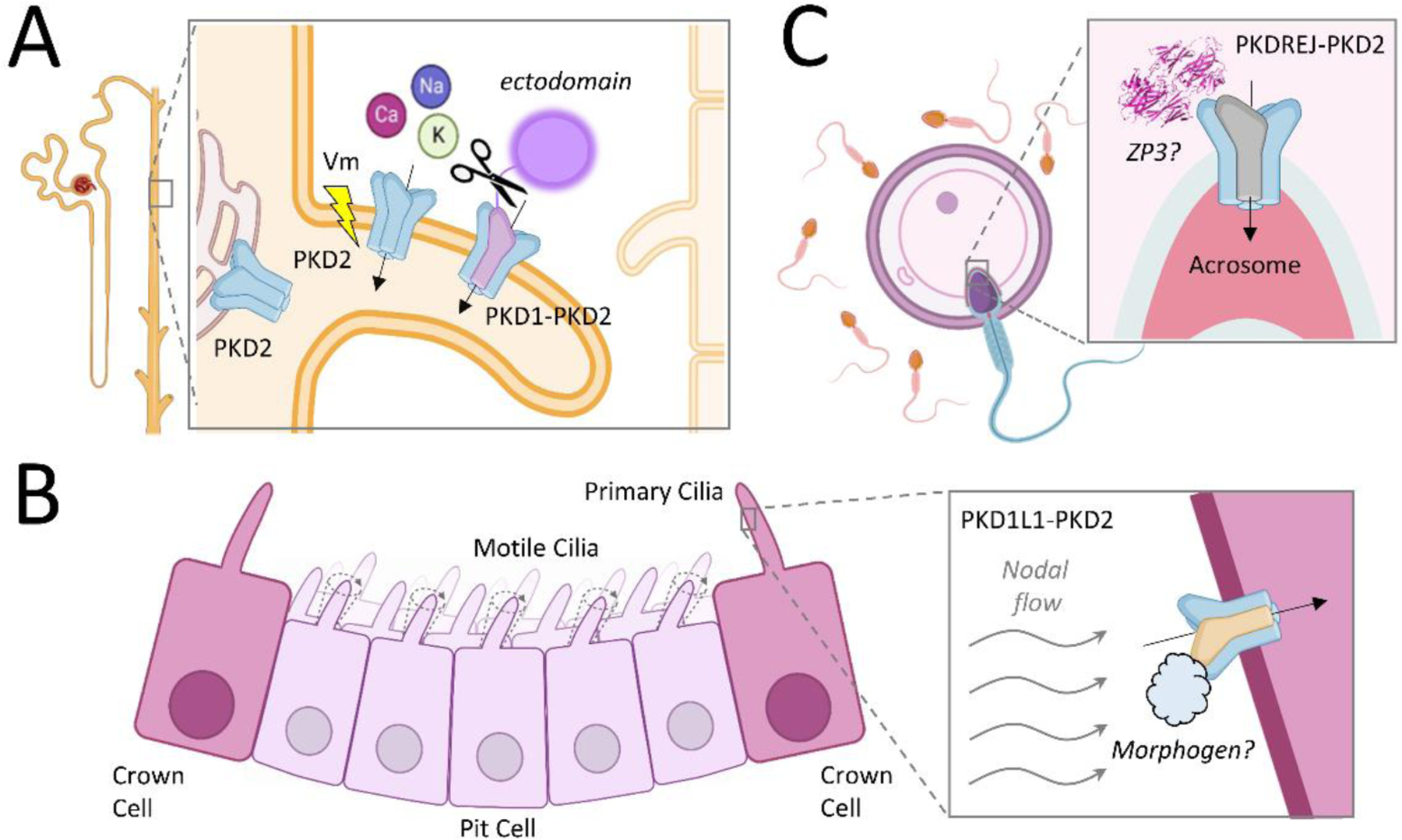
Polycystin subunits can form hetero- and homotetrameric ion channels in the membranes of various compartments of the cell. Homotetrameric polycystin channels are voltage- and calcium-modulated, whereas heterotetrameric versions are proposed to be ligand- or autoproteolytically regulated. Their importance is underscored by variants associated with autosomal dominant polycystic kidney disease and by vital roles in fertilization and embryonic development. The diversity in polycystin assembly and subcellular distribution allows for a multitude of sensory functions by this class of channels. In this review, we highlight their recent structural and functional characterization, which has provided a molecular blueprint to investigate the conformational changes required for channel opening in response to unique stimuli. We consider each polycystin channel type individually, discussing how they contribute to sensory cell biology, as well as their impact on the physiology of various tissues.

Keywords: ADPKD; TRP channel; autosomal dominant polycystic kidney disease; ciliopathies; gating mechanism; polycystin; primary cilia.


Figures

Figure 1
Figure 1
Topologies of polycystin channel subunits. The eight members are divided into PKD2-related (a) and PKD1-related (b) channel subunits. The Uniprot IDs of the human polycystin subunits are P98161 (PKD1), Q8TDX9 (PKD1L1), Q7Z442 (PKD1L2), Q7Z443 (PKD1L3), Q9NTG1 (PKDREJ), Q13563 (PKD2), Q9P0L9 (PKD2L1), and Q9NZM6 (PKD2L2). Abbreviations: CTD, C-terminal domain; GPS, G protein–coupled receptor proteolytic site; LDL-A, low density lipoprotein A; PD, pore domain; PLAT, polycystin-1 lipoxygenase and alpha toxin (domain); REJ, receptor for egg jelly domain; TOP, tetragonal opening for polycystins (domain); VSD, voltage sensor domain; SUEL, sea urchin egg lectin; WSC, cell Wall integrity and Stress Component protein.
Figure 2
Figure 2
Assembly and proposed gating mechanism of homomeric polycystin channels. (a) Transmembrane view of a single PKD2 subunit with identified structural domains and location of ADPKD-causing missense variants (13, 50). (b) Transmembrane view of overlaid PKD2 (gray; PDB 5T4D) and PKD2L1 (tan; PDB 5Z1W) channels, with expanded views of the VSD and PD (13, 15). (c) Extracellular and transmembrane view of domain-swapped PKD2 channels compared to nondomain-swapped Kv11 Eag1 channel. Abbreviations: ADPKD, autosomal dominant polycystic kidney disease; Kv, member of voltage-gated potassium channel family; PD, pore domain; PDB, protein data bank; TOP, tetragonal opening for polycystins domain; VSD, voltage sensor domain; VSDL, voltage sensor-like domain.
Figure 3
Figure 3
Assembly and proposed gating mechanisms of heteromeric polycystin channels. (a) Transmembrane view of a single PKD1 subunit with identified structural domains and location of ADPKD-causing missense variants (16). (b) Extracellular and transmembrane views of structural overlaid PKD1-PKD2 (PDB 6A70) and PKD1L3-PKD2L1 (PDB 7D7E) heteromeric channels, with an expanded view of the PKD1-S11 transmembrane segment’s positively charged residues lining the ion conducting pathway (16). (c) Transmembrane view of the PKD1L3-PKD2L1 channel with an expanded view of the pore Ca2+-binding site captured in the apo (PDB 7D7E) and Ca2+-occupied (PDB 7D7F) states (143). (d) Transmembrane view of the third PKD2L1 subunit (PKD2L1 DIII VSD) of the Ca2+-occupied PKD1L3-PKD2L1 channel complex. Abbreviations: ADPKD, autosomal dominant polycystic kidney disease; CTMD, C-terminal transmembrane domain; NTMD, N-terminal transmembrane domain; PDB, protein data bank; PLAT, polycystin-1 lipoxygenase and alpha toxin (domain); TOP, tetragonal opening for polycystins (domain); VSD, voltage sensor domain.
Figure 4
Figure 4
Subcellular localization of polycystin complexes in various human tissues. (a) Heteromeric PKD1-PKD2 and homomeric PKD2 channels within the primary cilia, endoplasmic reticulum, and apical plasma membranes of the collecting duct epithelium of the kidney nephron. (b) Heteromeric PKD1L1-PKD2 location within the (nonmotile) primary cilia of crown cells within the embryonic node. (c) PKDREJ-PKD2 heteromeric channels localize to the acrosomal crest of sperm.

References

    1. Alexander SP, Mathie A, Peters JA, Veale EL, Striessnig J, et al. 2021. The concise guide to pharmacology 2021/22: ion channels. Br. J. Pharmacol 178(Suppl. 1):S157–245 - PubMed
    1. Hughes J, Ward CJ, Peral B, Aspinwall R, Clark K, et al. 1995. The polycystic kidney disease 1 (PKD1) gene encodes a novel protein with multiple cell recognition domains. Nat. Genet 10:151–60 - PubMed
    1. Mochizuki T, Wu G, Hayashi T, Xenophontos SL, Veldhuisen B, et al. 1996. PKD2, a gene for polycystic kidney disease that encodes an integral membrane protein. Science 272:1339–42 - PubMed
    1. Brasier JL, Henske EP. 1997. Loss of the polycystic kidney disease (PKD1) region of chromosome 16p13 in renal cyst cells supports a loss-of-function model for cyst pathogenesis. J. Clin. Investig 99:194–99 - PMC - PubMed
    1. Veldhuisen B, Saris JJ, de Haij S, Hayashi T, Reynolds DM, et al. 1997. A spectrum of mutations in the second gene for autosomal dominant polycystic kidney disease (PKD2). Am. J. Hum. Genet 61:547–55 - PMC - PubMed

Publication types

LinkOut - more resources