

Myxoid glioneuronal tumor: Histopathologic, neuroradiologic, and molecular features in a single center series
- PMID: 36764090
- PMCID: PMC9929589
- DOI: 10.1016/j.neo.2023.100885
Myxoid glioneuronal tumor: Histopathologic, neuroradiologic, and molecular features in a single center series
Abstract
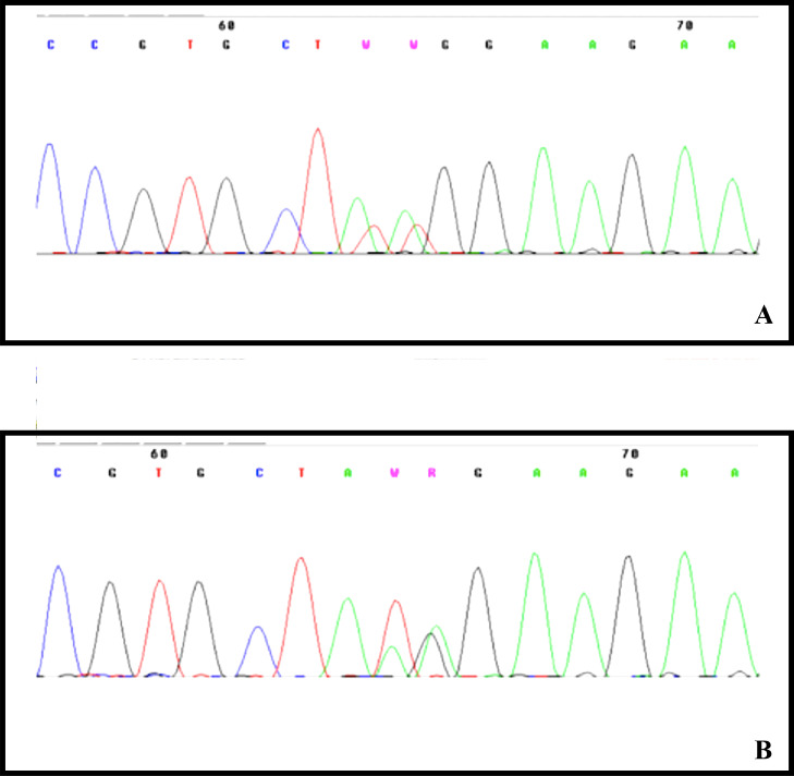
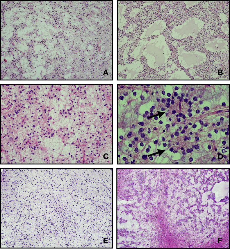
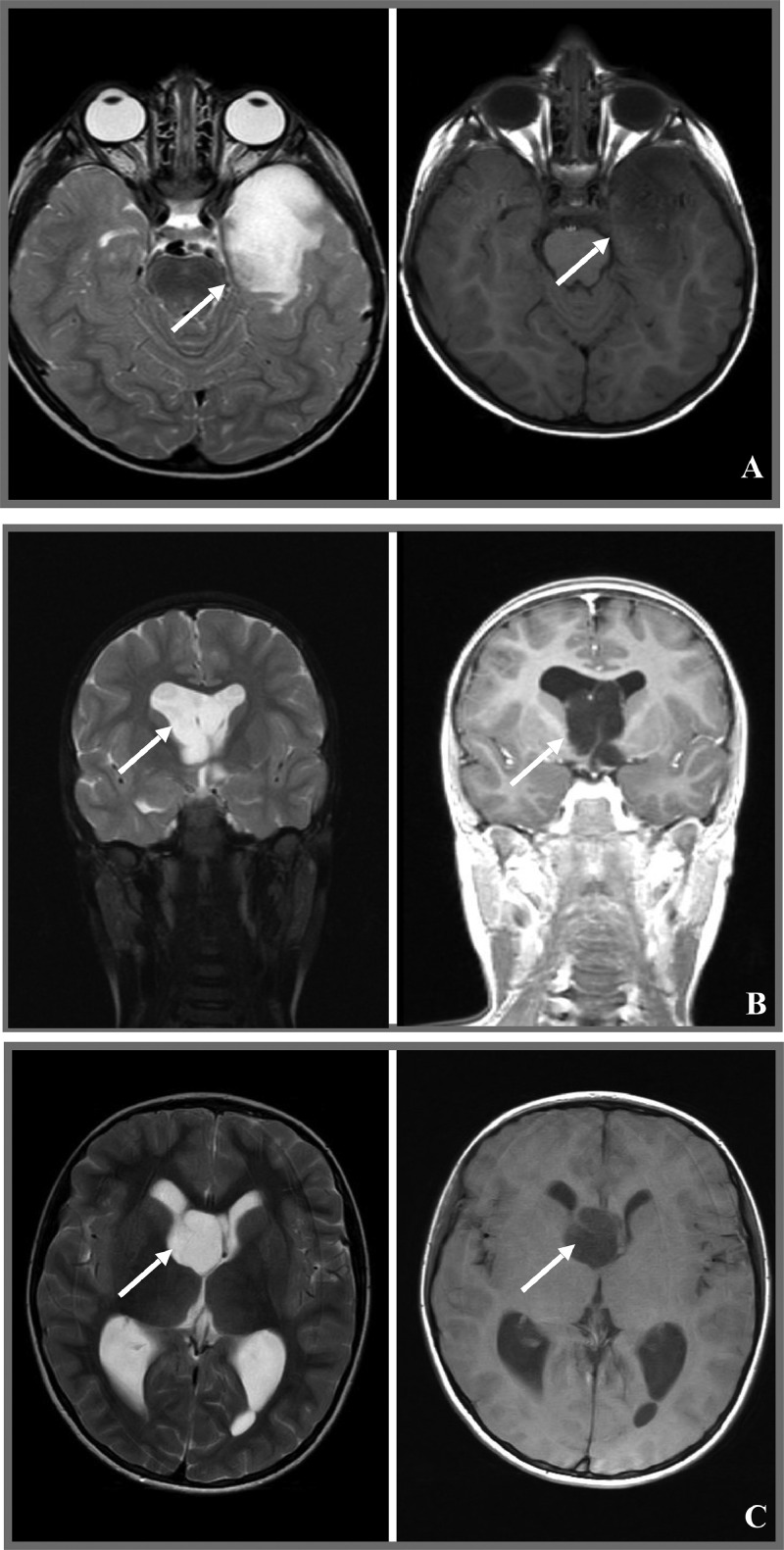
Background: Myxoid glioneuronal tumor (MGT) is a benign glioneuronal neoplasm recently introduced in the World Health Organization (WHO) classification of the central nervous system (CNS) tumors. MGTs are typically located in the septum pellucidum, foramen of Monro or periventricular white matter of the lateral ventricle. They were previously diagnosed as dysembryoplastic neuroepithelial tumors (DNT), showing histological features almost indistinguishable from classical cortical DNT. Despite that, MGTs have been associated with a specific dinucleotide substitution at codon 385 in the platelet-derived growth factor receptor alpha (PDGFRA) gene, replacing a lysine residue with either leucine or isoleucine (p. LysK385Leu/Iso). This genetic variation has never been described in any other CNS tumor.
Materials and methods: Thirty-one consecutive tumors, previously diagnosed as DNTs at the Meyer Children's Hospital IRCCS between January 2010 and June 2021 were collected for a comprehensive study of their clinical, imaging, pathological features, and molecular profile.
Results: In six out of the thirty-one tumors we had previously diagnosed as DNTs, we identified the recurrent dinucleotide mutation in the PDGFRA. All six tumors were typically located within the periventricular white matter of the lateral ventricle and in the septum pellucidum. We then renamed these lesions as MGT, according to the latest WHO CNS classification. In all patients we observed an indolent clinical course, without recurrence.
Conclusion: MGT represent a rare but distinct group of neoplasm with a typical molecular profiling, a characteristic localization, and a relative indolent clinical course.
Keywords: Dysembryoplastic neuroepithelial tumors; Myxoid glioneuronal tumor; PDGFRA; Septum pellucidum.
Copyright © 2023. Published by Elsevier Inc.
Conflict of interest statement
Declaration of Competing Interest None.
Figures

References
-
- Solomon D.A., Korshunov A., Sill M., Jones D.T.W., Kool M., Pfister S.M., Fan X., Bannykh S., Hu J., Danielpour M., Li R., Johnston J., Cham E., Cooney T., Sun P.P., Oberheim Bush N.A., McDermott M., Van Ziffle J., Onodera C., Grenert J.P., Bastian B.C., Villanueva-Meyer J.E., Pekmezci M., Bollen A.W., Perry A. Myxoid glioneuronal tumor of the septum pellucidum and lateral ventricle is defined by a recurrent PDGFRA p.K385 mutation and DNT-like methylation profile. Acta Neuropathol. 2018;136(2):339–343. doi: 10.1007/s00401-018-1883-2. - DOI - PMC - PubMed
-
- WHO Classification of Tumours Editorial Board . 5th ed. Vol. 6. WHO Classification of Tumours Series; WHO; Geneva, Switzerland: 2021. (Central Nervous System Tumours. Lyon (France): International Agency for Research on Cancer).
-
- Lucas C.G., Villanueva-Meyer J.E., Whipple N., Oberheim Bush N.A., Cooney T., Chang S., McDermott M., Berger M., Cham E., Sun P.P., Putnam A., Zhou H., Bollo R., Cheshier S., Poppe M.M., Fung K.M., Sung S., Glenn C., Fan X., Bannykh S., Hu J., Danielpour M., Li R., Alva E., Johnston J., Van Ziffle J., Onodera C., Devine P., Grenert J.P., Lee J.C., Pekmezci M., Tihan T., Bollen A.W., Perry A. Solomon DAMyxoid glioneuronal tumor, PDGFRA p.K385-mutant: clinical, radiologic, and histopathologic features. Brain Pathol. 2020;30(3):479–494. doi: 10.1111/bpa.12797. - DOI - PMC - PubMed
-
- Thom M., Toma A., An S., Martinian L., Hadjivassiliou G., Ratilal B., Dean A., McEvoy A., Sisodiya S.M., Brandner S. One hundred and one dysembryoplastic neuroepithelial tumors: an adult epilepsy series with immunohistochemical, molecular genetics, and clinical correlations and a review of the literature. J. Neuropathol. Exp. Neurol. 2011;70(10):859–878. doi: 10.1097/NEN.0b013e3182302475. - DOI - PubMed
MeSH terms
Substances
LinkOut - more resources
Full Text Sources
Medical
Miscellaneous
