. 2023 Apr:60:102626.
doi: 10.1016/j.redox.2023.102626. Epub 2023 Feb 3.

STC2 activates PRMT5 to induce radioresistance through DNA damage repair and ferroptosis pathways in esophageal squamous cell carcinoma

Affiliations

STC2 activates PRMT5 to induce radioresistance through DNA damage repair and ferroptosis pathways in esophageal squamous cell carcinoma

Kan Jiang et al. Redox Biol. 2023 Apr.

Abstract

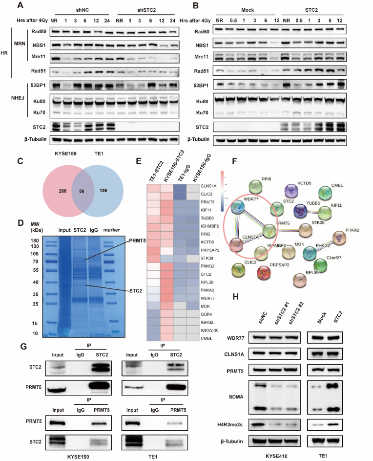
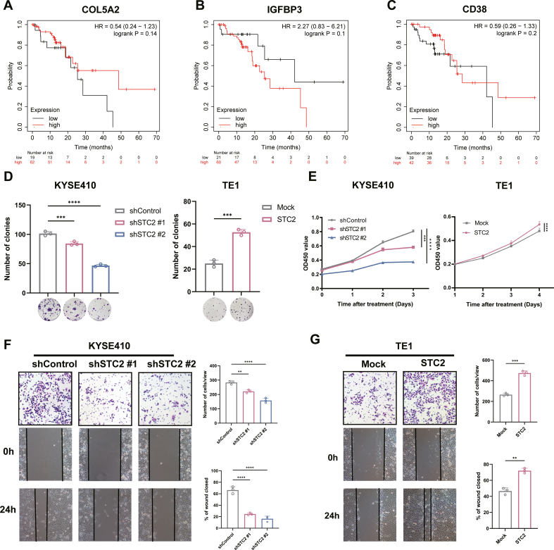
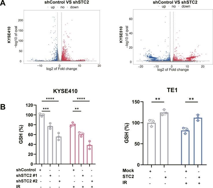
Radioresistance is the major reason for the failure of radiotherapy in esophageal squamous cell carcinoma (ESCC). Previous evidence indicated that stanniocalcin 2 (STC2) participates in various biological processes of malignant tumors. However, researches on its effect on radioresistance in cancers are limited. In this study, STC2 was screened out by RNA-sequencing and bioinformatics analyses as a potential prognosis predictor of ESCC radiosensitivity and then was determined to facilitate radioresistance. We found that STC2 expression is increased in ESCC tissues compared to adjacent normal tissues, and a higher level of STC2 is associated with poor prognosis. Also, STC2 mRNA and protein expression levels were higher in radioresistant cells than in their parental cells. Further investigation revealed that STC2 could interact with protein methyltransferase 5 (PRMT5) and activate PRMT5, thus leading to the increased expression of symmetric dimethylation of histone H4 on Arg 3 (H4R3me2s). Mechanistically, STC2 can promote DDR through the homologous recombination and non-homologous end joining pathways by activating PRMT5. Meanwhile, STC2 can participate in SLC7A11-mediated ferroptosis in a PRMT5-dependent manner. Finally, these results were validated through in vivo experiments. These findings uncovered that STC2 might be an attractive therapeutic target to overcome ESCC radioresistance.

Keywords: DNA damage Repair; Esophageal squamous cell carcinoma; Ferroptosis; PRMT5; Radioresistance; STC2.


Conflict of interest statement

Declaration of competing interest The authors declare no conflict of interest.

Figures

Fig. 1
Fig. 1
STC2 was identified as a potential prognosis predictor of ESCC radiosensitivity. a Volcano plot of differentially expressed genes (DEGs) between radioresistant cells and their normal parental cells. b Venn diagram of the co-upregulated DEGs among 2 RNA-sequencing datasets (TE1R/TE1, KYSE150R/KYSE150) and 3 datasets from the GEO database (GSE100942, GSE149609, GSE164158). c STC2 mRNA expression level in adjacent normal tissues and ESCC from the database. d Immunohistochemistry of STC2 expression in 24 ESCC tissues and their adjacent non-tumor tissues. Scale bar: 20 μm e Kaplan-Meier curves of ESCC survival based on the expression level of STC2 according to the TCGA database. f-g qRT-PCR and western blotting were performed to detect STC2 expression in radioresistant cancer cells compared with their normal cancer cells. ***p < 0.001; ****p < 0.0001.
Fig. 2
Fig. 2
STC2 facilitates radioresistance and promotes DNA damage repair (DDR) of ESCC after IR exposure. a Examination of STC2 protein levels in different ESCC cell lines. b The protein level of STC2 was detected in STC2-KD and -overexpressing cells transfected with the related lentivirus. c Colony formation assays and survival fraction of cells after irradiation with 0, 2, 4, and 8 Gy d The values of D0, Dq, N in each group. e Representative images of γH2AX-positive nuclei in STC2-KD and -overexpressing groups at different times post IR. The 0h means no IR. γH2AX foci in red, nuclear counterstaining with 4′,6-diamidino-2-phenylindole in blue. Scale bar: 20 μm f Comet assay was carried out in STC2-KD and -overexpressing cells at the indicated time points after IR treatment. Scale bar: 20 μm *p < 0.05; **p < 0.01; ***p < 0.001; ****p < 0.0001. (For interpretation of the references to colour in this figure legend, the reader is referred to the Web version of this article.)
Fig. 3
Fig. 3
STC2 promotes IR-induced HR and NHEJ and PRMT5 are identified as a target of STC2. a-b WB analysis of homologous recombination (HR) and non-homologous end joining (NHEJ) related proteins influenced by STC2 expression following 4Gy IR at different time points. NR means no IR. c Venn diagram of differential expressed gene between the anti-IgG group and anti-STC2 group in 2 overexpressing cells. d Coomassie blue-stained gel with protein. e The heatmap of top 20 proteins with significant fold change. f Protein-protein network of these top 20 significant genes. g Co-IP experiments were performed using either an STC2 antibody to pull down PRMT5 or a PRMT5 antibody to pull down STC2 in TE1-STC2 and KYSE150-STC2 cells. h The protein levels of WDR77, CLNS1A, PRMT5, SDMA and H4R3me2s were determined by western blotting in KYSE410-shSTC2 cells and TE1-STC2 cells. . (For interpretation of the references to colour in this figure legend, the reader is referred to the Web version of this article.)
Fig. 4
Fig. 4
STC2 enhances DNA damage repair of ESCC by activating PRMT5 a-b Mock and overexpressing-STC2 cells were treated with the indicated drugs. Representative pictures and summary of γH2AX foci formation after 12h when exposed to IR (4 Gy) Scale bar: 20 μm. c-d Neutral comet assay performed after exposure to irradiation (12h). Scale bar: 20 μm e Dose responses of survival factions of mock and overexpressing-STC2 cells (KYSE150, TE1) treated with indicated drugs. f Western blot assay of involved proteins in overexpressing-STC2 KYSE150 and TE1 cells treated with indicated drugs. *p < 0.05; **p < 0.01; ***p < 0.001; ****p < 0.0001.
Fig. 5
Fig. 5
STC2 is investigated to participate in ferroptosis with or without IR a Differential gene expression between shSTC2 cells and control cancer cells from RNA-Seq. b Kyoto Encyclopedia of Genes and Genomes (KEGG) analysis of differentially expressed proteins between STC2-depleted and control KYSE410 cells. c The heatmap of ferroptosis-related gene enriched in KEGG. d Transmission electron microscopy of STC2-depleted and control cells. e Representative images of intracellular levels of Fe2+ in STC2-depleted and -overexpressed cells. f The relative MDA level of STC2 depleted and overexpressing cells with or without IR (4Gy). g The relative total ROS levels were assayed via DCFH-DA fluorescence with or without IR (4Gy). h The relative lipid ROS levels were assayed via C11-BODIPY fluorescence with or without IR (4Gy). *p < 0.05; **p < 0.01; ***p < 0.001; ****p < 0.0001.
Fig. 6
Fig. 6
STC2 inhibits ferroptosis by targeting SLC7A11. a The relationship between STC2 and SLC7A11 in esophageal cancer (EC) was extracted from the GEPIA website. b The correlation between STC2 and SLC7A11 in ESCC was confirmed in the TCGA dataset. c The relationship between STC2 and SLC3A2 in EC was extracted from the GEPIA website. d The correlation between STC2 and SLC3A2 in ESCC was confirmed in the TCGA dataset. e Western blot of SLC7A11, SLC3A2, and GPX4 in KYSE410-shSTC2 cells and TE1-STC2 cells with or without IR (4Gy). f-h The relative MDA level of TE1-STC2 cells treated with RSL3, Erastin, and sulfasalazine. i-k The relative lipid ROS level of TE1-STC2 cells treated with RSL3, Erastin, and sulfasalazine. *p < 0.05; **p < 0.01; ***p < 0.001; ****p < 0.0001.
Fig. 7
Fig. 7
STC2 inhibits ferroptosis in a PRMT5-dependent manner. a-b PRMT5 inhibitor GSK332659 decreased the protein level of ATF4, SL7A11 and SLC3A2 in a concentration-dependent and time-dependent manner. c-d Lipid ROS of STC2-overexpressing cells treated with GSK332659(20 μM) when exposed to IR (4Gy). e The relative MDA level of cells treated with GSK332659(20 μM) and subjected to IR (4Gy). f The protein expression of ATF4, SLC7A11 and SLC3A2 were measured in cells treated with GSK332659(20 μM) and exposed to IR. *p < 0.05; **p < 0.01; ***p < 0.001; ****p < 0.0001.
Fig. 8
Fig. 8
STC2 facilitates radioresistance in ESCC in vivo. a Schematic representation of the xenograft study design and experimental workflow. b General view of tumor mass in each indicated group at 15 days after treatment. c Growth curves of xenograft tumors in each group. d Tumor weights were measured in each group. e Representative immunohistochemistry images of the expressions of STC2 and H4R3me2s protein in the xenograft tumors. Scale bar: 20 μm.
Fig. 9
Fig. 9
The mechanism of the study.
Fig. S1
Fig. S1
Fig. S2
Fig. S2
Fig. S3
Fig. S3
Fig. S4
Fig. S4
Fig. S5
Fig. S5

References

    1. Sung H., Ferlay J., Siegel R.L., Laversanne M., Soerjomataram I., Jemal A., Bray F. Global cancer statistics 2020: GLOBOCAN estimates of incidence and mortality worldwide for 36 cancers in 185 countries. CA A Cancer J. Clin. 2021;71:209–249. - PubMed
    1. Chen W., Zheng R., Baade P.D., Zhang S., Zeng H., Bray F., Jemal A., Yu X.Q., He J. Cancer statistics in China, 2015. CA A Cancer J. Clin. 2016;66:115–132. - PubMed
    1. Zhou M., Wang H., Zeng X., Yin P., Zhu J., Chen W., Li X., Wang L., Wang L., Liu Y., Liu J., Zhang M., Qi J., Yu S., Afshin A., Gakidou E., Glenn S., Krish V.S., Miller-Petrie M.K., Mountjoy-Venning W.C., Mullany E.C., Redford S.B., Liu H., Naghavi M., Hay S.I., Wang L., Murray C.J.L., Liang X. Mortality, morbidity, and risk factors in China and its provinces, 1990-2017: a systematic analysis for the Global Burden of Disease Study 2017. Lancet. 2019;394:1145–1158. - PMC - PubMed
    1. Eyck B.M., van Lanschot J.J.B., Hulshof M., van der Wilk B.J., Shapiro J., van Hagen P., van Berge Henegouwen M.I., Wijnhoven B.P.L., van Laarhoven H.W.M., Nieuwenhuijzen G.A.P., Hospers G.A.P., Bonenkamp J.J., Cuesta M.A., Blaisse R.J.B., Busch O.R., Creemers G.M., Punt C.J.A., Plukker J.T.M., Verheul H.M.W., Spillenaar Bilgen E.J., van der Sangen M.J.C., Rozema T., Ten Kate F.J.W., Beukema J.C., Piet A.H.M., van Rij C.M., Reinders J.G., Tilanus H.W., Steyerberg E.W., van der Gaast A., Group C.S. Ten-year outcome of neoadjuvant chemoradiotherapy plus surgery for esophageal cancer: the randomized controlled CROSS trial. J. Clin. Oncol. 2021;39:1995–2004. - PubMed
    1. van Hagen P., Hulshof M.C., van Lanschot J.J., Steyerberg E.W., van Berge Henegouwen M.I., Wijnhoven B.P., Richel D.J., Nieuwenhuijzen G.A., Hospers G.A., Bonenkamp J.J., Cuesta M.A., Blaisse R.J., Busch O.R., ten Kate F.J., Creemers G.J., Punt C.J., Plukker J.T., Verheul H.M., Spillenaar Bilgen E.J., van Dekken H., van der Sangen M.J., Rozema T., Biermann K., Beukema J.C., Piet A.H., van Rij C.M., Reinders J.G., Tilanus H.W., van der Gaast A., Group C. Preoperative chemoradiotherapy for esophageal or junctional cancer. N. Engl. J. Med. 2012;366:2074–2084. - PubMed

MeSH terms

Substances