. 2023 Mar 2;186(5):1039-1049.e17.
doi: 10.1016/j.cell.2023.01.013. Epub 2023 Feb 9.

A molecular device for the redox quality control of GroEL/ES substrates

Affiliations

A molecular device for the redox quality control of GroEL/ES substrates

Emile Dupuy et al. Cell. .

Abstract

Hsp60 chaperonins and their Hsp10 cofactors assist protein folding in all living cells, constituting the paradigmatic example of molecular chaperones. Despite extensive investigations of their structure and mechanism, crucial questions regarding how these chaperonins promote folding remain unsolved. Here, we report that the bacterial Hsp60 chaperonin GroEL forms a stable, functionally relevant complex with the chaperedoxin CnoX, a protein combining a chaperone and a redox function. Binding of GroES (Hsp10 cofactor) to GroEL induces CnoX release. Cryoelectron microscopy provided crucial structural information on the GroEL-CnoX complex, showing that CnoX binds GroEL outside the substrate-binding site via a highly conserved C-terminal α-helix. Furthermore, we identified complexes in which CnoX, bound to GroEL, forms mixed disulfides with GroEL substrates, indicating that CnoX likely functions as a redox quality-control plugin for GroEL. Proteins sharing structural features with CnoX exist in eukaryotes, suggesting that Hsp60 molecular plugins have been conserved through evolution.

Keywords: TPR; chaperones; chaperonin; protein folding; proteostasis; redox; thioredoxin.


Conflict of interest statement

Declaration of interests The authors declare no competing interests.

Figures
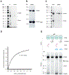
Figure 1.
Figure 1.. CnoX interacts stably with GroEL.
(A) GroEL co-elutes with CnoX when CnoX is pulled down from wild-type cell extracts using α-CnoX antibodies. Both proteins are absent when the experiment is repeated with extracts prepared from the ΔcnoX mutant. The image of sodium dodecyl sulphate–polyacrylamide gel electrophoresis (SDS-PAGE), stained with Coomassie blue, is representative of >3 replicates. * indicates the light and heavy chains of the antibodies. (B) Purified CnoXN-Strep and GroEL form a complex that can be isolated using streptavidin affinity purification. Two fractions are shown. (C) Purified CnoX and GroEL form a complex that can be isolated using size-exclusion chromatography. (D) Formation of a complex between FM-CnoX and GroEL can be monitored using fluorescence anisotropy. The noncooperative model gives an adequate fit to these data, with a Kd of 310 ± 10 nM. (E) CnoX and unfolded CS co-elute with GroEL from a gel filtration column. Addition of GroES triggers the release of CnoX from GroEL, while CS remains bound to GroEL. Size-exclusion chromatography was performed in the presence of ADP (50 μM), and fractions were analyzed by SDS-PAGE. The results are representative of >3 experiments.
Figure 2.
Figure 2.. CryoEM shows that the TPR domain of CnoX binds GroEL.
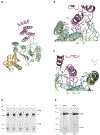
(A, B) CryoEM 2D class averages of the GroEL-CnoX complex reconstituted in vitro at a 10:1 molar ratio (scale bar: 100 Å). (C, D) Side and top view of the structure of the GroEL-CnoX complex shown as a solvent accessible surface. The equatorial, intermediate, and apical domains of GroEL are shown in slate, orange, and light cyan, respectively, and CnoX is shown in pink.
Figure 3.
Figure 3.. The C-terminal α-helix of CnoX binds a shallow cleft in the apical domain of GroEL.
(A) Ribbon representation of a single GroEL-CnoX protomer. CnoX binds GroEL via its C-terminal α-helix. The intermediate and apical domains of GroEL are shown in orange and light cyan, respectively. CnoX is shown in pink. For comparison, the GroEL-CnoX structure is shown superimposed on the structure of T-state GroEL (yellow; Protein Data Bank [PDB]: 1GRL). (B, C) Close-up views of the GroEL-CnoX binding interface. CnoX binds GroEL through the following hydrogen-bond and electrostatic interactions (CnoX-GroEL): R255-E304, R277-G298, R277-T299, Y284-E304, and Y284 C-term-R345. For comparison, the GroEL-CnoX structure is shown superimposed on the structure of T-state GroEL (yellow; PDB: 1GRL). (D) GroEL co-elutes with CnoX (lane 1) but not with CnoXΔCter (lane 2), CnoXR277L (lane 3), CnoXY284L (lane 4), or CnoXC-His (lane 5) when CnoX is pulled down from cell extracts using α-CnoX antibodies. In these experiments, CnoX and its variants were expressed in ΔcnoX cells. The SDS-PAGE gel, stained with Coomassie blue, is representative of >3 replicates. * indicates the light and heavy chains of the antibodies. (E) GroEL§, a GroEL variant with mutations in the CnoX-binding site (G298A/T299L/V300K/E304L/I305K/M307K/R345L), does not elute together with CnoX from a size-exclusion chromatography column (right), in contrast to wild-type GroEL (left). Three consecutive elution fractions are shown for each chromatography column.
Figure 4.
Figure 4.. CnoX functions as a molecular plugin to rescue GroEL substrates from oxidative damage.
(A) CnoX co-elutes with GroEL when the chaperonin is pulled down from wild-type cell extracts using specific antibodies. High-molecular-weight complexes corresponding to dithiothreitol (DTT)-sensitive mixed disulfides are detected by α-CnoX antibodies. These complexes are not detected when the experiment is repeated using extracts from cells expressing a CnoX mutant lacking the two cysteine residues, CnoXno_cys. (B) Obligate GroEL substrates trapped in mixed-disulfide complexes with CnoX and pulled down using α-GroEL antibodies were identified by liquid chromatography with tandem MS (LC-MS/MS). (C) Model: 1. CnoX forms a stable complex with GroEL via its C-terminal α-helix in a nucleotide-independent manner. Positioned on the apical domain of GroEL, CnoX interacts with incoming substrates for GroEL, acting as a redox quality-control plugin. 2. If the substrate that reaches GroEL for folding presents oxidized cysteine residues (to a sulfenic acid or in a disulfide bond), CnoX reacts with the substrate via the cysteines of its thioredoxin domain, and a mixed disulfide is formed. Interactions between the substrate and the GroEL cavity occur. 3. Cytoplasmic reducing pathways reduce the mixed disulfide, releasing the substrate in a reduced, folding-competent state. 4. GroES binding triggers CnoX release from GroEL and encapsulation of the substrate within the folding cage for folding.
Figure 5.
Figure 5.. Unfolded CS is present in the cavity of GroEL when in a mixed disulfide with CnoX.
(A) Co-overexpression of CnoXN-Strep, CS, and GroEL leads to the formation of a ternary complex that was purified by affinity chromatography (left) and size-exclusion chromatography (right). (B) The ternary complex was treated with disuccinimidyl sulfoxide (DSSO) —a crosslinker with an amine-reactive N-hydroxysuccinimide ester at each end of a 7-carbon spacer arm— and subjected to proteolytic digestion with trypsin. The resulting peptide mixture was analyzed by LC-MS/MS. Seven crosslinked peptides between GroEL residues and CS were detected. (C) Four of the GroEL residues that crosslink to CS face the inside of the cavity.

References

    1. Anfinsen CB (1973). Principles that govern the folding of protein chains. Science 181, 223–230. - PubMed
    1. Jahn TR, and Radford SE (2005). The Yin and Yang of protein folding. The FEBS journal 272, 5962–5970. - PubMed
    1. Ellis RJ (2001). Macromolecular crowding: obvious but underappreciated. Trends in biochemical sciences 26, 597–604. - PubMed
    1. Hartl FU, Bracher A, and Hayer-Hartl M (2011). Molecular chaperones in protein folding and proteostasis. Nature 475, 324–332. 10.1038/nature10317. - DOI - PubMed
    1. Goloubinoff P, Christeller JT, Gatenby AA, and Lorimer GH (1989). Reconstitution of active dimeric ribulose bisphosphate carboxylase from an unfolded state depends on two chaperonin proteins and Mg-ATP. Nature 342, 884–889. - PubMed

Publication types