. 2023 Mar;299(3):103001.
doi: 10.1016/j.jbc.2023.103001. Epub 2023 Feb 9.

Structure of VanS from vancomycin-resistant enterococci: A sensor kinase with weak ATP binding

Affiliations

Structure of VanS from vancomycin-resistant enterococci: A sensor kinase with weak ATP binding

Kimberly C Grasty et al. J Biol Chem. 2023 Mar.

Abstract

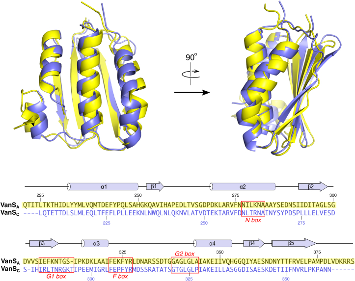
The VanRS two-component system regulates the resistance phenotype of vancomycin-resistant enterococci. VanS is a sensor histidine kinase that responds to the presence of vancomycin by autophosphorylating and subsequently transferring the phosphoryl group to the response regulator, VanR. The phosphotransfer activates VanR as a transcription factor, which initiates the expression of resistance genes. Structural information about VanS proteins has remained elusive, hindering the molecular-level understanding of their function. Here, we present X-ray crystal structures for the catalytic and ATP-binding (CA) domains of two VanS proteins, derived from vancomycin-resistant enterococci types A and C. Both proteins adopt the canonical Bergerat fold that has been observed for CA domains of other prokaryotic histidine kinases. We attempted to determine structures for the nucleotide-bound forms of both proteins; however, despite repeated efforts, these forms could not be crystallized, prompting us to measure the proteins' binding affinities for ATP. Unexpectedly, both CA domains displayed low affinities for the nucleotide, with KD values in the low millimolar range. Since these KD values are comparable to intracellular ATP concentrations, this weak substrate binding could reflect a way of regulating expression of the resistance phenotype.

Keywords: ATP binding; antibiotic resistance; histidine kinase; two-component system; vancomycin-resistant enterococci.


Conflict of interest statement

Conflict of interest The authors declare that they have no conflicts of interest with the contents of this article.

Figures

Figure 1
Figure 1
Structures of the VanSAand VanSCCA domains.A, schematic illustration of the VanS domain structure. B, ribbon diagram showing the VanSA CA-domain structure, colored using a “rainbow” scheme that ranges from blue at the N terminus to red at the C terminus. The numbering of the secondary-structure elements is also illustrated on the VanSA structure (this numbering is the same for both VanSA and VanSC). C, the VanSC CA-domain structure, with key structural motifs highlighted (these motifs are present in both VanSA and VanSC). These motifs include the N-box (red; the two conserved Asn residues are shown), the G1-box (yellow, including the noncanonical Asn residue found in both VanSA and VanSC), the F-box (magenta), and the G2-box (blue). Stereo versions of B and C can be found in Fig. S12. CA, catalytic ATP-binding domain.
Figure 2
Figure 2
Comparison of the VanSAand VanSCCA domains. Top, superposition of VanSA and VanSC, shown in two orthogonal orientations. VanSA is colored yellow, whereas VanSC is shown in slate blue. Below is shown the pairwise sequence alignment of the two CA domains. Secondary structural elements and conserved sequence motifs are indicated. CA, catalytic ATP-binding domain.
Figure 3
Figure 3
Nucleotide binding by the VanSAand VanSCCA domains. A–C, binding experiments using TNP–ATP. The insets show direct binding of TNP–ATP, whereas the main figures show competition assays in which TNP–ATP is displaced by ATP. Lines show fits of the appropriate binding expressions to the data. A, VanSA CA domain; B, VanSC CA domain; C, EnvZ CA domain. D, equilibrium dialysis binding experiments with ATP. Solid black lines represent fits of Equation 2 to the data. No attempt was made to fit a binding isotherm to the data for the N291D mutant of VanSC, since no evidence for binding was seen. Dotted lines in different colors show the theoretical binding curves for selected KD values, as calculated from Equation 2. Dissociation constants inferred from the binding experiments in this figure are given in Table 2. E, divergent stereo view of the nucleotide-binding site of VanSA (magenta), superimposed upon the ATP-bound structure of YycG from Bacillus subtilis (gray; Protein Data Bank ID: 3SL2). The magnesium ion in the YycG structure is shown as a yellow sphere. Selected amino-acid side chains in and around the nucleotide-binding site are shown. CA, catalytic ATP-binding domain.
Figure 4
Figure 4
Comparison of VanSAand VanSCwith other prokaryotic CA domains.A, superposition of the VanSA and VanSC CA domains with 12 other CA domains, as performed using the program MULTIPROT (35). Details of the comparison structures can be found in Fig. S8. The different proteins adopt highly similar conformations, with the exception of the region between the G1-box and helix 4, which includes the ATP lid. This region lies at the lower left in this panel. B, comparison of the nucleotide conformations for the eight structures containing bound nucleotide. A structure cartoon for CpxA (light gray) is included to provide structural context. C, comparison of the region between the G1-box and helix α4. All structures were superimposed using MULTIPROT and are shown in the same orientation; the cartoon at the lower right illustrates the secondary structure elements involved. Color coding in this panel is the same as that used in A. Structures containing bound nucleotide are identified with an asterisk. The entire ATP lid is disordered in SrrB, which is therefore omitted from C. CA, catalytic ATP-binding domain.

References

    1. Levine D.P. Vancomycin: a history. Clin. Infect. Dis. 2006;42:S5–S12. - PubMed
    1. Joshi S., Shallal A., Zervos M. Vancomycin-resistant Enterococci: epidemiology, infection prevention, and control. Infect Dis. Clin. North Am. 2021;35:953–968. - PubMed
    1. Tacconelli E., Carrara E., Savoldi A., Harbarth S., Mendelson M., Monnet D.L., et al. Discovery, research, and development of new antibiotics: the WHO priority list of antibiotic-resistant bacteria and tuberculosis. Lancet Infect. Dis. 2018;18:318–327. - PubMed
    1. Rice L.B. Federal funding for the study of antimicrobial resistance in nosocomial pathogens: no ESKAPE. J. Infect Dis. 2008;197:1079–1081. - PubMed
    1. Loll P.J., Axelsen P.H. The structural biology of molecular recognition by vancomycin. Annu. Rev. Biophys. Biomol. Struct. 2000;29:265–289. - PubMed

Publication types

MeSH terms

LinkOut - more resources