Review
. 2023 Mar;16(3):408-425.
doi: 10.1016/j.jcmg.2022.12.026. Epub 2023 Feb 8.

Excessive Trabeculation of the Left Ventricle: JACC: Cardiovascular Imaging Expert Panel Paper

Affiliations
Review

Excessive Trabeculation of the Left Ventricle: JACC: Cardiovascular Imaging Expert Panel Paper

Steffen E Petersen et al. JACC Cardiovasc Imaging. 2023 Mar.

Abstract

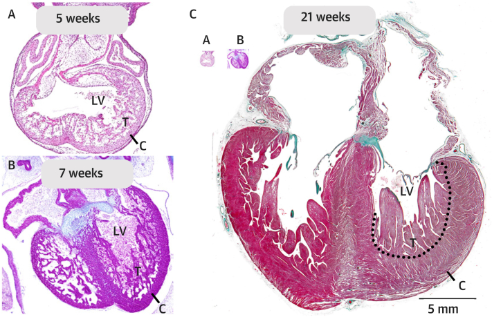
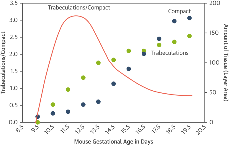
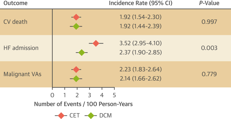
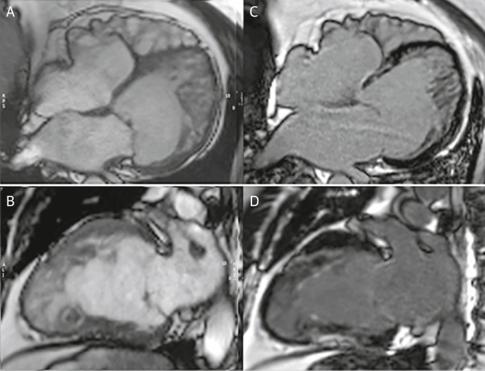
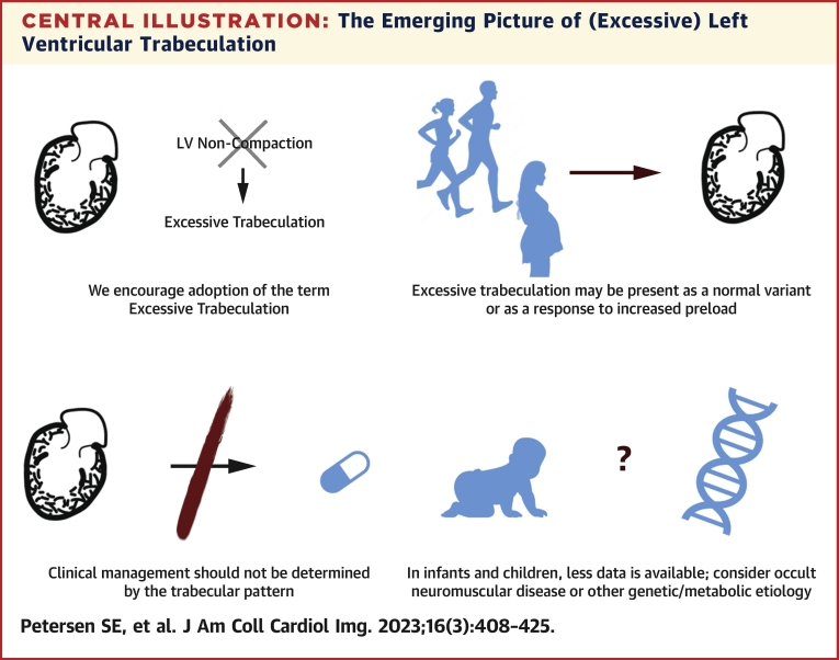
Excessive trabeculation, often referred to as "noncompacted" myocardium, has been described at all ages, from the fetus to the adult. Current evidence for myocardial development, however, does not support the formation of compact myocardium from noncompacted myocardium, nor the arrest of this process to result in so-called noncompaction. Excessive trabeculation is frequently observed by imaging studies in healthy individuals, as well as in association with pregnancy, athletic activity, and with cardiac diseases of inherited, acquired, developmental, or congenital origins. Adults with incidentally noted excessive trabeculation frequently require no further follow-up based on trabecular pattern alone. Patients with cardiomyopathy and excessive trabeculation are managed by cardiovascular symptoms rather than the trabecular pattern. To date, the prognostic role of excessive trabeculation in adults has not been shown to be independent of other myocardial disease. In neonates and children with excessive trabeculation and normal or abnormal function, clinical caution seems warranted because of the reported association with genetic and neuromuscular disorders. This report summarizes the evidence concerning the etiology, pathophysiology, and clinical relevance of excessive trabeculation. Gaps in current knowledge of the clinical relevance of excessive trabeculation are indicated, with priorities suggested for future research and improved diagnosis in adults and children.

Keywords: cardiovascular imaging; clinical management; diagnosis; etiology; left ventricular noncompaction; prognosis.


Conflict of interest statement

Funding Support and Author Disclosures Dr Petersen has received support from the National Institute for Health and Care Research Barts Biomedical Research Centre; and has consulted with Circle Cardiovascular Imaging Inc. Dr Friedrich has received support from the McGill Health Centre Foundation; has served as an advisor to and is a shareholder of Circle Cardiovascular Imaging Inc; and is a founder and shareholder of Area19 Medical Inc. All other authors have reported that they have no relationships relevant to the contents of this paper to disclose.

Figures

None
Graphical abstract
Figure 1
Figure 1
Developmental Anatomy of the Human LV (A) At 5 weeks gestation, Carnegie stage 14 is shown; a substantial trabeculated (T) wall has developed; and the compact wall (C) is thin. (B) At 7 weeks gestation, Carnegie stage 19 is shown, and the ventricular wall remains much trabeculated. (C) At 21 weeks gestation, the heart (and fetus) has grown tremendously, notice the images of the 2 embryonic hearts of A and B are inserted to scale. The compact wall is now much thicker than in the embryonic stages and so is the trabecular layer. This illustrates that a decrease of the trabecular layer, that is, compaction, is not required for the formation of a thick compact wall. LV = left ventricle.
Figure 2
Figure 2
How Development Shapes LV Wall Morphology Developmental changes to the proportions of trabecular and compact myocardium (red line) are driven by different rates of positive growth, not compaction, of the trabeculations (green dots) and compact tissue (blue dots) that vary by gestational age. Such differing rates of growth of different parts of the body is termed “allometric growth” and is a frequent driver of morphologic change in development and in nature. The graphs are redrawn based on data from a mouse model. Abbreviation as in Figure 1.
Figure 3
Figure 3
Prognosis of CET and DCM Prognostic comparison between dilated cardiomyopathy (DCM) (green diamonds) and cardiomyopathy with excessive trabeculation (CET) (red diamonds) based on previously published meta-analyses., Because conditions of greater preload and afterload are associated with excessive trabeculation, excessive trabeculation as a cause of HF admission should not be inferred from meta-analyses. CV = cardiovascular; HF = heart failure; VA = ventricular arrhythmia.
Figure 4
Figure 4
Case Report 1 A 63-year-old man presented with a history of nonsustained ventricular tachycardia and paroxysmal atrial fibrillation treated with atrial ablation. CMR was performed for further evaluation. Cine images at end-diastole in 4-chamber (A) (Video 1) and 2-chamber long-axis views (B) (Video 2) show excessive trabeculation with biventricular dilation with an EF of 48%. Scattered areas of late gadolinium enhancement were present with a nonischemic pattern (C and D). Genetic testing showed a MYH7 allelic variant. Stress perfusion cardiac magnetic resonance (not shown) showed diffuse perfusion abnormalities in multiple myocardial segments. Although excessive trabeculation is present, the presentation of ventricular dilatation, low EF, and nonischemic myocardial scar and genetic abnormality is the same as in dilated cardiomyopathy. Patient treatment is based on the symptoms and the prognostic risks of arrhythmia, stroke, and contractile impairment. CMR = cardiac magnetic resonance; EF = ejection fraction.
Figure 5
Figure 5
Case Report 2 (A) Fetal echocardiogram at 25 weeks in a child with hydrops fetalis demonstrated severely depressed biventricular systolic dysfunction with excessive trabeculation (Video 3). (B) Supraventricular tachycardia in this fetus developed 1 week later. Forty-eight hours after delivery, left ventricular EF reduced to 33% (Video 4). (C) Five years later, the same patient demonstrated progressive left ventricular dilatation with an left ventricular end-diastolic diameter Z score of +2.7 and borderline EF of 50% and global longitudinal strain of −17% (Video 5). The working diagnosis was cardiomyopathy with excessive trabeculation. Neonates and children with excessive trabeculation have been understudied, with low rates of longitudinal follow-up. Such patients should routinely undergo follow-up, with close clinical evaluation and potentially neuromuscular disease testing. If familial disease is suspected, genetic testing may be indicated. Abbreviation as in Figure 4.
Figure 6
Figure 6
Case Report 3 (A) A 3-month-old boy presented with biventricular systolic dysfunction with excessive trabeculation (Video 6). (B) At 6 months of age, this evolved to a restrictive phenotype requiring placement of a left ventricular assist device (Berlin heart) (Video 7). At 12-months of age (not shown), the patient underwent orthotopic heart transplantation. The clinical management and significance of excessive trabeculation and associated disorders in young patients is not well established. As in this example, the evolution of myocardial dysfunction may not be predictable on baseline examination. For patients with myocardial dysfunction in particular, close clinical follow-up is suggested.
Figure 7
Figure 7
Case Report 4 Images of a 38-year-old master triathlete with history of catheter ablation for persistent atrial fibrillation. Echocardiography at preparticipation screening documented the presence of mildly reduced left ventricular systolic function, mild eccentric aortic regurgitation, and a severely dilated LV with excessive trabeculation (positive Jenni and Chin criteria) that resulted in the diagnosis of so-called left ventricular noncompaction. CMR confirmed the presence of a severely dilated LV with mildly impaired systolic function and excessive trabeculation (A) according to the Petersen criteria (noncompacted [trabecular] to compact layer ratio: 3.1). Severe eccentric aortic regurgitation was present (B and C) in addition to a thrombus within the left atrial appendage (A and D). Six months after surgical aortic valve replacement (E) and left atrial appendage occlusion, significant left ventricular reverse remodeling occurred with near-complete normalization of left ventricular function, volumes, and diameters (end-diastolic frame [F]; end-systolic frame [G]), further leading to a reduction of Petersen criteria for excessive trabeculation (noncompacted [trabecular] to compact layer ratio: 2.4). Moderate-to-severe left ventricular dilatation must be carefully investigated in athletes, irrespective of extent of ventricular trabeculation. If the excessive trabeculation had been part of a cardiomyopathy, arguably, these substantial improvements in left ventricular volumes and function would not have been observed. RF = regurgitant fraction; RVol = regurgitant volume; other abbreviations as in Figures 1 and 4.
Central Illustration
The Emerging Picture of (Excessive) Left Ventricular Trabeculation LV = left ventricular.

Comment in

  • Discourage LVNC or Revise the Criteria of LVNC?
    Wang J, Han Y, Chen Y. Wang J, et al. JACC Cardiovasc Imaging. 2023 Jun;16(6):868. doi: 10.1016/j.jcmg.2023.03.024. JACC Cardiovasc Imaging. 2023. PMID: 37286273 No abstract available.
  • Reply: Discourage LVNC or Revise the Criteria of LVNC?
    Petersen SE, Jensen B, Aung N, Friedrich MG, McMahon CJ, Mohiddin SA, Pignatelli RH, Ricci F, Anderson RH, Bluemke DA. Petersen SE, et al. JACC Cardiovasc Imaging. 2023 Jun;16(6):869. doi: 10.1016/j.jcmg.2023.03.023. JACC Cardiovasc Imaging. 2023. PMID: 37286274 No abstract available.

References

    1. Kawel N., Nacif M., Arai A.E., et al. Trabeculated (noncompacted) and compact myocardium in adults: the multi-ethnic study of atherosclerosis. Circ Cardiovasc Imaging. 2012;5(3):357–366. - PMC - PubMed
    1. Oechslin E., Jenni R. Left ventricular noncompaction: from physiologic remodeling to noncompaction cardiomyopathy. J Am Coll Cardiol. 2018;71(7):723–726. - PubMed
    1. Jenni R., Oechslin E., Schneider J., Attenhofer Jost C., Kaufmann P.A. Echocardiographic and pathoanatomical characteristics of isolated left ventricular non-compaction: a step towards classification as a distinct cardiomyopathy. Heart. 2001;86(6):666–671. - PMC - PubMed
    1. Gati S., Chandra N., Bennett R.L., et al. Increased left ventricular trabeculation in highly trained athletes: do we need more stringent criteria for the diagnosis of left ventricular non-compaction in athletes? Heart. 2013;99(6):401–408. - PubMed
    1. Gati S., Papadakis M., Papamichael N.D., et al. Reversible de novo left ventricular trabeculations in pregnant women: implications for the diagnosis of left ventricular noncompaction in low-risk populations. Circulation. 2014;130(6):475–483. - PubMed

MeSH terms