. 2023 Feb 10;14(2):104.
doi: 10.1038/s41419-023-05624-3.

A vector-encoded bispecific killer engager to harness virus-activated NK cells as anti-tumor effectors

Affiliations

A vector-encoded bispecific killer engager to harness virus-activated NK cells as anti-tumor effectors

Alessia Floerchinger et al. Cell Death Dis. .

Abstract

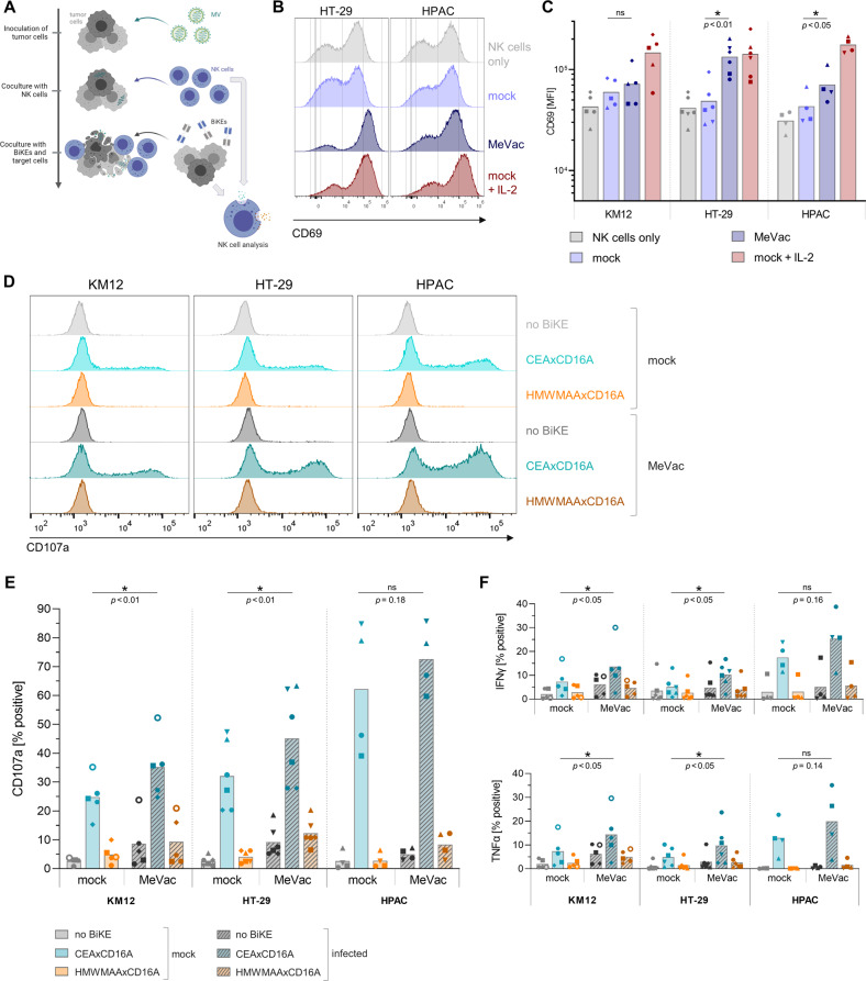
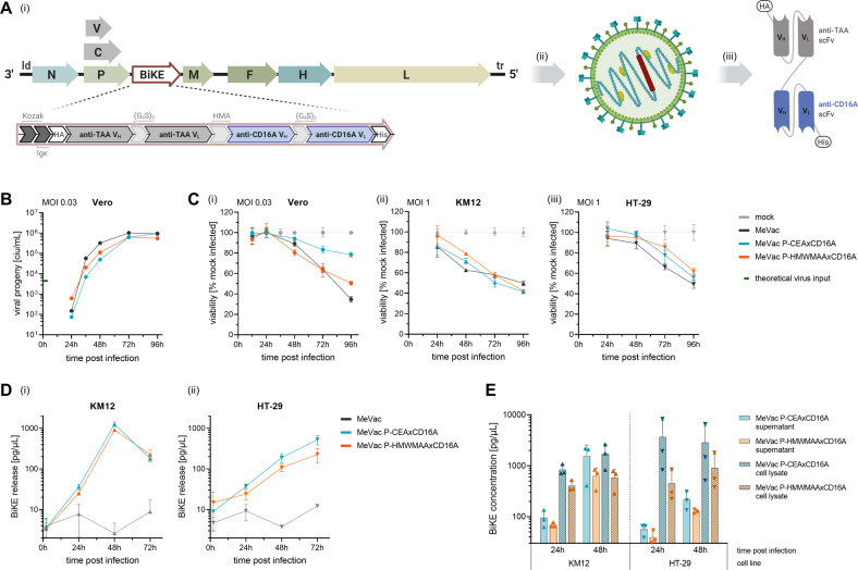
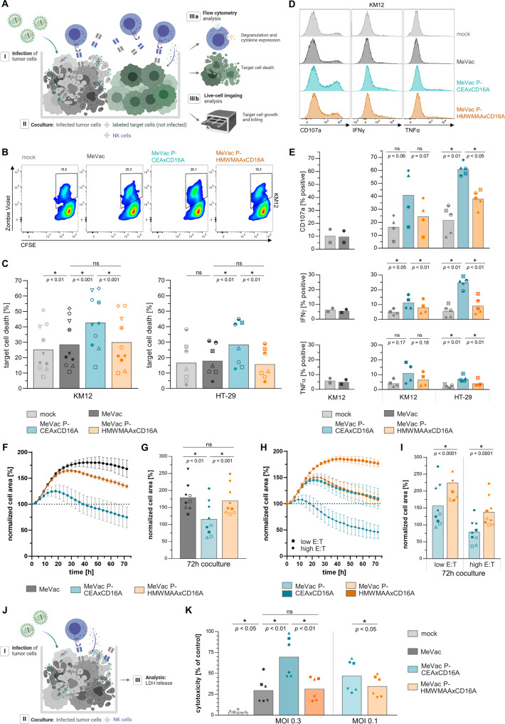
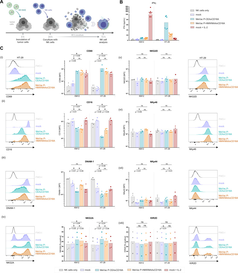
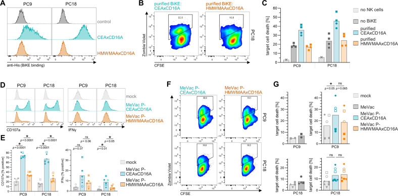
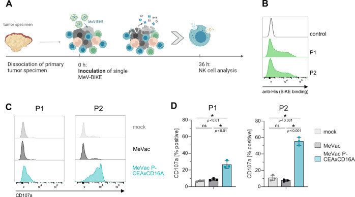
Treatment with oncolytic measles vaccines (MV) elicits activation of immune cells, including natural killer (NK) cells. However, we found that MV-activated NK cells show only modest direct cytotoxic activity against tumor cells. To specifically direct NK cells towards tumor cells, we developed oncolytic measles vaccines encoding bispecific killer engagers (MV-BiKE) targeting CD16A on NK cells and carcinoembryonic antigen (CEA) as a model tumor antigen. MV-BiKE are only slightly attenuated compared to parental MV and mediate secretion of functional BiKE from infected tumor cells. We tested MV-BiKE activity in cocultures of colorectal or pancreatic cancer cells with primary human NK cells. MV-BiKE mediate expression of effector cytokines, degranulation and specific anti-tumor cytotoxicity by NK cells. Experiments with patient-derived pancreatic cancer cultures indicate that efficacy of MV-BiKE may vary between individual tumors with differential virus permissiveness. Remarkably, we confirmed MV-BiKE activity in primaryhuman colorectal carcinoma specimens with autochthonous tumor and NK cells.This study provides proof-of-concept for MV-BiKE as a novel immunovirotherapy to harness virus-activated NK cells as anti-tumor effectors.


Conflict of interest statement

GU acts as CMO, CSO, and COO of CanVirex, a company developing oncolytic viruses as cancer immunotherapeutics. All other authors declare no competing interests.

Figures

Fig. 1
Fig. 1. NK cell activation and effector functions in MV therapy combined with bispecific killer engagers.
NK cell activation upon exposure to colorectal cancer (KM12, HT-29) or pancreatic ductal adenocarcinoma (HPAC) cells infected with recombinant measles vaccine strain virus (MV) was assessed in vitro. NK cell degranulation and cytokine expression were monitored in response to MV therapy with subsequent BiKE treatment. A Schematic outline of combination therapy experiments. Colorectal cancer (KM12, HT-29) or pancreatic ductal adenocarcinoma (HPAC) cells were inoculated with MV at a multiplicity of infection (MOI) of 1 or subjected to mock infection one day prior to coculture with NK cells isolated from healthy donor PBMCs. After 12 h co-incubation, i.e., 36 h post inoculation, expression of the early activation marker CD69 was analyzed by flow cytometry. NK cell only control and IL-2 stimulated positive control were included for comparison. In further experiments, BiKEs were purified from supernatants of infected Vero producer cells (vBiKEs). Non-infected target cells and vBiKEs were added to the coculture and incubated for 4 h. NK cell degranulation and cytokine expression were analyzed by flow cytometry. B Exemplary histograms depict expression levels of CD69 on live CD45+ CD56+ NK cells after coculture with infected cells compared to control conditions. C CD69 mean fluorescence intensity (MFI) is summarized for n ≥ 4 donors for each cell line with different symbols representing individual donors. D NK cell degranulation (CD107a) in response to MeVac and vBiKE monotherapy or combination therapy is shown in exemplary histograms for one donor. E Percentages of CD107a+ NK cells, F IFNγ+ and TNFα+ NK cells in different conditions are summarized for n ≥ 4 donors per cell line with different symbols representing individual donors. Statistical analysis was performed by paired t-test for each target cell line.
Fig. 2
Fig. 2. Oncolytic measles virus encoding BiKEs.
A Schematic of recombinant MV-BiKE. (i) The BiKE transgene is encoded downstream of the P open reading frame within the MV genome. Hemagglutinin (HA) and hexa histidine (His6) tags enable purification and detection. Kozak and Igκ leader sequences mediate efficient translation and secretion. (G4S)3, glycine-serine peptide linker; HMA, human muscle aldolase linker; scFv, single chain variable fragment with variable heavy (VH) and variable light (VL) chain; TAA, tumor-associated antigen. (ii) Transgene-encoding virus can be rescued via a reverse genetics system and triggers (iii) BiKE secretion upon infection. BiKEs included in this study target either human carcinoembryonic antigen (CEA) as model tumor antigen or human high molecular weight melanoma-associated antigen (HMWMAA) as a non-relevant control. B Replication kinetics of MV-BiKE. Vero producer cells were inoculated with MeVac P-BiKE or unmodified MeVac at MOI 0.03. Viral progeny at designated time points were determined via serial dilution titration assay to obtain a multistep growth curve. C Direct cytotoxic effect of MV-BiKE. Cells were infected with respective viruses at MOI 0.03 for Vero (i) or at MOI 1 for colorectal cancer cell lines KM12 (ii) and HT-29 (iii). Cell viability was determined via XTT assay and normalized to mock-infected control. D, E BiKE expression kinetics on infected colorectal cancer cells. KM12 and HT-29 cells were inoculated with MeVac P-BiKE or MeVac at MOI 1. Supernatants and cell lysates were collected at indicated time points. BiKE concentrations were determined via ELISA using anti-His6 and anti-HA antibodies and purified vBiKEs as standard. D Kinetics of BiKE concentrations in culture supernatants after infection with MV-BiKE. E Concentrations of cell-associated BiKE in tumor cell lysates and BiKE release into culture supernatants. In CE mean values of technical triplicates are shown. Error bars indicate SD.
Fig. 3
Fig. 3. Efficacy of MV-BiKE immunovirotherapy against infected and bystander colorectal cancer cells.
A Schematic outline of in vitro coculture experiments. MV-BiKE infected colorectal cancer cells were cocultured with NK cells and non-infected bystander tumor cells labeled with CFSE or tagRFP. NK cell degranulation, cytokine expression and killing of CSFE+ target cells were analyzed by flow cytometry. The tagRFP+ tumor cell area was monitored for 72 h using live-cell imaging. Recombinant MV encoding the tumor-targeting CEAxCD16A BiKE are compared to mock-infected control, MV without transgene and MV encoding an HMWMAAxCD16A control BiKE with irrelevant specificity. BE Flow cytometry analysis of MV-BiKE efficacy. B Exemplary pseudocolor plots for KM12 depict CFSE+ target cell viability based on Zombie Violet labeling of dead cells. C Mean bystander target cell death per treatment is shown for n = 10 donors (KM12) and n = 7 donors (HT-29) with individual donors represented by different symbols. D NK cell degranulation and cytokine expression in response to MV-BiKE therapy. Exemplary histograms for KM12 target cells illustrate CD107a surface levels and intracellular cytokine accumulation. E Mean percentages of CD107a+, IFNγ+ and TNFα+ NK cells, respectively, are summarized for n = 4 donors (KM12) and n = 5 donors (HT-29) with individual donors represented by symbols as in C. Mock controls compared to MeVac are shown separately for two donors (left panels). FI Live-cell imaging analysis of MV-BiKE efficacy against HT-29. Dotted line indicates 100%. F Exemplary time course plots show the kinetics of bystander tumor cell area during coculture. Mean and SD for three donors in one out of three independent experiments is shown (compare Fig. S4). G Normalized cell area after 72 h coculture is summarized for n = 9 donors. H Time course plots as in (F) illustrate efficacy of MV-BiKE treatment at higher and lower E:T ratios (shown in darker and lighter color, respectively), with quantification for n = 9 donors in three independent experiments in (I). J Schematic depicting the coculture of MV-BiKE infected HT-29 cells with NK cells for analysis of LDH release into culture supernatants. K Cytotoxicity in LDH release assays is summarized for n = 6 donors. Statistical analysis was performed by paired t-test for comparison of two groups (I) and one-way ANOVA with Šidák’s multiple comparisons test for more than two paired groups (C, E, G, K).
Fig. 4
Fig. 4. NK cell characterization in response to MV or MV-BiKE therapy.
NK cell surface markers and IFNγ release were analyzed after coculture with infected colorectal cancer cells. MV-BiKE infections were compared to NK cell only controls, mock or IL-2 stimulation. A Schematic outline of the experiment. B IFNγ concentrations in coculture supernatants were quantified by ELISA. Results are shown for KM12 and HT-29 tumor cells with technical triplicates for n = 2 donors, represented by different symbols. C Characterization of NK cell surface markers by flow cytometry. Exemplary histograms from one donor are depicted including Fluorescence Minus One + Isotype (FMO-I) controls for each marker. Mean fluorescence intensity (MFI) or the percentage of cells expressing the respective marker are summarized for n = 4 donors. Different symbols represent individual donors. For CD16, the mean of three technical replicates is shown per donor. For additional data on MV alone or combined with purified vBiKEs, refer to Fig. S6. Statistical analysis was performed by one-way ANOVA with Šidák’s multiple comparisons test.
Fig. 5
Fig. 5. MV-BiKE efficacy against patient-derived pancreatic cancer cultures.
A BiKE binding to pancreatic cancer cultures. BiKEs were purified from supernatants of Vero producer cells infected with respective viruses (vBiKEs). Cancer cells were incubated with CEAxCD16A vBiKEs or the HMWMAAxCD16A control. Binding was assessed via flow cytometry based on BiKE labeling with anti-His6 antibodies. Histograms for PC9 and PC18 are depicted. B, C NK cell cytotoxicity upon vBiKE treatment. Allogenic, IL-2 pre-stimulated NK cells were co-incubated with CFSE-labeled PC9 or PC18 cultures (E:T = 5:1) in presence of vBiKEs (5 ng/µL) for 12 h with subsequent analysis of target cell death via flow cytometry. B Pseudocolor plots exemplarily illustrate gating and CFSE+ tumor cell viability for PC18 based on Zombie Violet staining of dead cells. C Target cell death for n = 2 donors with technical duplicates per donor is shown. DG NK cell activity upon infection of patient-derived cultures with MV-BiKE. Infected cancer cells were cocultured with CFSE-labeled, non-infected target cells and allogenic, IL-2 pre-stimulated NK cells for 12 h (compare Fig. 3A). NK cell degranulation, cytokine expression, and target cell killing were analyzed by flow cytometry. D NK cell degranulation (CD107a) and intracellular IFNγ accumulation in response to MV-BiKE therapy are depicted in exemplary histograms. E Mean percentages of CD107a+ and IFNγ+ NK cells are shown for n = 6 donors with different symbols representing individual donors. F Pseudocolor plots display the viability of non-infected CFSE+ target cells in coculture with respective MV-BiKE infected cancer cells and NK cells. G Mean percentages of target cell death in designated cocultures are summarized for n = 5 donors (PC9, top) and n = 7 donors (PC18, bottom) with donors represented by symbols as in (E). Statistical analysis was performed by one-way ANOVA with Šidák’s multiple comparisons test.
Fig. 6
Fig. 6. MV-BiKE activity in primary colorectal cancer.
NK cell degranulation was analyzed after infection of single cell suspensions of patient-derived colorectal carcinoma specimens. MV-BiKE infections were compared to MV infection and mock controls. A Schematic outline of ex vivo experiments. B BiKE binding to single cell suspensions of patient-derived colorectal carcinoma. Cell suspensions were incubated with CEAxCD16A vBiKEs. Binding was assessed via flow cytometry (gated on all viable cells) based on BiKE labeling with anti-His6 antibodies. Histograms for patient 1 (P1) and patient 2 (P2) are depicted. C, D NK cell granulation upon MV-BiKE treatment. Colorectal carcinoma single cell suspensions were infected and NK cell degranulation was analyzed by flow cytometry. C NK cell degranulation (CD107a) in response to MV-BiKE therapy is depicted in exemplary histograms. D Mean percentages of CD107a+ NK cells are shown for n = 3 technical replicates of n = 2 patients (P1, left; P2, right) with different symbols representing individual patients.

Similar articles

Cited by

References

    1. Harrington K, Freeman DJ, Kelly B, Harper J, Soria JC. Optimizing oncolytic virotherapy in cancer treatment. Nat Rev Drug Disco. 2019;18:689–706. - PubMed
    1. Russell SJ, Peng KW. Measles virus for cancer therapy. Curr Top Microbiol Immunol. 2009;330:213–41. - PMC - PubMed
    1. Engeland CE, Ungerechts G. Measles Virus as an Oncolytic Immunotherapy. Cancers (Basel) 2021;13:544. - PMC - PubMed
    1. Aref S, Bailey K, Fielding A. Measles to the Rescue: A Review of Oncolytic Measles Virus. Viruses [Internet]. 2016 Oct [cited 2019 Jul 11];8. Available from: https://www.ncbi.nlm.nih.gov/pmc/articles/PMC5086626/. - PMC - PubMed
    1. Pidelaserra-Martí G, Engeland CE. Mechanisms of measles virus oncolytic immunotherapy. Cytokine Growth Factor Rev. 2020;56:28–38. - PubMed

Publication types