. 2023 Feb 10;14(1):747.
doi: 10.1038/s41467-023-36331-4.

Barrier properties of Nup98 FG phases ruled by FG motif identity and inter-FG spacer length

Affiliations

Barrier properties of Nup98 FG phases ruled by FG motif identity and inter-FG spacer length

Sheung Chun Ng et al. Nat Commun. .

Abstract

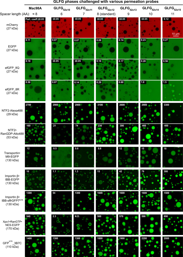
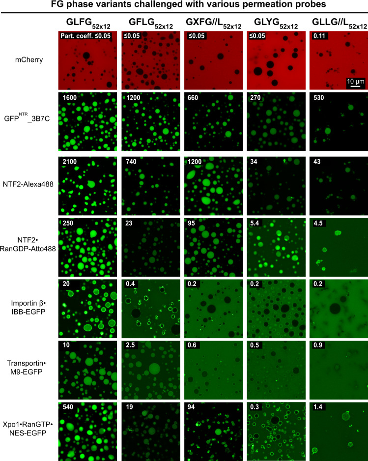
Nup98 FG repeat domains comprise hydrophobic FG motifs linked through uncharged spacers. FG motifs capture nuclear transport receptors (NTRs) during nuclear pore complex (NPC) passage, confer inter-repeat cohesion, and condense the domains into a selective phase with NPC-typical barrier properties. We show that shortening inter-FG spacers enhances cohesion, increases phase density, and tightens such barrier - all consistent with a sieve-like phase. Phase separation tolerates mutating the Nup98-typical GLFG motifs, provided domain-hydrophobicity remains preserved. NTR-entry, however, is sensitive to (certain) deviations from canonical FG motifs, suggesting co-evolutionary adaptation. Unexpectedly, we observed that arginines promote FG-phase-entry apparently also by hydrophobic interactions/ hydrogen-bonding and not just through cation-π interactions. Although incompatible with NTR·cargo complexes, a YG phase displays remarkable transport selectivity, particularly for engineered GFPNTR-variants. GLFG to FSFG mutations make the FG phase hypercohesive, precluding NTR-entry. Extending spacers relaxes this hypercohesion. Thus, antagonism between cohesion and NTR·FG interactions is key to transport selectivity.


Conflict of interest statement

The authors declare no competing interests.

Figures

Fig. 1
Fig. 1. Inter-GLFG spacer length determines the phase separation propensity.
a, b Sequences (partial) of the wild-type Tetrahymena thermophila MacNup98A (“Mac98A”) FG domain (a) and a sequence-regularized variant, GLFG52×12 (b). The latter served as a template for variant design in this study. GLFG52×12 is composed of 52 repeat units, each containing one GLFG motif (green) and one eight-amino-acid spacer (length of each repeat unit = 12 AA). For space economy, only the N-terminal ~130 residues of each are shown (see Supplementary Note 1 for complete sequences). c Based on GLFG52×12, a variant with one residue in each inter-GLFG spacer replaced by Asp was constructed (GLFG//D52×12). The sequence is listed with that of Mac98A and GLFG52×12. For each, the N-terminal sequence up to the fourth FG motif is shown. For GLFG52×12 and GLFG//D52×12, the C-terminal sequences follow the same design strategy as the N-terminal sequences shown (see Supplementary Note 1 for complete sequences). Each FG domain or variant was expressed, purified, and dissolved at a concentration of 1 mM in 4 M guanidinium hydrochloride. As a test of phase separation property, this stock of each was diluted 100-fold quickly with assay buffer (50 mM Tris/HCl pH 7.5, 150 mM NaCl, 5 mM DTT) to allow phase separation. The dilution was centrifuged. SDS samples of the obtained pellets (FG phase), if there were, and supernatants (soluble content) were loaded for SDS-PAGE at an equal ratio, followed by Coomassie blue-staining for quantification. Saturation concentration (Csat) of each was taken as the concentration of the supernatant. For GLFG//D52×12, no phase separation was detected at the assay protein concentration (=10 μM), and the exact Csat was not determined. d Based on GLFG52×12, variants with the same number (=52) of GLFG motifs but varying inter-GLFG spacer lengths were constructed. N-terminal sequences up to the fourth FG motif are shown (see Supplementary Note 1 for complete sequences). For each variant, a 1 mM stock in 4 M guanidinium hydrochloride was prepared, and phase separation was analysed as described above. c, d Each of the assays was performed twice on independent samples with similar results, and representative images are shown.
Fig. 2
Fig. 2. Variation of inter-GLFG spacer length impacts intra-FG phase density.
a Mac98A FG domain and indicated variants (the former has irregular spacer length with an average of about 8 AA), were dissolved at a concentration of 1 mM (except that GLFG52×15 was dissolved at a concentration of 2 mM, because of its higher saturation concentration) in 4 M guanidinium hydrochloride, and phase separation was initiated by a rapid 50-fold dilution with assay buffer. Each was further diluted fourfold in 12 μM Hoechst 33342. Samples were analysed by confocal laser-scanning microscopy (CLSM). The numbers in orange indicate the fluorescence intensities of the Hoechst dye inside the FG phases. The fluorescence intensities were relative to that of GLFG52×12 (arbitrarily set to 100). The above assay was performed twice on independent samples with similar results, and representative images are shown. b Phase separation of Mac98A FG domain and its variants was initiated similarly as in a and the samples were analysed by optical diffraction tomography (ODT) independently (in the absence of Hoechst). Panels show maps of refractive index (RI). For each, ten independent FG particles were analysed. Representative images and mean values (±S.D. between the ten FG particles) of RI, mass density (Cdense), FG motif concentration and solvent % are shown. *Note: Mac98A FG domain has <52 FG motifs but also contains FG-like motifs/ hydrophobic residues in spacers (Supplementary Table 2), which are not counted here. c Csat shown in Fig. 1d is plotted against the spacer length (L) of variants. Mean values of Csat (n = 2) are plotted and fitted to a simple exponential function (dashed line) with the R-squared value indicated. d Cdense obtained from ODT is plotted against L. For each, ten independent FG particles were analysed, and data are presented as mean ± S.D. (in molar concentration). e Gibbs free energy for phase separation (ΔG) of each variant was calculated by the equation in red (Eq.1 in the main text; with T = 298 K) and is plotted against L. The data are presented as mean values. Note: Csat of GLFG52×10 was extrapolated from the equation derived in c (=0.008 μM), and the corresponding ΔG was computed accordingly.
Fig. 3
Fig. 3. Variation of inter-GLFG spacer length impacts the transport selectivity of barrier.
FG phases assembled from the Mac98A FG domain and indicated variants were prepared as described in Fig. 2. They were challenged with the indicated permeation probes (for probe descriptions, see main text). In each case, the calculated molecular weight of the complex is indicated. Scanning settings/ image brightness were adjusted individually to cover the large range of signals. The numbers in white refer to the partition coefficients of the fluorescent species into the FG phases (fluorescence ratios in the central regions of the particles to that in the surrounding buffer). N.D.: Not determined due to difficulties in defining the central region. Each of the above assays was performed twice on independent samples with similar results, and representative images/mean values are shown. The same applies to Figs. 5–10.
Fig. 4
Fig. 4. Phase separation property is dependent on the overall hydrophobicity.
a GLFG52×12 serves as a template for variant design in this experiment. Canonical FG motifs are coloured green and mutations are coloured purple. Note that the inter-GLFG spacers of GLFG52×12 do not contain Phe and Leu. All the variants contain 52 repeat units, and each unit is 12-amino-acid long. The N-terminal sequence of each variant up to the fourth FG motif is shown; the rest of the sequence (~620 residues/48 repeat units) follows the same design strategy as shown (see Supplementary Note 1 for complete sequences). Phase separation of the variants was analysed as in Fig. 1c at [Variant] = 10 μM. No phase separation was observed for GAFG52×12, GLLG52×12, and GAYG52×12 under these conditions, and the exact saturation concentrations were not determined. Each of the assays was performed twice on independent samples with similar results, and representative images are shown. b Phase separation of GLFG52×12 and its variants was initiated and the resulting dense phases were analysed by optical diffraction tomography (ODT). Panels show maps of refractive index (RI). For each, ten independent FG particles were analysed. Representative images and mean values (±S.D. between the ten FG particles) of RI, mass density (Cdense) and motif concentration are shown. c Cdense for each FG phase variant determined by ODT. For each, ten independent particles were analysed, and the data are presented as mean ± S.D. between the ten FG particles (in molar concentration).
Fig. 5
Fig. 5. FG domain variants with non-canonical Fx motifs phase-separate into barriers that allow entry of NTR-cargo complexes.
FG or FG-like phases assembled from the indicated variants were challenged with the indicated probes. Scanning settings/ image brightness were adjusted individually due to the large range of signals. The numbers in white refer to the partition coefficients of the fluorescent species into the phases.
Fig. 6
Fig. 6. Spatial arrangement of Phe and Leu in FG domain is crucial for NTR-binding but not for engineered GFPNTR-variants.
FG or FG-like phases assembled from the indicated variants were challenged with the indicated probes. Scanning settings/ image brightness were adjusted individually due to the large range of signals. The numbers in white refer to the partition coefficients of the fluorescent species into the phases.
Fig. 7
Fig. 7. Arginines promote FG phase entry not just through cation-π interactions.
Indicated condensed phases were challenged with sffrGFP4, which is an engineered FG-philic GFP variant, and “sffrGFP4 25 R→K”, in which all the surface FG-philic arginine residues were replaced by FG-phobic lysines. Note that the GLLG//L phase, which lacks aromatic residues, still allows the partition of the Arg-rich sffrGFP4, indicating interactions other than cation-π promoted the entry.
Fig. 8
Fig. 8. An engineered GFP variant that binds specifically to GLFG motifs and NPCs.
a A rational mutation (M225F) was introduced to GFPNTR_3B7C, and the resulting variant was named TetraGFPNTR. Indicated FG or FG-like phases were challenged with GFPNTR_3B7C and TetraGFPNTR. Note that except for the canonical GLFG phase, the mutation reduces the partition coefficients into all other phases. b Panels show confocal scans of digitonin-permeabilized HeLa cells that had been incubated for 5 min with 250 nM of either GFPNTR_3B7C or TetraGFPNTR. Live images were taken without fixation or washing steps, i.e., they show directly an NPC-binding in relation to the free concentration and non-specific aggregation with nuclear or cytoplasmic structures. While 3B7C still showed some weak binding to nuclear structures other than NPCs, TetraGFPNTR stains NPCs more crisply, illustrating that TetraGFPNTR is a more specific NPC binder. This experiment was performed twice on independent samples with similar results, and representative images are shown. c Plot profiles corresponding to lines across nuclei marked in b. Fluorescence (GFP) intensities are normalized to the maximum (arbitrarily set to 100) of each line.
Fig. 9
Fig. 9. Exchanging GLFG to FSFG motifs results in a hypercohesive FG phase that precludes entry of NTR-cargo complexes.
a Based on a framework of GLFG52×12, FSFG52×12 was constructed as described above. The FG phase assembled from FSFG52×12 was challenged with the indicated probes. Scanning settings/ image brightness were adjusted individually due to the large range of signals. N.D.: Not determined due to difficulties in defining the central region. Note that all probes only arrested at the periphery of the phase. b Based on FSFG52×12, two mutants with a reduced number (=35 or 36) of FSFG motifs were constructed. “Mutant 1” corresponds to a sequence where every third FSFG motif in FSFG52×12 was mutated to SSSG (FG-phobic). “Mutant 2” is similar, but every fifth and sixth FSFG motif were mutated. For space economy, only the N-terminal ~144 residues of each are shown. The C-terminal sequences follow the same design strategy as the region shown (see Supplementary Note 1 for complete sequences). Corresponding regions of GLFG52×12 and FSFG52×12 are shown for comparison. c FG phases assembled from the above variants were challenged with the indicated probes as in a. d GLFG29×12 and FSFG29×12: corresponding to the N-terminal 29 repeats of GLFG52×12 and FSFG52×12, respectively, were tested for phase separation at a concentration of 20 μM. Each of the assays was performed twice on independent samples with similar results, and representative images are shown. Note that the FSFG motifs lead to stronger phase separation propensity than the GLFG motifs (see also Supplementary Fig. 2).
Fig. 10
Fig. 10. Hypercohesion of FSFG motifs can be balanced by longer inter-FG spacers.
a Based on FSFG52×12, two variants with the same number (52) of FSFG motifs but longer inter-FSFG spacers were constructed (FSFG52×15 and FSFG52×18). b Upper panels: condensed phases assembled from the indicated variants were stained with Hoechst 33342. The numbers in orange indicate the fluorescence intensities of the Hoechst dye inside the FG phases. The fluorescence intensities are relative to that of GLFG52×12 (arbitrarily set to 100). The above assay was performed twice on independent samples with similar results, and representative images are shown. Lower panels: the condensed phases were analysed by optical diffraction tomography (ODT) independently in the absence of Hoechst. Panels show maps of refractive index (RI). For each, ten independent FG particles were analysed. Representative images and mean values (±S.D. between the ten FG particles) of RI, mass density (Cdense), and FG motif concentration are shown. c Condensed phases assembled from FSFG52×12 and its two variants with longer spacers were challenged with the indicated probes. Note that lengthening the spacers relaxes the permeation selectivity.

Similar articles

Cited by

References

    1. Hoelz A, Glavy JS, Beck M. Toward the atomic structure of the nuclear pore complex: when top down meets bottom up. Nat. Struct. Mol. Biol. 2016;23:624–630. - PMC - PubMed
    1. Hampoelz B, Andres-Pons A, Kastritis P, Beck M. Structure and assembly of the nuclear pore complex. Annu. Rev. Biophys. 2019;48:515–536. - PubMed
    1. Lin DH, Hoelz A. The structure of the nuclear pore complex (an update) Annu. Rev. Biochem. 2019;88:725–783. - PMC - PubMed
    1. Bley CJ, et al. Architecture of the cytoplasmic face of the nuclear pore. Science. 2022;376:eabm9129. - PMC - PubMed
    1. Petrovic S, et al. Architecture of the linker-scaffold in the nuclear pore. Science. 2022;376:eabm9798. - PMC - PubMed

Publication types

MeSH terms

Substances