. 2023 Feb 10;8(1):4.
doi: 10.1038/s41525-022-00343-8.

Bi-allelic ATG4D variants are associated with a neurodevelopmental disorder characterized by speech and motor impairment

Collaborators, Affiliations

Bi-allelic ATG4D variants are associated with a neurodevelopmental disorder characterized by speech and motor impairment

Marie Morimoto et al. NPJ Genom Med. .

Abstract

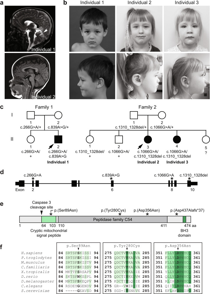
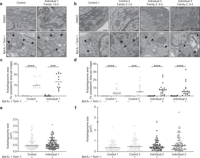
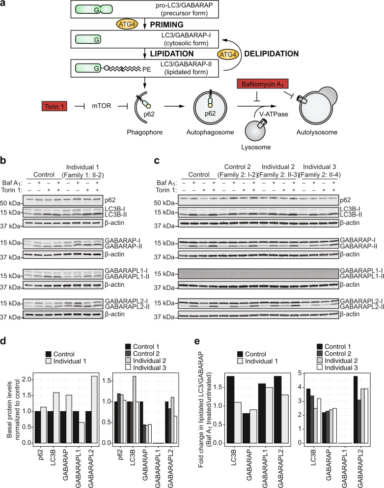
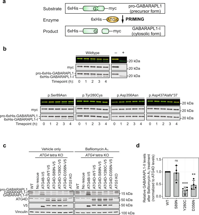
Autophagy regulates the degradation of damaged organelles and protein aggregates, and is critical for neuronal development, homeostasis, and maintenance, yet few neurodevelopmental disorders have been associated with pathogenic variants in genes encoding autophagy-related proteins. We report three individuals from two unrelated families with a neurodevelopmental disorder characterized by speech and motor impairment, and similar facial characteristics. Rare, conserved, bi-allelic variants were identified in ATG4D, encoding one of four ATG4 cysteine proteases important for autophagosome biogenesis, a hallmark of autophagy. Autophagosome biogenesis and induction of autophagy were intact in cells from affected individuals. However, studies evaluating the predominant substrate of ATG4D, GABARAPL1, demonstrated that three of the four ATG4D patient variants functionally impair ATG4D activity. GABARAPL1 is cleaved or "primed" by ATG4D and an in vitro GABARAPL1 priming assay revealed decreased priming activity for three of the four ATG4D variants. Furthermore, a rescue experiment performed in an ATG4 tetra knockout cell line, in which all four ATG4 isoforms were knocked out by gene editing, showed decreased GABARAPL1 priming activity for the two ATG4D missense variants located in the cysteine protease domain required for priming, suggesting that these variants impair the function of ATG4D. The clinical, bioinformatic, and functional data suggest that bi-allelic loss-of-function variants in ATG4D contribute to the pathogenesis of this syndromic neurodevelopmental disorder.


Conflict of interest statement

The authors declare no competing interests.

Figures

Fig. 1
Fig. 1. Bi-allelic variants in ATG4D segregate with a neurodevelopmental disorder in three individuals from two unrelated families.
a Neuroimaging of Individual 1 showing mild cerebellar atrophy disproportionately involving the superior cerebellar hemispheres and vermis at 5 years 3 months (arrowhead in T2-weighted sagittal view image, left panel). There were no remarkable neuroimaging findings for Individual 2 at 1 year 3 months (arrow in T1-weighted sagittal view image, right panel). b Photographs of the affected individuals showing a common facial gestalt characterized by almond-shaped eyes, a depressed nasal bridge, and a prominent Cupid’s bow. Age at the time of the photos is 5 years 3 months for Individual 1, 4 years 8 months for Individual 2, and 3 years 5 months for Individual 3. Written consent was obtained for the publication of photographs. c The pedigrees of two unrelated families with at least one affected individual exhibiting a neurodevelopmental disorder show segregation of compound heterozygous ATG4D variants with disease. d Schematic of the ATG4D gene showing the relative location of each variant (asterisks). The schematic is to scale, while the variant position is approximate. e Schematic of the ATG4D cysteine protease and its functional domains including the peptidase family C54 domain (dark gray), a cryptic mitochondrial signal peptide (light green), a BH3 domain (green), and a caspase 3 cleavage site (arrowhead). The location of the predicted amino acid changes is indicated (asterisks). The schematic and variant position are to scale. f Alignment of missense variants in ATG4D across multiple species including human (Homo sapiens), chimpanzee (Pan troglodytes), mouse (Mus musculus), dog (Canine familiaris), African clawed frog (Xenopus laevis), zebrafish (Danio rerio), fruit fly (Drosophila melanogaster), nematode (Caenhabdoritis elegans), and budding yeast (Saccharomyces cerevisiae). The amino acid residue of interest is indicated by the arrowhead and the level of conservation is indicated by the intensity of the color.
Fig. 2
Fig. 2. Transmission electron microscopy analyses of autophagosome biogenesis in cultured cells from individuals with bi-allelic variants in ATG4D.
a, b Representative TEM images of cultured primary fibroblasts from Control (GM09503) and Individual 1 (a) or lymphoblastoid cell lines from Control 1 (CCL-104), Control 2 (Family 2: I-2, mother of Individuals 2 and 3), Individual 2 (Family 2: II-3), and Individual 3 (Family 2: II-4) (B) treated with vehicle (DMSO) or 100 nM Torin 1 and 100 nM Bafilomycin A1 for 3 h to induce the formation of autophagosomes and to prevent their degradation, respectively (arrowheads). cf Quantification of autophagosome area (c, d) and size (e, f) from the experiments represented in (a, b). The data are presented as dot plots with the median indicated by a horizontal line. Light gray data points represent measurements made on control cells; dark gray data points represent measurements made on affected cells. For the quantification of autophagosome area, 12 images (primary fibroblasts) or 19 images (lymphoblastoid cell lines) taken from each sample were assessed per condition. A Kruskal–Wallis test and Dunn’s multiple comparisons test were performed for relevant predefined dataset pairs. For the quantification of autophagosome size, data points represent individual autophagosomes measured from Torin 1- and Bafilomycin A1-treated conditions. For the primary fibroblasts, n = 98 and n = 152 autophagic structures were respectively measured for Control (GM09503) and Individual 1 (Family 1: II-2), and a two-tailed Mann–Whitney U test was performed. For the lymphoblastoid cell lines, n = 67, n = 78, n = 122, and n = 92 autophagomsomes were respectively measured for Control 1 (CCL-104), Control 2 (Family 2: I-2, mother of Individuals 2 and 3), Individual 2 (Family 2: II-3), and Individual 3 (Family 2: II-4), and a Kruskal–Wallis test and Dunn’s multiple comparisons test were performed for relevant predefined dataset pairs. Only statistically significant comparisons are shown. Magnification: ×4000. Scale bar: 500 nm. ****p < 0.0001; ***p < 0.001; Baf A1, Bafilomycin A1; DMSO, dimethyl sulfoxide; TEM, transmission electron microscopy.
Fig. 3
Fig. 3. Analysis of the induction of lipidated LC3/GABARAP subfamily proteins in cells from individuals with bi-allelic variants in ATG4D.
a ATG4 family members prime the LC3/GABARAP subfamily proteins required for autophagosome biogenesis by cleaving pro-LC3/GABARAP (precursor form) into LC3/GABARAP-I (cytosolic form) to expose a C-terminal glycine residue that can then be lipidated by conjugation to phosphatidylethanolamine (PE), resident in phagophore membranes, to form LC3/GABARAP-II (lipidated form). This lipidated form in the expanding phagophore membrane can bind p62 and other proteins for selective autophagy. ATG4 family members also cleave LC3/GABARAP-PE to delipidate and recycle LC3/GABARAP to its cytosolic form. Torin 1 is an autophagy inducer and Bafilomycin A1 is an autophagy inhibitor. b Immunoblot analysis of primary fibroblasts from Control (GM09503) and Individual 1 (Family 1: II-2) assessing p62, LC3B, GABARAP, GABARAPL1, and GABARAPL2 upon induction and/or inhibition of autophagy by treatment with 100 nM Torin 1 and/or 100 nM Bafilomycin A1 for 3 h. c Immunoblot analysis of lymphoblastoid cell lines from Control 1 (CCL-104), Control 2 (Family 2: I-2, mother of Individuals 2 and 3), Individual 2 (Family 2: II-3), and Individual 3 (Family 2: II-4) assessing p62, LC3B, GABARAP, GABARAPL1, and GABARAPL2 upon induction and/or inhibition of autophagy by treatment with 100 nM Torin 1 and/or 100 nM Bafilomycin A1 for 3 h. β-actin was used as a loading control. p62 and LC3B were assessed on the same gel. d, e Quantification of basal protein levels (d) and autophagic flux (e) from immunoblot analyses of p62, total LC3B, total GABARAP, total GABARAPL1, and total GABARAPL2 in primary fibroblasts from Control (GM09503) and Individual 1 (Family 1: II-2) and lymphoblastoid cell lines from Control 1 (CCL-104), Control 2 (Family 2: I-2, mother of Individuals 2 and 3), Individual 2 (Family 2: II-3), and Individual 3 (Family 2: II-4). Autophagic flux is presented as the fold change of the lipidated form of each LC3/GABARAP subfamily member after treatment with Bafilomycin A1 compared to before treatment. Baf A1, Bafilomycin A1; kDa, kilodaltons; mTOR, mammalian target of rapamycin.
Fig. 4
Fig. 4. Assessment of the priming activity of the ATG4D variants using an in vitro GABARAPL1 priming assay and a GABARAPL1 priming rescue experiment.
a A schematic of the recombinant proteins used for the in vitro GABARAPL1 priming assay. Both the pro-GABARAPL1 substrate and the N-terminally cleaved ATG4D (ΔN63 ATG4D) enzyme are tagged with 6xHis at the N-terminus for purification. The pro-GABARAPL1 substrate also has a C-terminal myc tag to visualize priming activity with increased sensitivity. b in vitro GABARAPL1 priming assays demonstrating the priming activity of wildtype, p.Ser89Asn, p.Tyr280Cys, p.Asp356Asn, and p.Asp437Alafs*37 ΔN63 ATG4D enzymes. Recombinant 6xHis-GABARAPL1-myc substrate was incubated with wildtype or variant ΔN63 ATG4D enzymes for the indicated time and analyzed by immunoblot. The efficient ATG4B enzyme was incubated with the G116A GABARAPL1 mutant that cannot be primed as a negative control (−) or wildtype GABARAPL1 as a positive control (+) for 1 h. Representative of n = 3 independent experiments. c GABARAPL1 priming rescue experiments demonstrating the priming activity of ATG4 tetra knockout cells expressing V5-tagged wildtype (WT) ATG4D and the three ATG4D missense variants (p.Ser89Asn (p.S89N), p.Tyr280Cys (p.Y280C), and p.Asp356Asn (p.D356N)) treated with 200 nM Bafilomycin A1 or vehicle only for 8 h. ATG4 tetra knockout cells lack the ability to prime pro-GABARAPL1 (precursor form) to GABARAPL1-I (cytosolic form) and, subsequently, also lack the ability to form GABARAPL1-II (lipidated form); this is demonstrated by the presence of only pro-GABARAPL1. ATG5 knockout cells have the ability to prime but lack the ability to lipidate the LC3/GABARAP subfamily members; this is demonstrated by the presence of only GABARAPL1-I. Expression of V5-tagged ATG4B was used as a positive control. All targets for each treatment were assessed on the same gel. A non-specific band for the ATG4D blot is marked by an asterisk. Representative of n = 4 independent experiments. d Quantification of relative GABARAPL1-II levels after treatment with 200 nM Bafilomycin A for 8 h is represented as the mean ± standard deviation of 4 independent experiments. GABARAPL1-II levels were normalized to ATG4D levels. A one-way ANOVA was performed to compare relevant predefined dataset pairs and a Dunnett’s multiple comparisons post hoc test was performed to correct for multiple comparisons. **p < 0.01; ***p < 0.001; h, hours; kDa, kilodaltons; KO, knockout; ns, not significant; WT, wildtype.

References

    1. Mizushima N, Levine B. Autophagy in human diseases. N. Engl. J. Med. 2020;383:1564–1576. doi: 10.1056/NEJMra2022774. - DOI - PubMed
    1. Galluzzi L, et al. Molecular definitions of autophagy and related processes. EMBO J. 2017;36:1811–1836. doi: 10.15252/embj.201796697. - DOI - PMC - PubMed
    1. Levine B, Kroemer G. Biological functions of autophagy genes: a disease perspective. Cell. 2019;176:11–42. doi: 10.1016/j.cell.2018.09.048. - DOI - PMC - PubMed
    1. Nakatogawa H. Mechanisms governing autophagosome biogenesis. Nat. Rev. Mol. Cell Biol. 2020;21:439–458. doi: 10.1038/s41580-020-0241-0. - DOI - PubMed
    1. Tsukada M, Ohsumi Y. Isolation and characterization of autophagy-defective mutants of Saccharomyces cerevisiae. FEBS Lett. 1993;333:169–174. doi: 10.1016/0014-5793(93)80398-E. - DOI - PubMed