. 2023 Feb 10;14(1):744.
doi: 10.1038/s41467-023-36454-8.

Intrafusal-fiber LRP4 for muscle spindle formation and maintenance in adult and aged animals

Affiliations

Intrafusal-fiber LRP4 for muscle spindle formation and maintenance in adult and aged animals

Rangjuan Cao et al. Nat Commun. .

Abstract

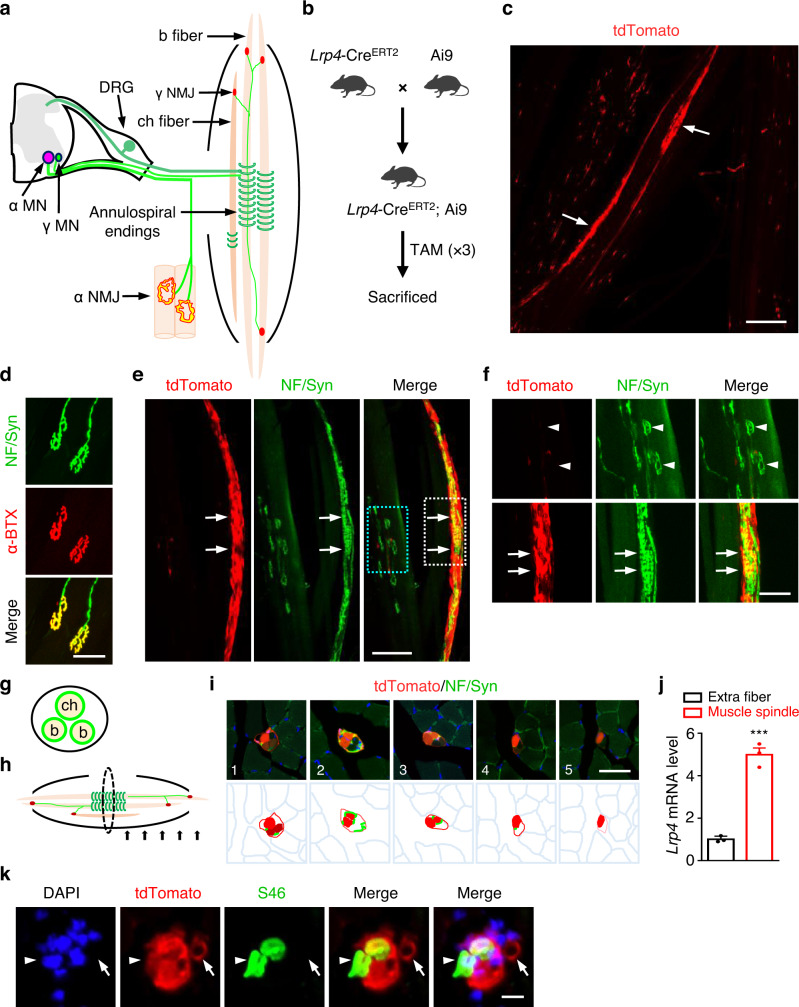
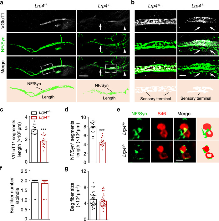
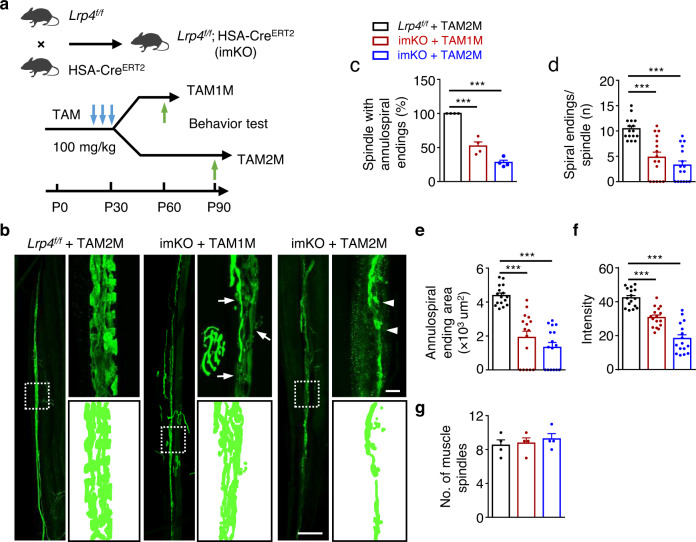
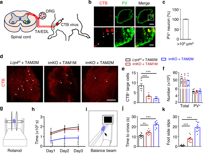
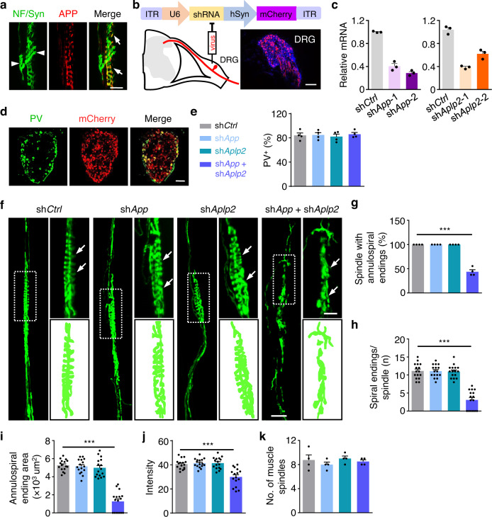
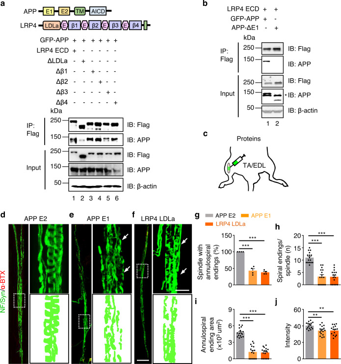
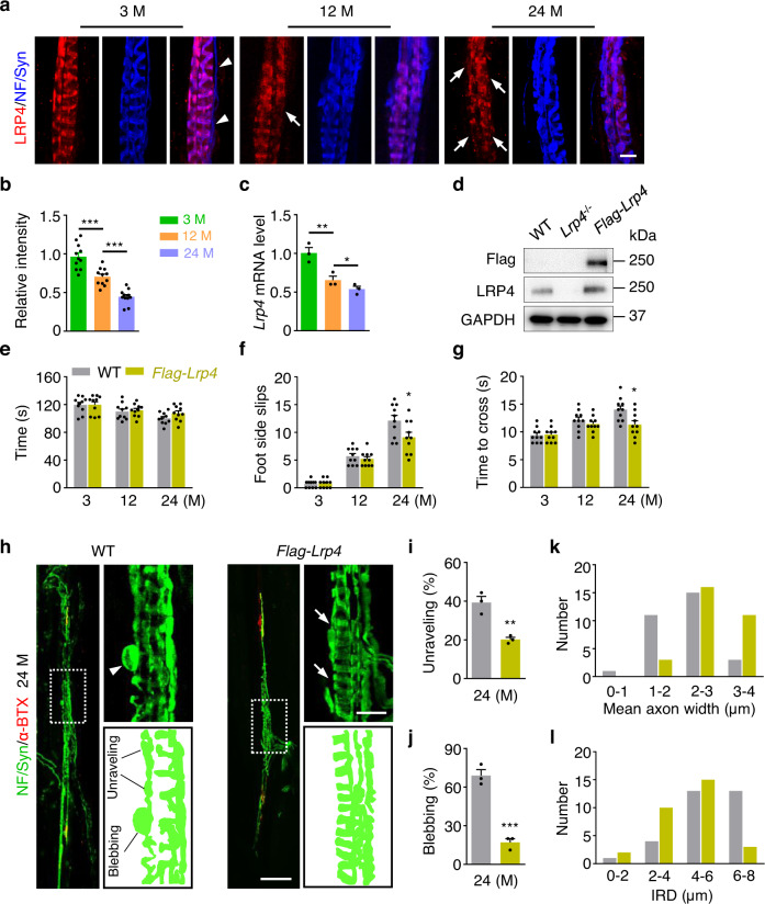
Proprioception is sensed by muscle spindles for precise locomotion and body posture. Unlike the neuromuscular junction (NMJ) for muscle contraction which has been well studied, mechanisms of spindle formation are not well understood. Here we show that sensory nerve terminals are disrupted by the mutation of Lrp4, a gene required for NMJ formation; inducible knockout of Lrp4 in adult mice impairs sensory synapses and movement coordination, suggesting that LRP4 is required for spindle formation and maintenance. LRP4 is critical to the expression of Egr3 during development; in adult mice, it interacts in trans with APP and APLP2 on sensory terminals. Finally, spindle sensory endings and function are impaired in aged mice, deficits that could be diminished by LRP4 expression. These observations uncovered LRP4 as an unexpected regulator of muscle spindle formation and maintenance in adult and aged animals and shed light on potential pathological mechanisms of abnormal muscle proprioception.


Conflict of interest statement

The authors declare no competing interests.

Figures

Fig. 1
Fig. 1. High expression of LRP4 in intrafusal fibers.
a Schematic diagram of muscle spindle and NMJ. γ MN, γ motor neuron; α MN, α motor neuron; DRG, dorsal root ganglia; b fiber, bag fiber; ch fiber, chain fiber. b Lrp4-CreERT2 crossed with reporter mice Ai9 and the schedule of tamoxifen administration. c tdTomato labeled a few fibers of leg muscles in Lrp4-CreERT2; Ai9 mice. Scale bar, 200 µm. This experiment was repeated three times independently with similar results. d Anti-NF/Syn antibodies labeled pretzel-like structures positive for CF568-labeled α-BTX. Scale bar, 50 µm. This experiment was repeated three times independently with similar results. e–i The enrichment of tdTomato in muscle spindle. e Representative images of muscle spindle in leg muscle of Lrp4-CreERT2; Ai9 mice at P14 stained by NF/Syn. Scale bar, 100 µm. f Enlarged images of blue rectangle and white rectangle in (e). Arrows, annulospiral endings; arrowheads, regular NMJs. Scale bar, 50 µm. This experiment was repeated three times independently with similar results. g The schematic diagram of transection view of a muscle spindle. h The schematic diagram of series transection view in (i). i Tdtomato was concentrated in the intrafusal fibers and capsules. Images of transection view of a muscle spindle in Lrp4-CreERT2; Ai9 leg muscle (P60) at different locations. The internal distance is 100 µm. Scale bar, 40 µm. This experiment was repeated three times independently with similar results. j Elevated expression of Lrp4 mRNA in muscle spindle. Tdtomato+ muscle spindles were isolated and proceed to quantitative RT-PCR analysis. The related Lrp4 mRNA was compared with the extrafusal fibers (t (4) = 12.046, ***p = 0.0003). Data are shown as mean ± SEM. n = 3 mice per group, unpaired two-tailed t test. k Representative images of transection view of Lrp4-CreERT2; Ai9 leg muscle (P60) stained by DAPI and S46. Scale bar, 10 µm. This experiment was repeated three times independently with similar results. Source data are provided as a Source Data file.
Fig. 2
Fig. 2. A critical role of LRP4 in spindle development at E18.5.
a–d Disrupted innervation of intrafusal fibers in Lrp4 mutant. a Representative images of TA muscle at E18.5 whole mount stained with VGluT1 (gray) and NF/Syn (green). Arrows, poorly organized axon terminals; arrowheads, axon terminal fragments. Scale bar, 100 µm. b Images enlarged from rectangles in (a). Disorganized boundaries of NF/Syn+ segments by Lrp4 knockout. Arrows, axons extended into VGluT1 negative region. Scale bar, 20 µm. c, d Reduced VGluT1+ or NF/Syn+ segments length in Lrp4−/−. Quantification of VGluT1+ segments (t (34) = 7.386, ***p < 0.0001) or NF/Syn+ (t (34) = 8.682, ***p < 0.0001) in both groups. n = 18 muscle spindles from 3 mice per group, unpaired two-tailed t test. e Representative transection images of TA muscle of E18.5 stained with NF/Syn (green) and S46 (Red), and bag fibers (S46+) were observed in control and Lrp4 mutant. Scale bar, 10 µm. Quantification of bag fiber number per spindle (f) and bag fiber size (g) in both groups. No difference was observed after Lrp4 knockout. n = 18 muscle spindles from 3 mice per group, unpaired two-tailed t test. Data are shown as mean ± SEM. Source data are provided as a Source Data file.
Fig. 3
Fig. 3. Reduced Egr3 levels in Lrp4 mutant mice.
a The induction of preliminary muscle spindle in Lrp4 mutant. Shown were TA muscles at E14.5 in indicated genotypes, whole-mount stained with PV (green), α-BTX (red), and Egr3 (magenta). Arrows, preliminary muscle spindle was induced and innervated by PV+ sensory axons. Scale bar, 50 µm. b, c Reduced innervation and Egr3 expression in Lrp4 mutant. Represent images of muscle spindle in TA whole mount stained with PV and Egr3 at E16.5 (b) or E18.5 (c). Arrows, spindle without Egr3 expression. Scale bar, 50 µm. d–g Quantification of data in (a–c). d Quantification of PV+ terminal length (E16.5, t (34) = 2.638, *p = 0.0125; E18.5, t (34) = 6.436, ***p < 0.0001). e Quantification of intensity of Egr3+ nuclei staining (E16.5, t (34) = 2.708, *p = 0.0105; E18.5, t (34) = 4.122, ***p = 0.0002). f Quantification of number of Egr3+ nuclei per spindle (E16.5, t (34) = 3.146, **p = 0.0034; E18.5, t (34) = 8.865, ***p < 0.0001). g Quantification of Egr3+ segment length (E16.5, t (34) = 2.053, *p = 0.0478; E18.5, t (34) = 4.879, ***p < 0.0001). n = 18 muscle spindles from 3 mice per group, unpaired two-tailed t test in (d–g). Data are shown as mean ± SEM. Source data are provided as a Source Data file.
Fig. 4
Fig. 4. Requirement of LRP4 for maintaining muscle spindles.
a Illustration of breading Lrp4f/f; HSA-CreERT2 (imKO for short) mice and the schematic of tamoxifen administration, behavior tests and tissue collection. b Disrupted annulospiral endings in tamoxifen treated imKO mice (P60, P90). Representative images of EDL muscles in indicated groups, whole mount stained with NF/Syn (green) to label axon and axon terminals. Muscle spindle was determined by morphology. Square, images enlarged in right panel. Arrows, axon fragmentations; arrowheads, degenerated axon terminals. Scale bars, 100 µm and 10 µm. c–g Quantification of data in (b). c Reduced percentage of muscle spindles with annulospiral endings in tamoxifen treated imKO mice; t (6) = 7.893, ***p = 0.0002; t (6) = 21.614, ***p < 0.0001. n = 4 mice per group, unpaired two-tailed t test. d Decreased average spiral endings per muscle spindle (t (30) = 4.915, ***p < 0.0001; t (30) = 7.265, ***p < 0.0001). n = 16 muscle spindles from 4 mice per group, unpaired two-tailed t test. e, f Reduction of annulospiral ending area per muscle spindle (t (30) = 6.215, ***p < 0.0001; t (30) = 9.347, ***p < 0.0001) and intensity of muscle spindle in each group (t (30) = 5.831, ***p < 0.0001; t (30) = 9.215, ***p < 0.0001). n = 16 muscle spindles from 4 mice per group, unpaired two-tailed t test. g No difference of total muscle spindle in EDL muscle in each group. n = 4 mice per group, unpaired two-tailed t test. Data are shown as mean ± SEM. Source data are provided as a Source Data file.
Fig. 5
Fig. 5. Disrupted sensory synapses and proprioceptive sensation in TAM-imKO mice.
a Schematic diagram of CTB virus injection and labeling. b Representative images of DRG sections stained with anti-CTB and anti-PV antibodies. CTB colocalized with PV positive neurons (asterisks). Scale bars, 200 µm and 50 µm. c Quantification of the percentage of CTB labeled large neurons (>1000 µm2) with PV staining, n = 4 sections from 4 mice (P60). d Representative images of DRG at indicated groups labeled by CTB. The positive neurons with soma size larger than 1000 µm2 were counted (asterisks). Scale bar, 300 µm. e Quantification of CTB labeled large neuron in (d); t (10) = 4.846, ***p = 0.0007; t (10) = 6.151, ***p = 0.0001, n = 6 mice per group, unpaired two-tailed t test. f Quantification of total neurons and PV+ neurons in each group. n = 6 mice per group. g Rotarod test. h Quantification of the time mice in each group fall off the rotating rod on the first, second and third testing day. Shorten time on rotating rod in imKO + TAM2M mice; ***p = 0.0001 for the first day, ***p < 0.0001 for the second and third day. n = 10 mice per group, unpaired two-tailed t test. i Balance beam walking task. j Quantification of the time mice consuming during the beam walking task in each test. imKO mice took longer time during beam walking task two month later after tamoxifen treatment; t (18) = 3.589, **p = 0.0021; t (18) = 9.823, ***p < 0.0001. n = 10 mice per group, unpaired two-tailed t test. k Quantification of the number of side slips during walking on the beam in control or tamoxifen treated mice. Elevated side slips after Lrp4 knockout; t (18) = 6.623, ***p < 0.0001; t (18) = 12.376, ***p < 0.0001. n = 10 mice per group, unpaired two-tailed t test. Data are shown as mean ± SEM. Source data are provided as a Source Data file.
Fig. 6
Fig. 6. Degenerated annulospiral endings after App and Aplp2 knocked out.
a EDL muscle of WT mouse was whole mount stained with NF/Syn (green) and APP (red). Arrows, APP and NF/Syn positive annulospiral endings; Arrowheads, only NF/Syn positive axons. Scale bar, 20 µm. b Construction and expression of AAV shRNA in DRG (P60). Scale bar, 100 µm. c Quantitative RT-PCR was performed to test the silence efficiency of App shRNAs and Aplp2 shRNAs one month after the injection. shApp-1 and shApp-2 were reduced to 0.405 ± 0.045 and 0.287 ± 0.026, respectively. shAplp2-1 and shAplp2-2 were decreased to 0.387 ± 0.016 and 0.62 ± 0.048, respectively. Data are shown as mean ± SEM, n = 3 mice per group. d Representative images of virus infected (red) DRG section stained with anti-PV (green). Scale bar, 100 µm. e Quantification of the percentage of PV+ neurons infected by mCherry-expressing virus. Data are shown as mean ± SEM, n = 4 mice per group. f Representative images of muscle spindle labeled by NF/Syn in shCtrl, shApp alone, shAplp2 alone and shApp plus shAplp2 groups. Arrows, annulospiral endings, rectangles, images enlarged in the right panel. Scale bars, 50 µm and 20 µm. g–k Quantification of data in (f). g Reduced percentage of muscle spindle with annulospiral endings in shApp + shAplp2; F (3, 12) = 174.2, ***p < 0.0001. n = 4 mice per group, one-way ANOVA. h Less spiral endings per spindle by the silence of App and Aplp2; F (3, 60) = 57.34, ***p < 0.0001. i Decreased average area of annulospiral ending per muscle spindle in shApp + shAplp2 group; F (3, 60) = 72.03, ***p < 0.0001. j Reduced intensity per muscle spindle after the silence of App and Aplp2; F (3,60) = 18.78, ***p < 0.0001. n = 16 muscle spindles from 4 mice per group, one-way ANOVA in (h–j). k No comparable difference in muscle spindle number among groups. n = 4 mice, one-way ANOVA. Data are shown as mean ± SEM. Source data are provided as a Source Data file.
Fig. 7
Fig. 7. LRP4-APP trans interaction for muscle spindle structure.
a, b Requirement of APP E1 and LRP4 LDLa for binding. a co-IP of GFP-APP co-transfected with Flag labeled LRP4 ECD, ECD deleted with LDLa (ΔLDLa), Δβ1, Δβ2, Δβ3 and Δβ4. This experiment was repeated three times independently with similar results. b co-IP of LRP ECD co-transfected with GFP labeled APP or APP ΔE1. Asterisks, non-specificity bands. Uncropped blots in Source Data. This experiment was repeated three times independently with similar results. c Schematic diagram of recombinant proteins injection (P60). df Impaired annulospiral endings after the injection of recombinant APP E1 and LRP4 LDLa (P60). EDL muscles injected by APP E2 (d), APP E1 (e) and LRP4 LDLa (f) recombinant proteins were whole mount stained with NF/Syn (green) and α-BTX (red). Arrows, the equatorial region of muscle spindle; rectangles, enlarged regions at side. Scale bars, 100 µm and 20 µm. g–j Quantification of data in (d–f), APP E2 serves as control. g Reduced percentage of muscle spindle with annulospiral endings by APP E1 and LRP4 LDLa injection; t (6) = 9.642, ***p < 0.0001; t (6) = 21.139, ***p < 0.0001. n = 4 mice per group, unpaired two-tailed t test. h Less spiral endings per spindle after APP E1 and LRP4 LDLa injection; t (30) = 8.152, ***p < 0.0001; t (30) = 8.924, ***p < 0.0001. i Decreased innervation area of muscle spindle in APP E1 and LRP4 LDLa injected mice; t (30) = 10.108, ***p < 0.0001; t (30) = 12.279, ***p < 0.0001. j Reduced intensity after the injection of APP E1 and LRP4 LDLa; t (30) = 3.589, **p = 0.0012; t (30) = 3.398, **p = 0.0019. n = 16 muscle spindles from 4 mice, unpaired two-tailed t test in (h–j). Data are shown as mean ± SEM. Source data are provided as a Source Data file.
Fig. 8
Fig. 8. Restoring spindle structure and function in aged mice by LRP4 expression.
a Representative images of EDL muscles of 3 M, 12 M, and 24 M-old mice, whole mount stained with 555-anti-LRP4 (red) and NF/Syn (blue). Arrowheads, non-annulospiral axons had no LRP4 expression. Arrows, LRP4 negative annulospiral endings in aged mice. Scale bar, 10 µm. b Quantification of the relative intensity of LRP4; t (20) = 9.063, ***p < 0.0001; t (20) = 4.34, ***p = 0.0003. n = 11 muscle spindles from 3 mice per group, unpaired two-tailed t test. c Quantitative RT-PCR tested the relative expression of Lrp4 mRNA in 3 M, 12 M or 24 M mice; t (4) = 5.861, **p = 0.004; t (4) = 4.006, *p = 0.016. n = 3 mice per group, unpaired two-tailed t test. d Expression of Flag-LRP4 in skeletal muscle was confirmed by Western blotting. Uncropped blots in Source Data. e Quantification of longest running time on rotating rod between WT and Flag-Lrp4 mice at indicated age. f Quantification of side slips in beam walking task of WT and Flag-Lrp4 mice at indicated age; t (18) = 2.251, *p = 0.037. g Quantification of time required for beam walking task; t (18) = 2.793, *p = 0.012. n = 10 mice per group, unpaired two-tailed t test in (e–g). h Representative images of muscle spindle in EDL muscles of 24 M-old mice, whole mount stained with NF/Syn (green) and α-BTX (red). Arrowhead, bleb of axon; arrows, annulospiral endings. Scale bars, 100 µm and 20 µm. i Quantification of unraveling muscle spindle in each group; t (4) = 5.584, **p = 0.005. j Quantification of the incidence of axon with large blebs in each group; t (4) = 9.55, ***p = 0.0007. n = 3 mice per group, unpaired two-tailed t test in (i) and (j). The distribution of mean axon width (k) and inter-rotational distance (l) in aged WT and Flag-Lrp4 mice. Data are shown as mean ± SEM, n = 3 mice per group. Source data are provided as a Source Data file.

Similar articles

Cited by

References

    1. Li L, Xiong WC, Mei L. Neuromuscular junction formation, aging, and disorders. Annu Rev. Physiol. 2018;80:159–188. - PubMed
    1. Wu H, Xiong WC, Mei L. To build a synapse: signaling pathways in neuromuscular junction assembly. Development. 2010;137:1017–1033. - PMC - PubMed
    1. Lin S, Landmann L, Ruegg MA, Brenner HR. The role of nerve- versus muscle-derived factors in mammalian neuromuscular junction formation. J. Neurosci. 2008;28:3333–3340. - PMC - PubMed
    1. Cohen LA. Analysis of position sense in human shoulder. J. Neurophysiol. 1958;21:550–562. - PubMed
    1. McCloskey DI. Kinesthetic sensibility. Physiol. Rev. 1978;58:763–820. - PubMed

Publication types

MeSH terms

Substances

LinkOut - more resources