. 2023 Feb 10;13(1):2389.
doi: 10.1038/s41598-023-29576-y.

Spiders' digestive system as a source of trypsin inhibitors: functional activity of a member of atracotoxin structural family

Affiliations

Spiders' digestive system as a source of trypsin inhibitors: functional activity of a member of atracotoxin structural family

Oscar Bento Silva Neto et al. Sci Rep. .

Abstract

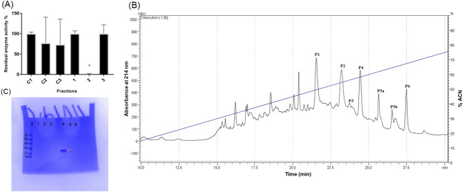
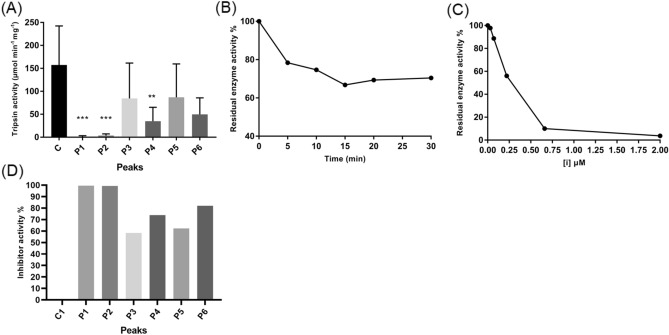
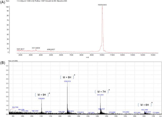
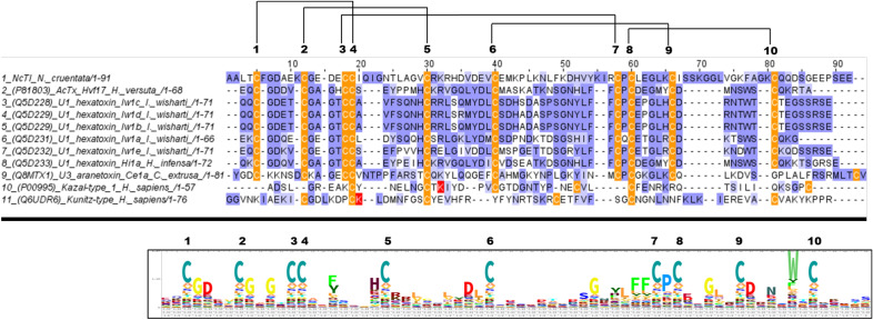
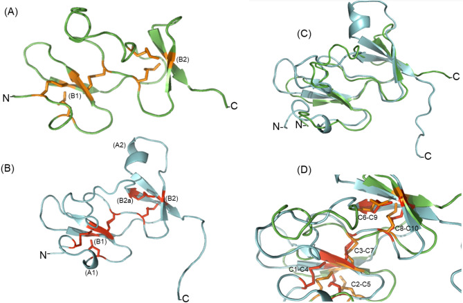
Spiders are important predators of insects and their venoms play an essential role in prey capture. Spider venoms have several potential applications as pharmaceutical compounds and insecticides. However, transcriptomic and proteomic analyses of the digestive system (DS) of spiders show that DS is also a rich source of new peptidase inhibitor molecules. Biochemical, transcriptomic and proteomic data of crude DS extracts show the presence of molecules with peptidase inhibitor potential in the spider Nephilingis cruentata. Therefore, the aims of this work were to isolate and characterize molecules with trypsin inhibitory activity. The DS of fasting adult females was homogenized under acidic conditions and subjected to heat treatment. After that, samples were submitted to ion exchange batch and high-performance reverse-phase chromatography. The fractions with trypsin inhibitory activity were confirmed by mass spectrometry, identifying six molecules with inhibitory potential. The inhibitor NcTI (Nephilingis cruentata trypsin inhibitor) was kinetically characterized, showing a KD value of 30.25 nM ± 8.13. Analysis of the tertiary structure by molecular modeling using Alpha-Fold2 indicates that the inhibitor NcTI structurally belongs to the MIT1-like atracotoxin family. This is the first time that a serine peptidase inhibitory function is attributed to this structural family and the inhibitor reactive site residue is identified. Sequence analysis indicates that these molecules may be present in the DS of other spiders and could be associated to the inactivation of prey trypsin (serine peptidase) ingested by the spiders.


Conflict of interest statement

The authors declare no competing interests.

Figures

Figure 1
Figure 1
Purification of Nephilingis cruentata MMD inhibitors. (A) Bovine trypsin inhibitory assay with the eluted fractions from the ionic-exchange batch separation and respective controls. (C1): control assay of bovine trypsin with 25 mM of ammonium bicarbonate at pH 8.5; (C2): control assay of bovine trypsin 25 mM of ammonium bicarbonate containing 1 M NaCl at pH 8.5; (C3): control assay of bovine trypsin with 25 mM of ammonium bicarbonate at pH 3.5; (1): Fraction eluted with 25 mM of ammonium bicarbonate at pH 8.5; (2): Fraction eluted with 25 mM of ammonium bicarbonate with 1 M NaCl at pH 8.5; (3): Fraction eluted with 25 mM of ammonium bicarbonate at pH 3.5. Inhibitory activity was measured using bovine trypsin 12 ng/µl as enzyme source and (0.1 mM) ZFRMCA as substrate (N = 3). (B) Chromatographic separation profile (UV at 214 nm) of fraction 2 (inhibitory active fraction against bovine trypsin), using a C18 column. Elution was performed by a linear gradient of 5% to 90% of solution B in 35 min. (C) SDS-PAGE in a 4–20% polyacrylamide gel of chromatographic peaks (P1-P6). Lane S molecular mass marker (97–14 kDa); lanes respectively: 1: P1; 2: P2; 3: P3; 4: P4; 5: P5; and 6: P6. The red arrow shows a single band detected by Coomassie blue G-250R. The approximate mass for the band on line 4 is 28.78 kDa calculated by gel relative migration.
Figure 2
Figure 2
Inhibitory activity assays of NcTI and RP-HPLC purified inhibitory compounds. Inhibitory activity of peaks of the reverse phase chromatography profile as indicated in Fig. 1B. (A) Assay of inhibitory activity of peaks (P1–P6) of the chromatography profile (N = 3), C (control) of bovine trypsin without inhibitor compounds. **, ***Statistical analysis was performed using an unpaired Student T-Test. (B) Assay of time effect on inhibitory activity of P4 (NcTI). (C) Titration of inhibitor NcTI of Nephilingis cruentata in different concentrations (µM), was realized by the pre-incubation with trypsin (12 µg/µl) at 30 °C for 20 min. Trypsin residual activity was determined using ZFRMCA as substrate (N = 3). (D) Inhibitory activity of peaks (P1–P6) in percentage of inhibition (%), C (control) is 0% of inhibitory activity.
Figure 3
Figure 3
Mass spectrometry analysis of NcTI, Peak 4. (A) Mass spectrum of peak P4 obtained by MALDI-TOF, positive linear mode using sinapinic acid as a matrix, peak with a mass of 10,039.0455 Da. (B) Mass spectrometry profile obtained by LC–MS of P4, in positive mode. Ions of the ionic envelope of NcTI m/z = 1239.9110, 1102.4051, and 1417.0741 correspond to the average mass of 9912.3680 Da by deconvolution.
Figure 4
Figure 4
Alignment of NcTI and its best hits sequences from the NCBI database. Cys residue is in red, signal peptide in green. Blosum 62 score shows the conserved residues, and lines that link the residues Cys indicate disulfide bonds, according to the tridimensional prediction for NcTI. 100% 1-seq represents NcTI while the other sequence labels of the alignment represents the sequences from Nephila pilipes (2-seq, GFS71053.1); Trichonephila inaurata madagascariensis (3-seq, GFY62779.1); T. clavata (4-seq, GFQ63925.1); Caerostris darwini (5-seq, GIX87444.1); C. extrusa (6-seq, GIY59696.1); Oedothorax gibbosus (7-seq, KAG8182432.1); Parasteatoda tepidariorum (8-seq, XP_015922804.1); Stegodyphus mimosarum (9-seq, KFM72188.1); Pseudogymnoascus sp. (10 seq, KFY00061.1); and Araneus ventricosus (11-seq, GBN80185.1).
Figure 5
Figure 5
Amino acid sequence alignment of NcTI with ACTX-Hvf17, and representatives of the MIT-like atracotoxin family. Cys residues are shown in orange, and Lys residues, representing the reactive site residues of the inhibitors of the Kazal-type and Kunitz-type, are shown in red. Blosum 62 score shows the conserved positions, and lines connecting the Cys residues indicate disulfide bonds. Uniprot accession numbers and percentage of identity in relation to NcTI. 100% 1-seq: NcTI of Nephilingis cruentata; 20.59% ACTX-Hvf17 of Hadronyche versuta (2-seq, P81803); 26.76% U1 Hexatoxin-Iw1c of Illawarra wisharti (3-seq, Q5D228); 26.76% U1 Hexatoxin Iw1d of I. wisharti (4-seq, Q5D229); 26.76% U1 Hexatoxin Iw1b of I. wisharti (5-seq,Q5D230); 22.73% U1 Hexatoxin Iw1a of I. wisharti (6-seq,Q5D231); 32.39% Hexatoxin Iw1e of I. wisharti (7-seq, Q5D232); 20.83% U1 Hexatoxin Hi1a of Hadronyche infensa (8-seq, Q5D233); 23.38% U3 Aranetoxin-Ce1 of Caerostris extrusa (9-seq,Q8MTX1); 17.86% Serine protease inhibitor Kazal-type 1 (10-seq, P00995); 15.69% Kunitz-type protease inhibitor 4 (11-seq, Q6UDR6). The logo shows a graphic representation of the amino acid sequence conservation in MIT-like atracotoxin family.
Figure 6
Figure 6
Comparison between the tridimensional structures predicted for NcTI and ACTX-Hvf17. (A) Tertiary structure predicted to ACTX-Hvf17. (B) Tertiary Structure predicted to NcTI. (C) Comparison of the structures of NcTI and ACTX-Hvf17. (D) Overlapping of disulfide bonds (C1–C4, C2–C5, C3–C7, C6–C9, and C8–C10) between the structures of NcTI and ACTX-Hvf17.
Figure 7
Figure 7
Molecular docking simulations between the inhibitor NcTI with the bovine trypsin (PDB:1S0Q). (A) Amino acid sequence alignment of NcTI with representative members of the MIT-like atracotoxin family. The residues in blue and the squares indicate the residues that are the possible candidates for the reactive site of the inhibitor. (B) Representation of the candidate loops to interact with the active site of trypsin with the NcTI structure colored in cyan, trypsin in purple and the trypsin residues His23, Phe24, and Ser177 are in green. (C) Molecular docking with NcTI-Lys49 as an active residue, showing the residue interaction with the trypsin residues Phe24, His23, and the catalytic Ser177. (D) Molecular docking with NcTI-Arg32 as an active residue. (E) Molecular docking with NcTI-Lys31 as an active residue. (F) Molecular docking with ACTX-Hvf17Arg31 as an active residue and its interaction with the trypsin Phe24, His23, and the catalytic Ser177.

Similar articles

Cited by

References

    1. Fuzita FJ, Pinkse MWH, Patane JSL, Verhaert PDEM, Lopes AR. High throughput techniques to reveal the molecular physiology and evolution of digestion in spiders. BMC Genomics. 2016;17:1–19. doi: 10.1186/s12864-016-3048-9. - DOI - PMC - PubMed
    1. Walter A, et al. Characterisation of protein families in spider digestive fluids and their role in extra-oral digestion. BMC Genomics. 2017;18:1–13. doi: 10.1186/s12864-017-3987-9. - DOI - PMC - PubMed
    1. King GF, Hardy MC. Spider-venom peptides: Structure, pharmacology, and potential for control of insect pests. Annu. Rev. Entomol. 2013;58:475–496. doi: 10.1146/annurev-ento-120811-153650. - DOI - PubMed
    1. Langenegger N, Nentwig W, Kuhn-Nentwig L. Spider venom: Components, modes of action, and novel strategies in transcriptomic and proteomic analyses. Toxins. 2019;11:611. doi: 10.3390/toxins11100611. - DOI - PMC - PubMed
    1. Daly NL, Craik DJ. Bioactive cystine knot proteins. Curr. Opin. Chem. Biol. 2011;15:362–368. doi: 10.1016/j.cbpa.2011.02.008. - DOI - PubMed

Publication types