Case Reports
. 2023 Feb 10;23(1):57.
doi: 10.1186/s12905-023-02182-w.

Cellular angiofibroma in the retroperitoneal space during pregnancy: A case report

Affiliations
Case Reports

Cellular angiofibroma in the retroperitoneal space during pregnancy: A case report

Hanae Abe et al. BMC Womens Health. .

Abstract

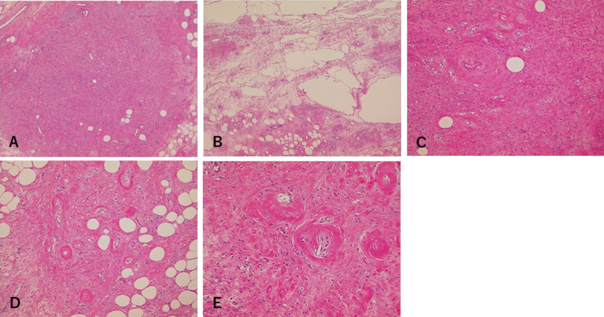
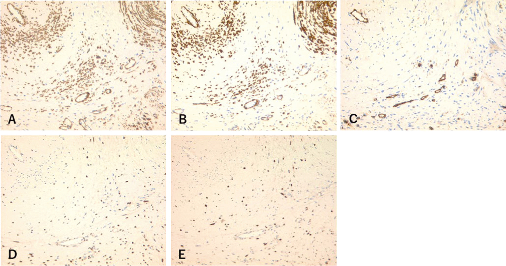
Background: Cellular angiofibroma (CA) is a rare, benign mesenchymal tumor first described by Nucci et al. (Am J Surg Pathol 21:636-644, 1997. 10.1097/00000478-199706000-00002). It affects both men and women, although it is more common in middle-aged women. CA is well circumscribed and usually observed on the body surface, primarily in distal genital regions. Aggressive angiomyxoma and angiomyofibroblastoma are clinically and histologically similar; therefore, it may be necessary to distinguish between CA and these similar tumors. We present a rare case of CA, with atypical features, in the retroperitoneal space during pregnancy.

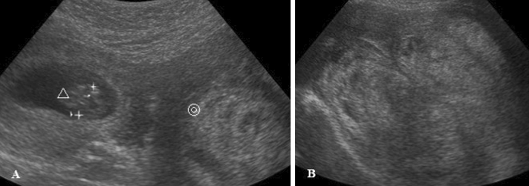
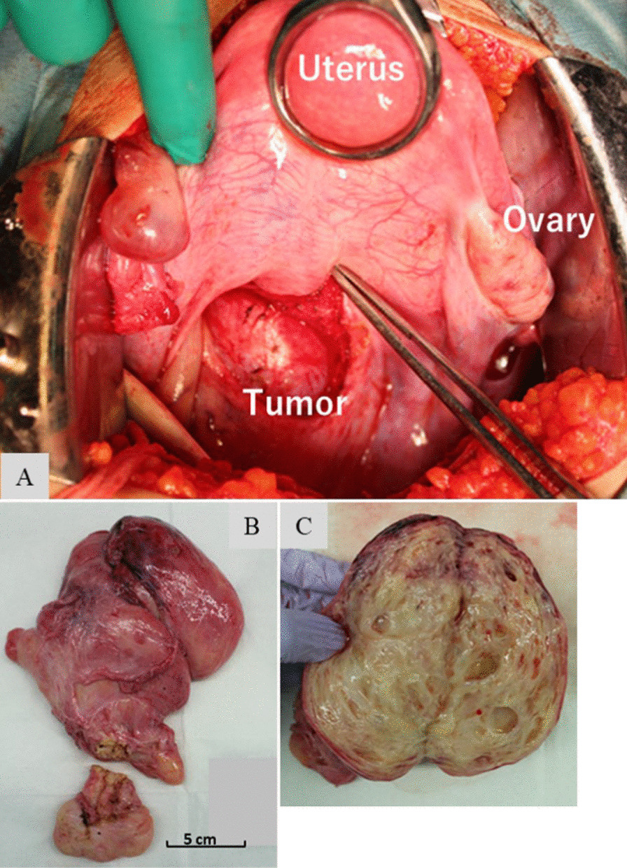
Case presentation: The presence of a 130 mm tumor was detected in a 19-year-old woman. The tumor, located in the retroperitoneal space, was found during first pregnancy examination. At 16 weeks of gestation, the woman developed nausea and fever, and it was diagnosed with acute pyelonephritis. After a few days, the amniotic membranes prematurely ruptured, leading to a miscarriage. The woman underwent a tumor resection, after miscarriage. This case presented with atypical features of CA. This included the young age of the patient, and presence of a tumor in the retroperitoneal space.

Conclusion: In this case, the diagnosis of CA was difficult due to the rarity of the disease and its atypical clinical features. From this experience, we recommend that the discussion on the efficacy of surgical treatment and pregnancy outcomes should be done based on individual case, and not generalized.

Keywords: Aggressive angiomyxoma; Angiomyofibroblastoma; Cellular angiofibroma; Intraperitoneal space; Pregnancy.


Conflict of interest statement

The authors declare that they have no competing interests.

Figures

Fig. 1
Fig. 1
Transabdominal ultrasonography at 9 weeks of gestation. △ Fetus, ◎ Tumor. A The uterine body was shifted superiorly by the tumor. B Tumor size was 130 mm. It had a hyperechoic region, mainly with a partially low region
Fig. 2
Fig. 2
Initial magnetic resonance imaging (MRI) findings. A MRI at 12 weeks of gestation. The tumor measured 115 mm × 150 mm × 160 mm. It showed high signal intensity bands, such as whorl-like formation on T2-weighted images. B Schematic illustration of MRI scan
Fig. 3
Fig. 3
Intraoperative findings and gross appearance. A The tumor grew caudally in the retroperitoneal space and occupied the posterior uterine to the rectovaginal regions. B Tumor size is 170 × 138 × 70 mm. C The tumor is yellowish and has multiple small cysts
Fig. 4
Fig. 4
Hematoxylin and eosin staining. No atypical cells or necrosis was observed. A (magnification ×40): Adipose cells and slight vascular hyperplasia. B (magnification ×40): Constituting the cyst with myxomatous change edematous liquefaction. C (magnification ×100): Spindle-shaped tumor cells were transferred from the blood vessel to the interstitial tissue. D (magnification ×100), E (magnification ×200): Vessel walls, which were small- to medium-sized, showed hyalinization
Fig. 5
Fig. 5
Immunohistochemical staining. A αSMA and B h-caldesmon are partially positive in the tumor cells, except for the blood vessels. C CD34 is partially positive in spindle cells. D ER and E PgR are positive in most tumor cells

References

    1. Nucci MR, Granter SR, Fletcher CDM. Cellular angiofibroma: a benign neoplasm distinct from angiomyofibroblastoma and spindle cell lipoma. Am J Surg Pathol. 1997;21:636–644. doi: 10.1097/00000478-199706000-00002. - DOI - PubMed
    1. Mandato VD, Santagni S, Cavazza A, et al. Cellular angiofibroma in women: a review of the literature. Diagn Pathol. 2015;10:144. doi: 10.1186/s13000-015-0361-6. - DOI - PMC - PubMed
    1. Khong TY, Mooney EE, Ariel I, et al. Sampling and definitions of placental lesion: Amsterdam placental workshop group consensus statement. Arch Pathol Lab Med. 2016;140(7):698–713. doi: 10.5858/arpa.2015-0225-CC. - DOI - PubMed
    1. Khmou M, Lamalmi N, Malihy A, et al. Cellular angiofibroma of the vulva: a poorly known entity, a case report and literature review. BMC Clin Pathol. 2016;16:8. doi: 10.1186/s12907-016-0030-z. - DOI - PMC - PubMed
    1. Flucke U, van Krieken JH, Mentzel T. Cellular angiofibroma: analysis of 25 cases emphasizing its relationship to spindle cell lipoma and mammary-type myofibroblastoma. Mod Pathol. 2011;24:82–89. doi: 10.1038/modpathol.2010.170. - DOI - PubMed

Publication types