Review
. 2023 Feb 10;17(1):7.
doi: 10.1186/s40246-023-00451-1.

SpliceAI-visual: a free online tool to improve SpliceAI splicing variant interpretation

Affiliations
Review

SpliceAI-visual: a free online tool to improve SpliceAI splicing variant interpretation

Jean-Madeleine de Sainte Agathe et al. Hum Genomics. .

Abstract

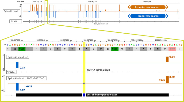
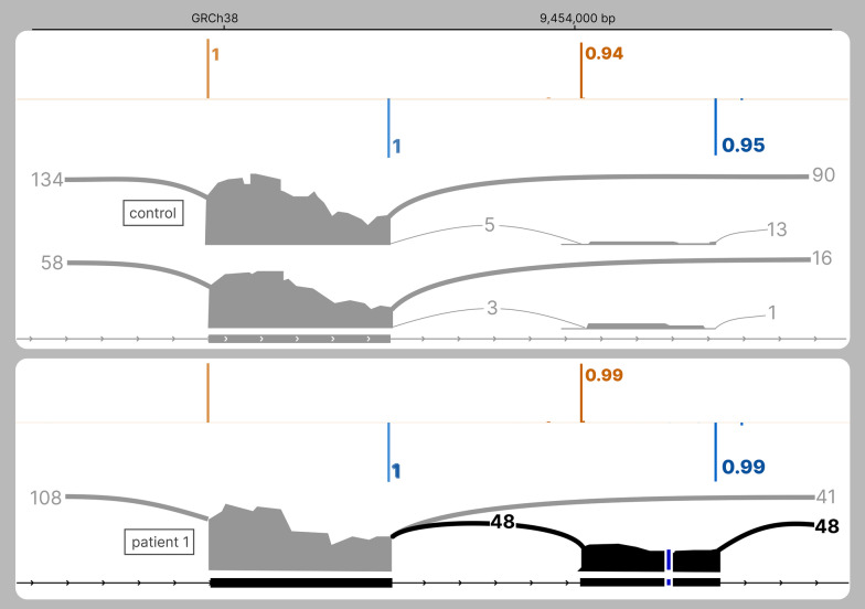
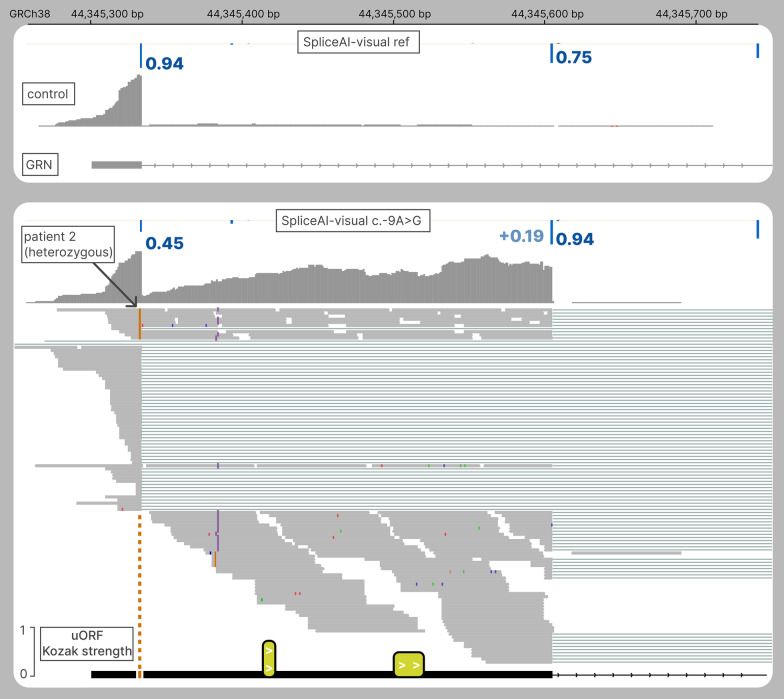
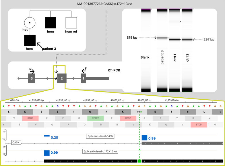
SpliceAI is an open-source deep learning splicing prediction algorithm that has demonstrated in the past few years its high ability to predict splicing defects caused by DNA variations. However, its outputs present several drawbacks: (1) although the numerical values are very convenient for batch filtering, their precise interpretation can be difficult, (2) the outputs are delta scores which can sometimes mask a severe consequence, and (3) complex delins are most often not handled. We present here SpliceAI-visual, a free online tool based on the SpliceAI algorithm, and show how it complements the traditional SpliceAI analysis. First, SpliceAI-visual manipulates raw scores and not delta scores, as the latter can be misleading in certain circumstances. Second, the outcome of SpliceAI-visual is user-friendly thanks to the graphical presentation. Third, SpliceAI-visual is currently one of the only SpliceAI-derived implementations able to annotate complex variants (e.g., complex delins). We report here the benefits of using SpliceAI-visual and demonstrate its relevance in the assessment/modulation of the PVS1 classification criteria. We also show how SpliceAI-visual can elucidate several complex splicing defects taken from the literature but also from unpublished cases. SpliceAI-visual is available as a Google Colab notebook and has also been fully integrated in a free online variant interpretation tool, MobiDetails ( https://mobidetails.iurc.montp.inserm.fr/MD ).


Conflict of interest statement

The authors declare that they have no competing interests.

Figures

Fig. 1
Fig. 1
The delta score (DS) pitfall: discrepancy between SpliceAI’s DS and SpliceAI raw scores (RS). SpliceAI-visual outputs of SCN1A deep intronic variant displayed in IGV. Above: SpliceAI raw scores for the reference allele of SCN1A; below: SpliceAI RS for the pathogenic deep intronic variant NM_001165963.4(SCN1A):c.4002 + 2461 T > C functionally attested to cause the retention of an intronic retention of 64pb[REF]. Orange: acceptor site prediction; Blue: donor site prediction. The variant position is highlighted in yellow
Fig. 2
Fig. 2
The delta score (DS) pitfall: discrepancy between SpliceAI’s DS and SpliceAI raw scores (RS). SpliceAI-visual outputs of MFGE8 deep intronic variant displayed in IGV. Above: SpliceAI RS for the reference allele of MFGE8; below: SpliceAI RS for the pathogenic variant NM_005928.4(MFGE8):c.871-803A > G functionally attested to cause the exonization of an intronic sequence containing a stop codon (red). Orange: acceptor site prediction; Blue: donor site prediction. The variant position is pointed by a dashed line
Fig. 3
Fig. 3
The delta score pitfall: SETD5 poison exon retention caused by an intronic substitution. RNAseq and SpliceAI-visual outputs displayed in IGV. Above: SpliceAI RS for the reference allele of SETD5, along with one control individual; below: SpliceAI RS for the pathogenic deep intronic variant NM_001080517.3:c.2476 + 198A > C, along with RNAseq of patient 1. Orange: acceptor site prediction; Blue: donor site prediction. The variant position is pointed out with a dashed line. Although the variant A > C is heterozygous, 95% of RNAseq reads carry the C, suggesting the causative role of this allele in the retention
Fig. 4
Fig. 4
The delta score pitfall: extending the 5’UTR of GRN. RNAseq and SpliceAI-visual outputs displayed in IGV. Above: SpliceAI RS for the reference allele of GRN along with RNAseq from one control; below: SpliceAI RS for NM_002087.4(GRN):c.-9A > G, along with RNAseq of patient 2. Bottom: two upstream Open Reading Frames in the intronic retention (yellow), height corresponding to the initiation strength of the AUG codon based on the Kozak context from TIS [20]
Fig. 5
Fig. 5
Scaling down the PVS1 criteria of a canonical splice site variant in CASK. Segregation, RT-PCR and SpliceAI RS of NM_003688.3(CASK):c.172 + 1G > A, hemizygous in patient 3. This variant leads to the complete in-frame retention of 18 bp (no wild-type 297 bp product was observed in patient 3 RT-PCR lane), as predicted by SpliceAI-visual. This 18-bp retention does not include stop codon and is predicted to insert 6 amino acids
Fig. 6
Fig. 6
Scaling down the PVS1 criteria of canonical splice site variants in KMT2D. Left: another a priori PVS1 variant NM_003482.4(KMT2D):c.5189-1G > C, present in 11 individuals in UK Biobank. This variant is predicted to result in an in-frame rescuing acceptor site, deleting 8 poorly conserved amino acids. Right: SpliceAI-visual outputs and BAM from one heterozygous from gnomAD of NM_003482.4(KMT2D):c.5782 + 1G > A. This variant is present in 3 individuals in gnomAD, which is not consistent with the penetrance of loss-of-function variants of KMT2D. Also, the mild rescuing DS of 0.28 is added to a nonzero RS on the reference allele (delta score pitfall) and is predicted to result in a complete rescue of this donor site, with the in-frame retention of 9 bp
Fig. 7
Fig. 7
Scaling down the PVS1 criteria of a canonical splice site variant in TTN. RNAseq and SpliceAI-visual outputs displayed in IGV showing the predicted exon skipping (top view), and the in-frame rescue (bottom view). Top tracks: SpliceAI RS for the reference allele of TTN along with RNAseq from 2 controls; bottom tracks: SpliceAI RS for the NM_001267550.2(TTN):c.31349-1G > C along with RNAseq of patient 4
Fig. 8
Fig. 8
Scaling down the PVS1 criteria of a putative frameshift in SETD5. SpliceAI-visual outputs displayed in IGV showing the predicted benign splicing outcome of this putative frameshift
Fig. 9
Fig. 9
SpliceAI-visual outputs displayed in IGV showing the predicted exon skipping resulting from the complex delins NM_001142800.2(EYS):c.2992_2992 + 6delinsTG. Top track: SpliceAI RS for the reference allele of EYS; bottom track: SpliceAI RS for the delins in EYS

References

    1. López-Bigas N, Audit B, Ouzounis C, Parra G, Guigó R. Are splicing mutations the most frequent cause of hereditary disease? FEBS Lett. 2005;579(9):1900–1903. doi: 10.1016/j.febslet.2005.02.047. - DOI - PubMed
    1. Jaganathan K, Kyriazopoulou Panagiotopoulou S, McRae JF, et al. Predicting splicing from primary sequence with deep learning. Cell. 2019;176(3):535–548.e24. doi: 10.1016/j.cell.2018.12.015. - DOI - PubMed
    1. Wai HA, Lord J, Lyon M, et al. Blood RNA analysis can increase clinical diagnostic rate and resolve variants of uncertain significance. Genet Med. 2020;22(6):1005–1014. doi: 10.1038/s41436-020-0766-9. - DOI - PMC - PubMed
    1. Ha C, Kim JW, Jang JH. Performance evaluation of SpliceAI for the prediction of splicing of NF1 variants. Genes. 2021;12(9):1308. doi: 10.3390/genes12091308. - DOI - PMC - PubMed
    1. Bychkov I, Galushkin A, Filatova A, et al. Functional analysis of the PCCA and PCCB gene variants predicted to affect splicing. IJMS. 2021;22(8):4154. doi: 10.3390/ijms22084154. - DOI - PMC - PubMed

Publication types