

SARS-CoV-2 Establishes a Productive Infection in Hepatoma and Glioblastoma Multiforme Cell Lines
- PMID: 36765590
- PMCID: PMC9913867
- DOI: 10.3390/cancers15030632
SARS-CoV-2 Establishes a Productive Infection in Hepatoma and Glioblastoma Multiforme Cell Lines
Abstract
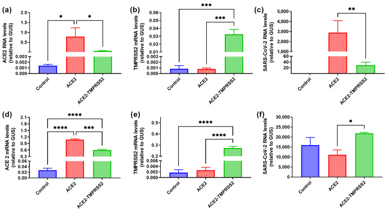
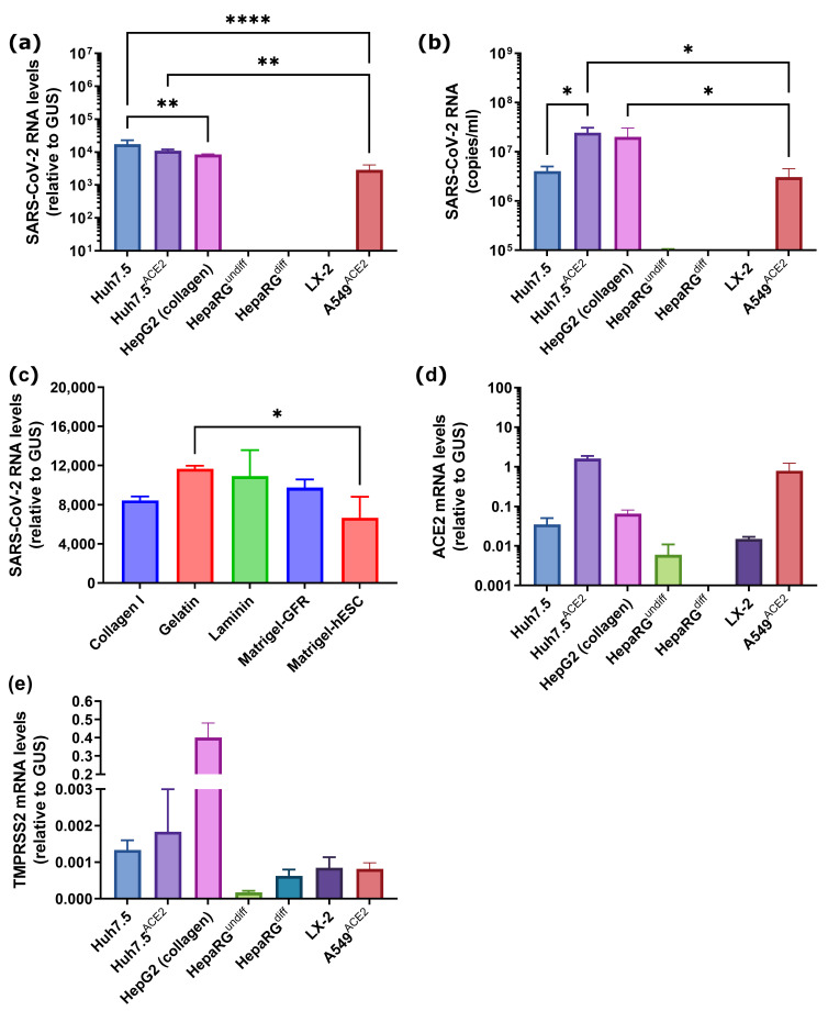
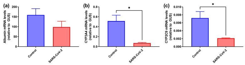
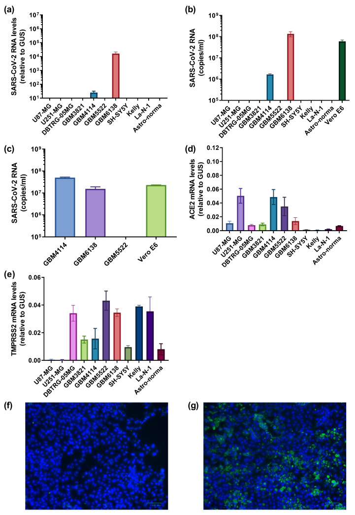
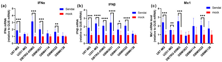
Severe acute respiratory syndrome associated coronavirus 2 (SARS-CoV-2) emerged at the end of 2019 and rapidly caused a pandemic that led to the death of >6 million people due to hypercoagulation and cytokine storm. In addition, SARS-CoV-2 triggers a wide array of pathologies, including liver dysfunction and neurological disorders. It remains unclear if these events are due to direct infection of the respective tissues or result from systemic inflammation. Here, we explored the possible infection of hepatic and CNS cell lines by SARS-CoV-2. We show that even moderate expression levels of the angiotensin-converting enzyme 2 (ACE2) are sufficient for productive infection. SARS-CoV-2 infects hepatoma Huh7.5 and HepG2 cells but not non-transformed liver progenitor or hepatocyte/cholangiocyte-like HepaRG cells. However, exposure to the virus causes partial dedifferentiation of HepaRG cells. SARS-CoV-2 can also establish efficient replication in some low-passage, high-grade glioblastoma cell lines. In contrast, embryonal primary astrocytes or neuroblastoma cells did not support replication of the virus. Glioblastoma cell permissiveness is associated with defects in interferon production. Overall, these results suggest that liver dysfunction during COVID-19 is not due to infection of these tissues by SARS-CoV-2. Furthermore, tumors may potentially serve as reservoirs for the virus during infection.
Keywords: SARS-CoV-2; glioblastoma; hepatoma; liver cells; permissiveness.
Conflict of interest statement
The authors declare no conflict of interest. The funders had no role in the design of the study; in the collection, analyses, or interpretation of data; in the writing of the manuscript; or in the decision to publish the results.
Figures
Similar articles
-
Susceptibility of neuroblastoma and glioblastoma cell lines to SARS-CoV-2 infection.Brain Res. 2021 May 1;1758:147344. doi: 10.1016/j.brainres.2021.147344. Epub 2021 Feb 5. Brain Res. 2021. PMID: 33556379 Free PMC article.
-
Antiviral Activity of Type I, II, and III Interferons Counterbalances ACE2 Inducibility and Restricts SARS-CoV-2.mBio. 2020 Sep 10;11(5):e01928-20. doi: 10.1128/mBio.01928-20. mBio. 2020. PMID: 32913009 Free PMC article.
-
Replication Kinetics, Cell Tropism, and Associated Immune Responses in SARS-CoV-2- and H5N1 Virus-Infected Human Induced Pluripotent Stem Cell-Derived Neural Models.mSphere. 2021 Jun 30;6(3):e0027021. doi: 10.1128/mSphere.00270-21. Epub 2021 Jun 23. mSphere. 2021. PMID: 34160239 Free PMC article.
-
Severe Acute Respiratory Syndrome Coronavirus 2 Impact on the Central Nervous System: Are Astrocytes and Microglia Main Players or Merely Bystanders?ASN Neuro. 2020 Jan-Dec;12:1759091420954960. doi: 10.1177/1759091420954960. ASN Neuro. 2020. PMID: 32878468 Free PMC article. Review.
-
Endothelial cell dysfunction, coagulation, and angiogenesis in coronavirus disease 2019 (COVID-19).Microvasc Res. 2021 Sep;137:104188. doi: 10.1016/j.mvr.2021.104188. Epub 2021 May 20. Microvasc Res. 2021. PMID: 34022205 Free PMC article. Review.
Cited by
-
Electrophysiological Impact of SARS-CoV-2 Envelope Protein in U251 Human Glioblastoma Cells: Possible Implications in Gliomagenesis?Int J Mol Sci. 2024 Jun 18;25(12):6669. doi: 10.3390/ijms25126669. Int J Mol Sci. 2024. PMID: 38928376 Free PMC article.
-
Soybean Trypsin Inhibitor Possesses Potency Against SARS-CoV-2 Infection by Blocking the Host Cell Surface Receptors ACE2, TMPRSS2, and CD147.Int J Mol Sci. 2025 Jul 9;26(14):6583. doi: 10.3390/ijms26146583. Int J Mol Sci. 2025. PMID: 40724833 Free PMC article.
-
Unwinding the SARS-CoV-2 Ribosomal Frameshifting Pseudoknot with LNA and G-Clamp-Modified Phosphorothioate Oligonucleotides Inhibits Viral Replication.Biomolecules. 2023 Nov 17;13(11):1660. doi: 10.3390/biom13111660. Biomolecules. 2023. PMID: 38002341 Free PMC article.
-
Metabolic Imprint of Poliovirus on Glioblastoma Cells and Its Role in Virus Replication and Cytopathic Activity.Int J Mol Sci. 2025 Jul 30;26(15):7346. doi: 10.3390/ijms26157346. Int J Mol Sci. 2025. PMID: 40806477 Free PMC article.
-
Natural-Target-Mimicking Translocation-Based Fluorescent Sensor for Detection of SARS-CoV-2 PLpro Protease Activity and Virus Infection in Living Cells.Int J Mol Sci. 2024 Jun 17;25(12):6635. doi: 10.3390/ijms25126635. Int J Mol Sci. 2024. PMID: 38928340 Free PMC article.
References
Grants and funding
LinkOut - more resources
Full Text Sources
Miscellaneous
