

Organ-on-a-Chip and Microfluidic Platforms for Oncology in the UK
- PMID: 36765593
- PMCID: PMC9913518
- DOI: 10.3390/cancers15030635
Organ-on-a-Chip and Microfluidic Platforms for Oncology in the UK
Abstract
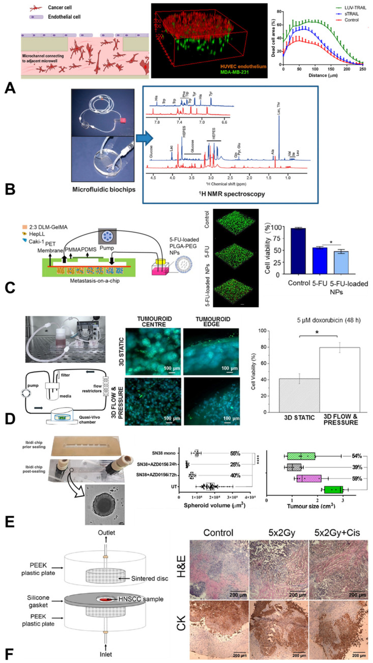
Organ-on-chip systems are capable of replicating complex tissue structures and physiological phenomena. The fine control of biochemical and biomechanical cues within these microphysiological systems provides opportunities for cancer researchers to build complex models of the tumour microenvironment. Interest in applying organ chips to investigate mechanisms such as metastatsis and to test therapeutics has grown rapidly, and this review draws together the published research using these microfluidic platforms to study cancer. We focus on both in-house systems and commercial platforms being used in the UK for fundamental discovery science and therapeutics testing. We cover the wide variety of cancers being investigated, ranging from common carcinomas to rare sarcomas, as well as secondary cancers. We also cover the broad sweep of different matrix microenvironments, physiological mechanical stimuli and immunological effects being replicated in these models. We examine microfluidic models specifically, rather than organoids or complex tissue or cell co-cultures, which have been reviewed elsewhere. However, there is increasing interest in incorporating organoids, spheroids and other tissue cultures into microfluidic organ chips and this overlap is included. Our review includes a commentary on cancer organ-chip models being developed and used in the UK, including work conducted by members of the UK Organ-on-a-Chip Technologies Network. We conclude with a reflection on the likely future of this rapidly expanding field of oncological research.
Keywords: cancer; mechanobiology; microfluidic; microphysiological system; oncology; organ-on-chip; pre-clinical model; spheroid; tumour cell.
Conflict of interest statement
M.M.K. and H.R.C.S. are Directors of the Queen Mary + Emulate Organs-on-Chips Centre, which is part-funded by Emulate Inc. Emulate Inc. were not involved in the preparation of this manuscript. The remaining authors declare that they have no conflict of interest. The funders had no role in the design of the study; in the collection, analyses, or interpretation of data; in the writing of the manuscript, or in the decision to publish the results.
Figures
Similar articles
-
A multi-organ-chip co-culture of liver and testis equivalents: a first step toward a systemic male reprotoxicity model.Hum Reprod. 2020 May 1;35(5):1029-1044. doi: 10.1093/humrep/deaa057. Hum Reprod. 2020. PMID: 32390056
-
Small Force, Big Impact: Next Generation Organ-on-a-Chip Systems Incorporating Biomechanical Cues.Front Physiol. 2018 Oct 9;9:1417. doi: 10.3389/fphys.2018.01417. eCollection 2018. Front Physiol. 2018. PMID: 30356887 Free PMC article. Review.
-
Organs-on-chips technologies - A guide from disease models to opportunities for drug development.Biosens Bioelectron. 2023 Jul 1;231:115271. doi: 10.1016/j.bios.2023.115271. Epub 2023 Mar 31. Biosens Bioelectron. 2023. PMID: 37060819 Review.
-
Fluidic circuit board with modular sensor and valves enables stand-alone, tubeless microfluidic flow control in organs-on-chips.Lab Chip. 2022 Mar 15;22(6):1231-1243. doi: 10.1039/d1lc00999k. Lab Chip. 2022. PMID: 35178541 Free PMC article.
-
From organoids to organoids-on-a-chip: Current applications and challenges in biomedical research.Chin Med J (Engl). 2025 Apr 5;138(7):792-807. doi: 10.1097/CM9.0000000000003535. Epub 2025 Feb 25. Chin Med J (Engl). 2025. PMID: 39994843 Free PMC article. Review.
Cited by
-
Air Pollution and Osteoporosis.Curr Osteoporos Rep. 2024 Dec;22(6):590-598. doi: 10.1007/s11914-024-00889-9. Epub 2024 Sep 20. Curr Osteoporos Rep. 2024. PMID: 39302569 Free PMC article. Review.
-
Modelling the Tumour Microenvironment, but What Exactly Do We Mean by "Model"?Cancers (Basel). 2023 Jul 26;15(15):3796. doi: 10.3390/cancers15153796. Cancers (Basel). 2023. PMID: 37568612 Free PMC article. Review.
-
Cancer-on-a-chip for precision cancer medicine.Lab Chip. 2025 Jul 8;25(14):3314-3347. doi: 10.1039/d4lc01043d. Lab Chip. 2025. PMID: 40376718 Free PMC article. Review.
-
Development of a dual-flow tissue perfusion device for modeling the gastrointestinal tract-brain axis.Biomicrofluidics. 2023 Oct 11;17(5):054104. doi: 10.1063/5.0168953. eCollection 2023 Sep. Biomicrofluidics. 2023. PMID: 37840538 Free PMC article.
-
Combination of Genomic Landsscape and 3D Culture Functional Assays Bridges Sarcoma Phenotype to Target and Immunotherapy.Cells. 2023 Sep 4;12(17):2204. doi: 10.3390/cells12172204. Cells. 2023. PMID: 37681936 Free PMC article. Review.
References
-
- United States Senate . United States Senate—117th Congress. United States Senate; Washington, DC, USA: 2022. S.5002—FDA Modernization Act 2.0.
-
- United States House of Representatives . United States House of Representatives—117th Congress. United States House of Representatives; Washington, DC, USA: 2022. H.R.2565—FDA Modernization Act of 2021.
Publication types
Grants and funding
LinkOut - more resources
Full Text Sources
