. 2023 Jan 22;15(3):687.
doi: 10.3390/cancers15030687.

Risk Group Stratification for Recurrence-Free Survival and Early Tumor Recurrence after Radiofrequency Ablation for Hepatocellular Carcinoma

Affiliations

Risk Group Stratification for Recurrence-Free Survival and Early Tumor Recurrence after Radiofrequency Ablation for Hepatocellular Carcinoma

Dong Ik Cha et al. Cancers (Basel). .

Abstract

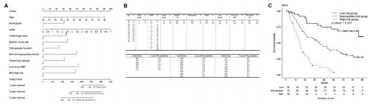
Purpose: Although the prognosis after radiofrequency ablation (RFA) for hepatocellular carcinoma (HCC) may vary according to different risk levels, there is no standardized follow-up protocol according to each patient's risk. This study aimed to stratify patients according to their risk of recurrence-free survival (RFS) and early (≤2 years) tumor recurrence (ETR) after RFA for HCC based on predictive models and nomograms and to compare the survival times of the risk groups derived from the models.

Methods: Patients who underwent RFA for a single HCC (≤3 cm) between January 2012 and March 2014 (n = 152) were retrospectively reviewed. Patients were classified into low-, intermediate-, and high-risk groups based on the total nomogram points for RFS and ETR, respectively, and compared for each outcome. Restricted mean survival times (RMSTs) in the three risk groups were evaluated for both RFS and ETR to quantitatively evaluate the difference in survival times.

Results: Predictive models for RFS and ETR were constructed with c-indices of 0.704 and 0.730, respectively. The high- and intermediate-risk groups for RFS had an 8.5-fold and 2.9-fold higher risk of events than the low-risk group (both p < 0.001), respectively. The high- and intermediate-risk groups for ETR had a 17.7-fold and 7.0-fold higher risk than the low-risk group (both p < 0.001), respectively. The RMST in the high-risk group was significantly lower than that in the other two groups 9 months after RFA, and that in the intermediate-risk group became lower than that in the low-risk group after 21 months with RFS and 24 months with ETR.

Conclusion: Our predictive models were able to stratify patients into three groups according to their risk of RFS and ETR after RFA for HCC. Differences in RMSTs may be used to establish different follow-up protocols for the three risk groups.

Keywords: carcinoma; hepatocellular; nomogram; predictive model; prognosis; radiofrequency ablation; survival times.


Conflict of interest statement

The authors declare no conflict of interest.

Figures

Figure 1
Figure 1
Patient inclusion process.
Figure 2
Figure 2
Nomogram to estimate recurrence-free survival (RFS) after percutaneous radiofrequency ablation for HCC, and nomogram-based low-, intermediate- and high-risk groups for RFS. (A) The included variables in the nomogram for RFS are age, Albumin-Bilirubin grade 2 (reference: grade 1), aspartate aminotransferase/platelet ratio index, Child-Pugh classification B (reference A), MoRAL score > 68, subcapsular location, non-rim hyperenhancement of the lesion (reference: no enhancement), enhancing capsule (reference: no enhancing capsule), low signal intensity (SI) of the lesion on hepatobiliary phase on gadoxetic acid-enhanced liver MRI (reference: iso or high SI), and microvascular invasion-high risk group. (B) The prognostic points of each variable are shown in the upper table, and the estimated RFS rate at 1, 2, and 5 years after RFS for HCC according to the total points are shown in the lower table. (C) For RFS, patients with nomogram scores in the lower quartile (<25%, 185.75 points) were classified as low-risk, those in the upper quartile (>25%, 242.56 points) as high-risk, and those in between (25–75%) were classified as intermediate-risk groups. The RFS rates of the low, intermediate, and high risk groups were 94.6% (95% CI 87.6–100%), 86.7% (95% CI 79.3–94.7%), and 68.0% (95% CI 54.6–84.7%) at 1 year; 89.0% (95% CI 79.4–99.8%), 63.9% (95% CI 53.9–75.8%), and 29.5% (95% CI 17.8–48.8%) at 2 years; and 71.7% (95% CI 58.2–88.2%), 38.5% (95% CI 28.7–51.6%), and 3.7% (95% CI 0.6–24%) at 5 years, respectively. The RFS rates among the three groups were significantly different (p < 0.001). Patients in the high-risk group had an 8.5-fold higher risk of recurrence or death than the low-risk group (95% CI 4.3–16.9, p < 0.001), and those in the intermediate-risk group had a 2.9-fold higher risk than the low-risk group (95% CI 1.6–5.5, p < 0.001). At 9 months, the restricted mean survival time (RMST) in the high-risk group was 8.192 months (95% CI 7.610–8.774 months), which was significantly different from the low-risk group (RMST 8.858 [95% CI 8.583–9.133 months], and the intermediate-risk group (RMST 8.877 [95% CI 8.736–9.018 months]. The RMST of the high-risk group was significantly lower than that of the other groups and was sustained until the last time point, which was 60 months. A significant difference between the intermediate- and low-risk groups occurred at 21 months after RFA (intermediate-risk group, RMST 18.549 months [95% CI 17.555–19.543 months]; and low-risk group, RMST 20.116 months [95% CI 19.056–21.177 months]; p = 0.035). RFS = recurrence-free survival, HCC = hepatocellular carcinoma, MoRAL = Model for tumor recurrence after living donor liver transplantation, SI = signal intensity, MRI = magnetic resonance imaging.
Figure 3
Figure 3
Nomogram to estimate early tumor recurrence after percutaneous radiofrequency ablation for HCC, and nomogram-based low-, intermediate- and high-risk groups for early tumor recurrence. (A) The included variables in the nomogram for early tumor recurrence are age, Albumin-Bilirubin grade 2 (reference: grade 1), MoRAL score > 68, non-rim hyperenhancement (reference: no enhancement), enhancing capsule (reference: no enhancing capsule), low signal intensity (SI) of the lesion on hepatobiliary phase on gadoxetic acid-enhanced liver MRI (reference: iso or high SI), and microvascular invasion-high risk group. (B) The prognostic points of each variable are shown in the upper table, and the estimated early tumor recurrence rate at 1 and 2 years after RFS for HCC according to the total points are shown in the lower table. (C) For early tumor recurrence, patients with nomogram scores in the lower quartile (<25%, 210.80 points) were classified as low-risk, those in the upper quartile (>25%, 289.66 points) as high-risk, and those in between (25–75%) were classified as intermediate-risk groups. The cumulative early tumor recurrence rates of the low-, intermediate-, and high-risk groups were 5.7% (95% CI 0.0–13.1%), 8.9% (95% CI 2.4–14.9%), and 39.5% (95% CI 21.8–53.2%) at 1 year, and 5.7% (95% CI 0.0–13.1%), 35.4% (95% CI 24–45.2%), and 63.2% (95% CI 44.1–75.7%) at 2 years. The cumulative early tumor recurrence rates among the three groups were significantly different (p < 0.001). Patients in the high-risk group had a 17.7-fold higher risk of recurrence or death than the low-risk group (95% CI 4.184–75.200, p < 0.001), and those in the intermediate-risk group had a 7.0-fold higher risk of recurrence or death than the low-risk group (95% CI 1.665–29.360, p < 0.001). At 9 months, restricted mean survival time (RMST) in the high-risk group, with an RMST of 8.203 months (95% CI 7.647–8.758 months), was significantly lower than that in other groups (low-risk group RMST 8.846 [95% CI 8.548–9.144 months], p = 0.045; and intermediate-risk group RMST 8.929 [95% CI 8.803–9.055 months], p = 0.012]. A significant difference between the intermediate- and low-risk groups occurred at 24 months after RFA (intermediate-risk group, RMST 21.077 months [95% CI 19.987–22.167 months]; and low-risk group, RMST 23.029 months [95% CI 21.694–24.363 months]; p = 0.026]. HCC = hepatocellular carcinoma, MoRAL = Model for tumor recurrence after living donor liver transplantation, SI = signal intensity, MRI = magnetic resonance imaging.

References

    1. European Association for the Study of the Liver Corrigendum to "EASL Clinical Practice Guidelines: Management of hepatocellular carcinoma. J. Hepatol. 2019;70:817. doi: 10.1016/j.jhep.2019.01.020. - DOI - PubMed
    1. Marrero J.A., Kulik L.M., Sirlin C.B., Zhu A.X., Finn R.S., Abecassis M.M., Roberts L.R., Heimbach J.K. Diagnosis, Staging, and Management of Hepatocellular Carcinoma: 2018 Practice Guidance by the American Association for the Study of Liver Diseases. Hepatology. 2018;68:723–750. doi: 10.1002/hep.29913. - DOI - PubMed
    1. Nault J.C., Sutter O., Nahon P., Ganne-Carrie N., Seror O. Percutaneous treatment of hepatocellular carcinoma: State of the art and innovations. J. Hepatol. 2018;68:783–797. doi: 10.1016/j.jhep.2017.10.004. - DOI - PubMed
    1. Rossi S., Ravetta V., Rosa L., Ghittoni G., Viera F.T., Garbagnati F., Silini E.M., Dionigi P., Calliada F., Quaretti P., et al. Repeated radiofrequency ablation for management of patients with cirrhosis with small hepatocellular carcinomas: A long-term cohort study. Hepatology. 2011;53:136–147. doi: 10.1002/hep.23965. - DOI - PubMed
    1. Shiina S., Tateishi R., Arano T., Uchino K., Enooku K., Nakagawa H., Asaoka Y., Sato T., Masuzaki R., Kondo Y., et al. Radiofrequency ablation for hepatocellular carcinoma: 10-year outcome and prognostic factors. Am. J. Gastroenterol. 2012;107:569–577. doi: 10.1038/ajg.2011.425. quiz 578. - DOI - PMC - PubMed

LinkOut - more resources