. 2023 Jan 25;15(3):731.
doi: 10.3390/cancers15030731.

Sialyl LewisX/A and Cytokeratin Crosstalk in Triple Negative Breast Cancer

Affiliations

Sialyl LewisX/A and Cytokeratin Crosstalk in Triple Negative Breast Cancer

Carlota Pascoal et al. Cancers (Basel). .

Abstract

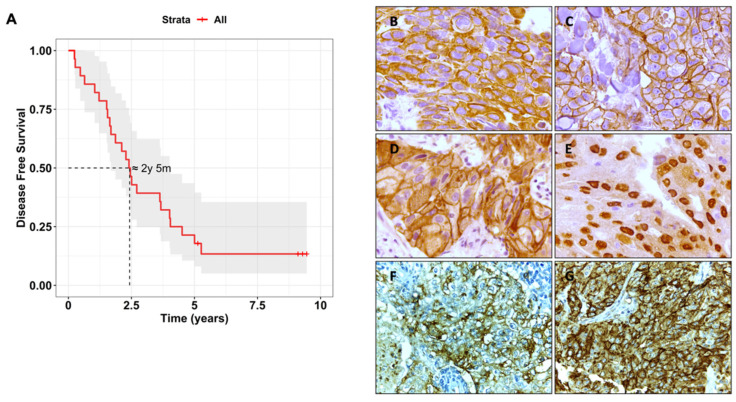
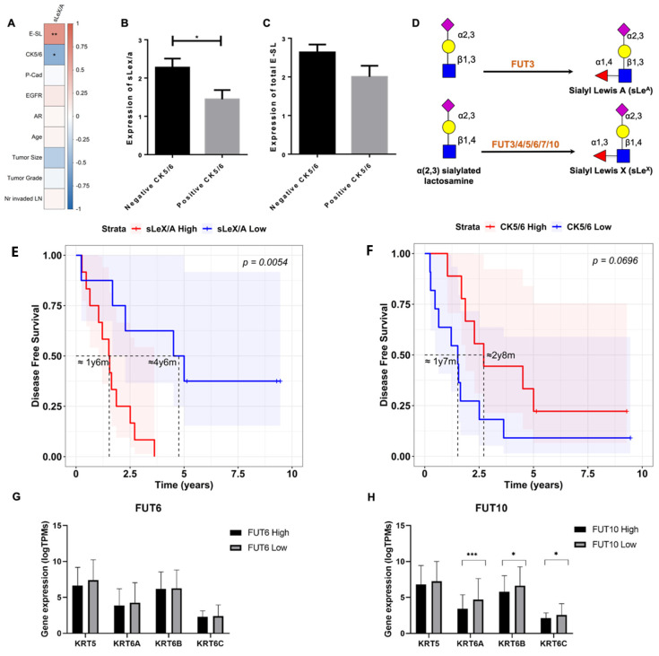
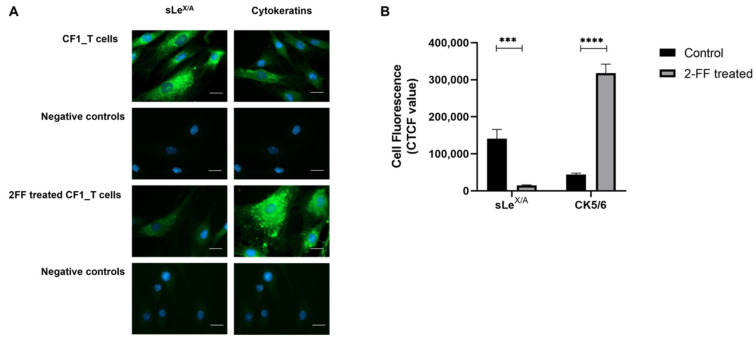
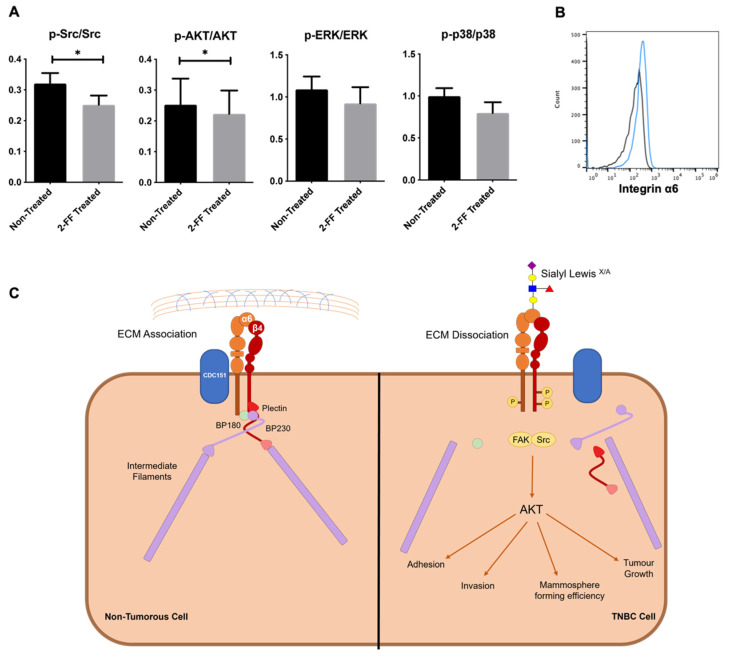
Triple-negative breast cancer (TNBC) encompasses multiple entities and is generally highly aggressive and metastatic. We aimed to determine the clinical and biological relevance of Sialyl-Lewis X and A (sLeX/A)-a fucosylated glycan involved in metastasis-in TNBC. Here, we studied tissues from 50 TNBC patients, transcripts from a TNBC dataset from The Cancer Genome Atlas (TCGA) database, and a primary breast cancer cell line. All 50 TNBC tissue samples analysed expressed sLeX/A. Patients with high expression of sLeX/A had 3 years less disease-free survival than patients with lower expression. In tissue, sLeX/A negatively correlated with cytokeratins 5/6 (CK5/6, which was corroborated by the inverse correlation between fucosyltransferases and CK5/6 genes. Our observations were confirmed in vitro when inhibition of sLeX/A remarkably increased expression of CK5/6, followed by a decreased proliferation and invasion capacity. Among the reported glycoproteins bearing sLeX/A and based on the STRING tool, α6 integrin showed the highest interaction score with CK5/6. This is the first report on the sLeX/A expression in TNBC, highlighting its association with lower disease-free survival and its inverse crosstalk with CK5/6 with α6 integrin as a mediator. All in all, sLeX/A is critical for TNBC malignancy and a potential prognosis biomarker and therapeutic target.

Keywords: aberrant glycosylation; cytokeratin expression; disease-free survival rate; intermediate filament proteins; sialyl LewisX/A (sLeX/A); triple-negative breast cancer (TNBC); α6 integrin.


Conflict of interest statement

The authors declare no conflict of interest.

Figures

Figure 1
Figure 1
Disease-free survival (DFS) of the triple-negative breast cancer (TNBC) patient cohort and respective immunohistochemical biomarker staining of respective TNBC tissues. (A) Kaplan–Meier curve of DFS of all TNBC patients enrolled in this study generated with the survminer (v0.4.9) and survival (v3.4-0) R packages. Representative microphotographs (400X magnification) of immunohistochemical staining against cytokeratin (CK) 5/6 (B), P-cadherin (C), Epidermal Growth Factor Receptor (EGFR) (D), Androgen Receptor (AR) (E), Sialyl-Lewis X and A (sLeX/A) (F) and E-selectin ligand (E-SL) (G) in TNBC tissue sections. Tissues were stained with hematoxylin, which colours nuclei. The staining with antibodies or E-Ig was followed by a horseradish peroxidase conjugated secondary antibody and visualised in brown.
Figure 2
Figure 2
sLeX/A/E-SL and CK5/6 expression in TNBC tissues are inversely correlated and influence DFS. (A) sLeX/A is positively correlated with E-SL staining (p = 0.002) and negatively correlated with CK5/6 expression (p = 0.019) in TNBC tissues. Non-parametric Spearman correlation was used to evaluate the association between features. CK5/6-positive TNBC have lower expression of sLeX/A (B) and E-SL (C) than the TNBC samples that do not express the CK5/6. TNBC cases were divided in two groups according to CK5/6 expression (score ≥ 1) or lack of expression (score = 0); (D) sLeX and sLeA structure and terminal step of addition of fucose by fucosyltransferases (FUT). (E) Patients with lower sLeX/A expression have a better 10 year disease-free survival than those with higher expression (p = 0.0054); (F) Contrastingly, patients with lower CK5/6 expression have worse 10 year DFS than those with higher expression (p = 0.0696); Kaplan–Meier curves show the DFS of patients with high (red) and low (blue) sLeX/A or CK5/6 expression (higher and lower expression values than the mean sLeX/A/CK5/6 expression, respectively) generated with the survminer (v0.4-9) and survival (v3.4-0) R packages. (G) FUT6 low gene expression group has increased KRT6A gene expression (p = 0.074), and (H) FUT10 low gene expression group has increased KRT6A (p = 0.002), -6B (p = 0.034) and -6C (p = 0.025), compared with the high expression groups. TNBC tissue genetic data from the TCGA database was analysed for gene expression. Samples were subdivided based on the median expression of FUT6 and FUT10 and correlated with genes involved in CK5/6 epitope expression (KRT5, KRT6A, -6B, -6C). Legend: * - p < 0.05, ** - p < 0.01, *** - p < 0.001.
Figure 3
Figure 3
Inhibition of fucosylation decreases sLeX/A expression and increases cytokeratin. CF1_T cells were treated or not with 2-fluorofucose (2-FF) and then labelled with HECA-452 mAb and anti-CK monoclonal antibody (mAb), as described in the materials and methods. (A) Fluorescence microscopy images (scale bar: 1 μm). The resulting fluorescence of labelling with HECA-452 and anti-CK is shown per column. The first row presents images of untreated cells stained with primary antibodies; on the second row there are the labelling controls in the absence of primary antibodies; the third row shows CF1_T cells treated with 2-FF and stained with primary antibody, and the fourth row exhibits labelling controls of 2-FF treated cells in the absence of primary antibodies. Images are representative of merging fluorescence where antibody labelling is shown in green, and nuclei were stained with 4′,6-diamidino-2-phenylindole (blue). (B) Corrected total cell fluorescence. The graph shows the calculated arbitrary values of corrected total cell fluorescence (CTCF) retrieved from images depicted in A as described in the materials and methods section. Legend: *** - p < 0.001, **** - p < 0.0001.
Figure 4
Figure 4
α6 integrin-associated pathways are affected upon 2-FF treatment. (A) Expression of phosphorylated proteins is affected by 2-FF. CF1_T cell line treated with 2-FF for 5 days were analysed regarding their expression of phosphorylated Src (p-Src), AKT (p-AKT), ERK1/2 (p-ERK), and respective total proteins by flow cytometry. Ratio between phosphorylated versus total protein expression is represented in the graphs (n = 3, p < 0.05 (*)). (B) CF1_T cells express α6 integrin. Flow cytometry analysis of CF1_T cells stained with anti-α6 integrin antibody, plus fluorescent secondary antibody. Representative histogram showing positive staining (blue line). Cells stained only with fluorescent secondary antibody and without anti-α6 integrin antibody were used as a negative control (dark line); (C) Proposed mechanism of the influence of sLeX/A decoration of α6integrin on cytokeratin expression. The crosstalk between α6 integrin and cytokeratins is established by the hemidesmosome (complex of α6β4 integrin, plectin, BP180, CD151, BP230). sLeX/A decoration activates integrin and the downstream signalling pathways, contributing to a more aggressive phenotype.

References

    1. Stagg J., Allard B. Immunotherapeutic approaches in triple-negative breast cancer: Latest research and clinical prospects. Ther. Adv. Med. Oncol. 2013;5:169–181. doi: 10.1177/1758834012475152. - DOI - PMC - PubMed
    1. Senkus E., Kyriakides S., Ohno S., Penault-Llorca F., Poortmans P., Rutgers E., Zackrisson S., Cardoso F. ESMO Guidelines Committee. Primary breast cancer: ESMO Clinical Practice Guidelines for diagnosis, treatment and follow-up. Ann. Oncol. Off. J. Eur. Soc. Med. Oncol. 2015;26((Suppl. 5)):v8–v30. doi: 10.1093/annonc/mdv298. - DOI - PubMed
    1. Cipriano É., Mesquita A. Emerging Therapeutic Drugs in Metastatic Triple-Negative Breast Cancer. Breast Cancer Basic Clin. Res. 2021;15 doi: 10.1177/11782234211002491. - DOI - PMC - PubMed
    1. Dent R., Trudeau M., Pritchard K.I., Hanna W.M., Kahn H.K., Sawka C.A., Lickley L.A., Rawlinson E., Sun P., Narod S.A. Triple-Negative Breast Cancer: Clinical Features and Patterns of Recurrence. Clin. Cancer Res. Off. J. Am. Assoc. Cancer Res. 2007;13:4429–4434. doi: 10.1158/1078-0432.CCR-06-3045. - DOI - PubMed
    1. Marra A., Trapani D., Viale G., Criscitiello C., Curigliano G. Practical classification of triple-negative breast cancer: Intratumoral heterogeneity, mechanisms of drug resistance, and novel therapies. NPJ Breast Cancer. 2020;6:54. doi: 10.1038/s41523-020-00197-2. - DOI - PMC - PubMed