Review
. 2023 Jan 28;15(3):808.
doi: 10.3390/cancers15030808.

The Evolution of Ki-67 and Breast Carcinoma: Past Observations, Present Directions, and Future Considerations

Affiliations
Review

The Evolution of Ki-67 and Breast Carcinoma: Past Observations, Present Directions, and Future Considerations

Brian S Finkelman et al. Cancers (Basel). .

Abstract

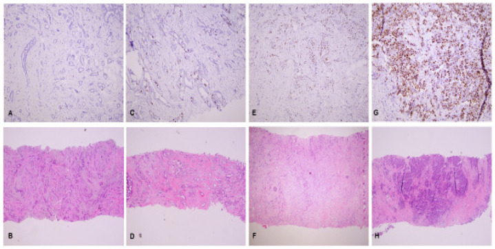
The 1983 discovery of a mouse monoclonal antibody-the Ki-67 antibody-that recognized a nuclear antigen present only in proliferating cells represented a seminal discovery for the pathologic assessment of cellular proliferation in breast cancer and other solid tumors. Cellular proliferation is a central determinant of prognosis and response to cytotoxic chemotherapy in patients with breast cancer, and since the discovery of the Ki-67 antibody, Ki-67 has evolved as an important biomarker with both prognostic and predictive potential in breast cancer. Although there is universal recognition among the international guideline recommendations of the value of Ki-67 in breast cancer, recommendations for the actual use of Ki-67 assays in the prognostic and predictive evaluation of breast cancer remain mixed, primarily due to the lack of assay standardization and inconsistent inter-observer and inter-laboratory reproducibility. The treatment of high-risk ER-positive/human epidermal growth factor receptor-2 (HER2) negative breast cancer with the recently FDA-approved drug abemaciclib relies on a quantitative assessment of Ki-67 expression in the treatment decision algorithm. This further reinforces the urgent need for standardization of Ki-67 antibody selection and staining interpretation, which will hopefully lead to multidisciplinary consensus on the use of Ki-67 as a prognostic and predictive marker in breast cancer. The goals of this review are to highlight the historical evolution of Ki-67 in breast cancer, summarize the present literature on Ki-67 in breast cancer, and discuss the evolving literature on the use of Ki-67 as a companion diagnostic biomarker in breast cancer, with consideration for the necessary changes required across pathology practices to help increase the reliability and widespread adoption of Ki-67 as a prognostic and predictive marker for breast cancer in clinical practice.

Keywords: Ki-67; abemaciclib; analytic validity; breast cancer; clinical validity; monarchE; predictive; prognostic; proliferation; standardization.


Conflict of interest statement

The authors declare no conflict of interest.

Figures

Figure 1
Figure 1
(AH): Ki-67 staining (10×) category fields based on the International Ki-67 Working Group recommendations, with corresponding hematoxylin and eosin (H&E), (4×) staining. (A,B): Negative/Very Low (<1%) Ki-67 staining, with corresponding (H&E); (C,D): Low (≤5%) Ki-67 staining, with corresponding (H&E); (E,F): Medium (6–29%) Ki-67 staining, with corresponding H&E; (G,H): High (≥30%) Ki-67 staining, with corresponding (H&E).
Figure 2
Figure 2
(A): Ki67-QC International Working Group whole section scoring protocol (global method): Specification of tissue fields and Ki-67 staining percentage in invasive tumor fields. * Fields are either non-invasive tissue (Field 0) or invasive carcinoma tissue (Fields 1–4). ** The percentage of Ki-67 staining in each invasive carcinoma field (fields 1–4) represents relative determinations of Ki-67 staining as a percentage of all the tissue on the glass slide (F0, F1, F2, F3, F4). Percentages should be considered based on the IKWG considerations of a low (≤5%) and high (≥30%) Ki-67 expression. Very low might be considered as <1%. *** Non-invasive tissue includes non-tumor tissue and in situ carcinoma. (B): Ki67-QC International Working Group whole section scoring protocol (global method): Calculation of overall Ki-67 staining percentage. * The percentage of each field (F0–F4) represents the area of that field relative to the area of the entire slide. For example, the percentage of invasive carcinoma field 1 (%F1) = %F1 = (area of F1/area F0 + area F1 + area F2 + area F3 + area F4 + area F5) × 100; ** For example, the relative percentage of invasive tumor nuclei in invasive carcinoma field 1 (%F1) = (% F1/%F1 + %F2 + %F3 + %F4) × 100; *** All invasive carcinoma fields may not be present (i.e., only fields of low and high Ki-67 staining may be present); however, in order to count a total of up to 400 cells, a total of four (4) invasive carcinoma fields must be considered. If a particular invasive carcinoma field is not present, then the count for that invasive carcinoma field would be allocated to another invasive carcinoma field that is present, per the IKWG Step 2 protocol: https://www.ki67inbreastcancerwg.org/wp-content/uploads/2018/12/Ki67-Phase-3b-WS-protocol-v1.pdf (accessed on 23 January 2023); **** In each field, count until either 100 invasive tumor nuclei have been counted, or all invasive tumor nuclei in the entire scoring field have been counted, whichever comes first. ***** The IKWG weighted Ki-67 score considers the effects of Ki-67 heterogeneity. IKWG unweighted Ki-67 = (total # of positive Ki-67 cells from all fields/total # of invasive tumor cells from all fields) × 100; IKWG weighted Ki-67 = ∑ (%F1, %F2, %F3, %F4) %F (# of positive Ki-67 cells in %F/# of invasive tumor cells in %F) × 100.
Figure 2
Figure 2
(A): Ki67-QC International Working Group whole section scoring protocol (global method): Specification of tissue fields and Ki-67 staining percentage in invasive tumor fields. * Fields are either non-invasive tissue (Field 0) or invasive carcinoma tissue (Fields 1–4). ** The percentage of Ki-67 staining in each invasive carcinoma field (fields 1–4) represents relative determinations of Ki-67 staining as a percentage of all the tissue on the glass slide (F0, F1, F2, F3, F4). Percentages should be considered based on the IKWG considerations of a low (≤5%) and high (≥30%) Ki-67 expression. Very low might be considered as <1%. *** Non-invasive tissue includes non-tumor tissue and in situ carcinoma. (B): Ki67-QC International Working Group whole section scoring protocol (global method): Calculation of overall Ki-67 staining percentage. * The percentage of each field (F0–F4) represents the area of that field relative to the area of the entire slide. For example, the percentage of invasive carcinoma field 1 (%F1) = %F1 = (area of F1/area F0 + area F1 + area F2 + area F3 + area F4 + area F5) × 100; ** For example, the relative percentage of invasive tumor nuclei in invasive carcinoma field 1 (%F1) = (% F1/%F1 + %F2 + %F3 + %F4) × 100; *** All invasive carcinoma fields may not be present (i.e., only fields of low and high Ki-67 staining may be present); however, in order to count a total of up to 400 cells, a total of four (4) invasive carcinoma fields must be considered. If a particular invasive carcinoma field is not present, then the count for that invasive carcinoma field would be allocated to another invasive carcinoma field that is present, per the IKWG Step 2 protocol: https://www.ki67inbreastcancerwg.org/wp-content/uploads/2018/12/Ki67-Phase-3b-WS-protocol-v1.pdf (accessed on 23 January 2023); **** In each field, count until either 100 invasive tumor nuclei have been counted, or all invasive tumor nuclei in the entire scoring field have been counted, whichever comes first. ***** The IKWG weighted Ki-67 score considers the effects of Ki-67 heterogeneity. IKWG unweighted Ki-67 = (total # of positive Ki-67 cells from all fields/total # of invasive tumor cells from all fields) × 100; IKWG weighted Ki-67 = ∑ (%F1, %F2, %F3, %F4) %F (# of positive Ki-67 cells in %F/# of invasive tumor cells in %F) × 100.
Figure 3
Figure 3
(A,B): Ki-67 immunohistochemical stain of breast cancer showing heterogeneous expression of Ki-67(10×). (A): Biopsy material; (B): Whole excision material.

Similar articles

Cited by

References

    1. Penault-Llorca F., Radosevic-Robin N. Ki67 assessment in breast cancer: An update. Pathology. 2017;49:166–171. doi: 10.1016/j.pathol.2016.11.006. - DOI - PubMed
    1. O Nielsen T., Leung S.C.Y., Rimm D.L., Dodson A., Acs B., Badve S., Denkert C., Ellis M.J., Fineberg S., Flowers M., et al. Assessment of Ki67 in Breast Cancer: Updated Recommendations from the International Ki67 in Breast Cancer Working Group. Gynecol. Oncol. 2020;113:808–819. doi: 10.1093/jnci/djaa201. - DOI - PMC - PubMed
    1. Tubiana M., Pejovic M.H., Chavaudra N., Contesso G., Malaise E.P. The long-term prognostic significance of the thymidine labelling index in breast cancer. Int. J. Cancer. 1984;33:441–445. doi: 10.1002/ijc.2910330404. - DOI - PubMed
    1. Dressler L.G., Seamer L., A Owens M., Clark G.M., McGuire W.L. Evaluation of a modeling system for S-phase estimation in breast cancer by flow cytometry. Cancer Res. 1987;47:5294–5302. - PubMed
    1. Gerdes J., Lelle R.J., Pickartz H., Heidenreich W., Schwarting R., Kurtsiefer L., Stauch G., Stein H. Growth fractions in breast cancers determined in situ with monoclonal antibody Ki-67. J. Clin. Pathol. 1986;39:977–980. doi: 10.1136/jcp.39.9.977. - DOI - PMC - PubMed