Review
. 2023 Jan 18;13(3):338.
doi: 10.3390/ani13030338.

Diagnostic Approach to Enteric Disorders in Pigs

Affiliations
Review

Diagnostic Approach to Enteric Disorders in Pigs

Andrea Luppi et al. Animals (Basel). .

Abstract

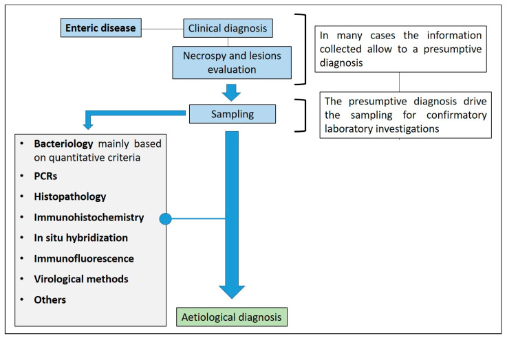
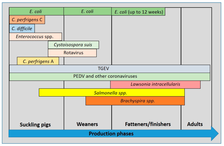
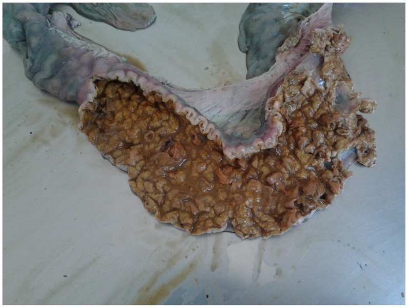
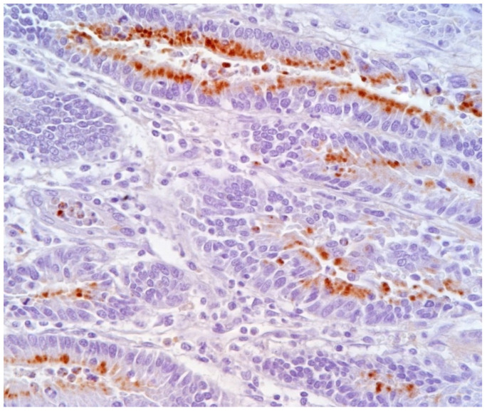
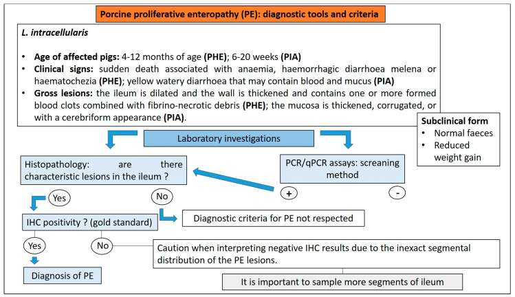
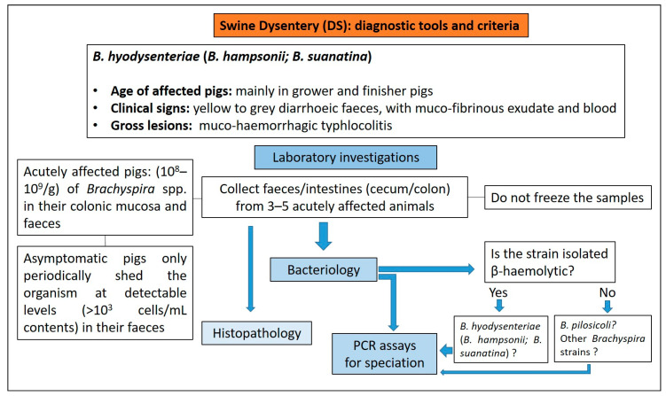
The diagnosis of enteric disorders in pigs is extremely challenging, at any age. Outbreaks of enteric disease in pigs are frequently multifactorial and multiple microorganisms can co-exist and interact. Furthermore, several pathogens, such as Clostridium perfrigens type A, Rotavirus and Lawsonia intracellularis, may be present in the gut in the absence of clinical signs. Thus, diagnosis must be based on a differential approach in order to develop a tailored control strategy, considering that treatment and control programs for enteric diseases are pathogen-specific. Correct sampling for laboratory analyses is fundamental for the diagnostic work-up of enteric disease in pigs. For example, histology is the diagnostic gold standard for several enteric disorders, and sampling must ensure the collection of representative and optimal intestinal samples. The aim of this paper is to focus on the diagnostic approach, from sampling to the aetiological diagnosis, of enteric disorders in pigs due to different pathogens during the different phases of production.

Keywords: diagnosis; enteric diseases; pig.


Conflict of interest statement

The authors declare that the research was conducted in the absence of any commercial or financial relationships that could be construed as a potential conflict of interest.

Figures

Figure 12
Figure 12
Diagnostic algorithm for the diagnosis of clostridiosis due to C. perfringens type A in pigs.
Figure 14
Figure 14
Diagnostic algorithm for the diagnosis of clostridiosis due to C. difficile in pigs.
Figure 20
Figure 20
Clinical presentation of proliferative enteropathy (modified from D’Annunzio et al., 2021) [81].
Figure 23
Figure 23
Ileum of pig affected by PE shows a hyperplastic epithelium of crypts (Haematoxylin and Eosin stain; 40×).
Figure 26
Figure 26
Pig with typical diarrhoea due to B. hyodysenteriae (A). Faeces are characterised by mucous, blood and muco-fibrinous exudate (B).
Figure 27
Figure 27
Colon of pig affected by SD shows oedema of the intestinal wall (A) and intestinal mucosa showing mucous, fibrin and blood (B).
Figure 1
Figure 1
Enteric disease and diagnostic algorithm.
Figure 2
Figure 2
Incidence of pathogens in enteric disease in pigs related to age (modified from Ségales et al., 2013) [6].
Figure 3
Figure 3
Intestine of pig. Main distribution of the aetiological agents and related lesions.
Figure 4
Figure 4
Enteric disease: sampling criteria in the diagnostic pathway.
Figure 5
Figure 5
Suckling piglet suffering from colibacillosis. Characteristic wet hair coat and inflammation of anus.
Figure 6
Figure 6
Suckling piglet affected by colibacillosis. The stomach shows hyperaemia of the fundus (arrow).
Figure 7
Figure 7
Suckling piglets suffering from colibacillosis. The small intestine is dilated, meteoric and hyperaemic.
Figure 8
Figure 8
Pig 45 days old with colibacillosis due to F4, STa, STb. The intestine is dilated and hyperaemic.
Figure 9
Figure 9
Pig 45 days old. Colibacillosis due to F4, STa, STb ETEC strain. Histological evaluation of jejunum shows bacterial layers attached to the brush border enterocytes (Haematoxylin and Eosin stain, 60×).
Figure 10
Figure 10
Diagnostic algorithm for the diagnosis of colibacillosis.
Figure 11
Figure 11
Diagnostic algorithm for the diagnosis of clostridiosis due to C. perfrigens type C in pigs.
Figure 13
Figure 13
Piglet 5 days of age. Marked oedema of the mesocolon in a case of clostridiosis due to C. difficile.
Figure 15
Figure 15
Diagnostic algorithm for the diagnosis of coccidiosis.
Figure 16
Figure 16
Diagnostic algorithm for the diagnosis of rotavirosis in pigs.
Figure 17
Figure 17
Extreme thinning of the small intestine of a 45-day-old pig affected by PED.
Figure 18
Figure 18
Fibrinous enteritis with involvement of colon (A) and small intestine (B) in pigs infected with S. Typhimurium.
Figure 19
Figure 19
Diagnostic algorithm for the diagnosis of salmonellosis in pigs.
Figure 21
Figure 21
Ileum of pig affected by PHE with a thickened wall and a blood clot in the lumen.
Figure 22
Figure 22
Ileum of pig. The picture shows a PIA complicated by secondary bacterial infections leading to necrotic enteritis, also known as regional ileitis.
Figure 24
Figure 24
Ileum of pig affected by PE. L. intracellularis organisms in the apical cytoplasm of hyperplastic epithelium lining intestinal glands (IHC, 40×) (Courtesy of Prof. G. Sarli).
Figure 25
Figure 25
Diagnostic algorithm for the diagnosis of PE in pigs.
Figure 28
Figure 28
Diagnostic algorithm for the diagnosis of SD in pigs.

References

    1. Vidal A., Martín-Valls G.E., Tello M., Mateu E., Martín M., Darwich L. Prevalence of enteric pathogens in diarrheic and non-diarrheic samples from pig farms with neonatal diarrhea in the North East of Spain. Vet. Microbiol. 2019;237:108419. doi: 10.1016/j.vetmic.2019.108419. - DOI - PMC - PubMed
    1. Sjölund M., Zoric M., Wallgren P. Financial impact on pig production: III; Proceedings of the Gastrointestinal Disorders: Proceedings of the 6th European Symposium of Porcine Health Management; Sorrento, Italy. 7–9 May 2014; p. 189.
    1. Thomson J.R., Friendship R.M. Digestive system. In: Zimmerman J.J., Karriker L.A., Ramirez A., Schwartz K.J., Stevenson G.W., Zhang J., editors. Disease of Swine. 11th ed. Wiley-Blackwell; Hoboken, NJ, USA: 2019. pp. 234–263.
    1. Arruda P.H.E., Gauger P. Optimizing Sample Selection, Collection, and Submission to Optimize Diagnostic Value. In: Zimmerman J.J., Karriker L.A., Ramirez A., Schwartz K.J., Stevenson G.W., Zhang J., editors. Disease of Swine. 11th ed. Wiley-Blackwell; Hoboken, NJ, USA: 2019. pp. 98–111.
    1. Looft T., Johnson T.A., Allen H.K., Bayles D.O., Alt D.P., Stedtfeld R.D., Sul W.J., Stedtfeld T.M., Chai B., Cole J.R., et al. In-feed antibiotic effects on the swine intestinal microbiome. Proc. Natl. Acad. Sci. USA. 2012;109:1691–1696. doi: 10.1073/pnas.1120238109. - DOI - PMC - PubMed

LinkOut - more resources