Pylephlebitis: A Systematic Review on Etiology, Diagnosis, and Treatment of Infective Portal Vein Thrombosis
- PMID: 36766534
- PMCID: PMC9914785
- DOI: 10.3390/diagnostics13030429
Pylephlebitis: A Systematic Review on Etiology, Diagnosis, and Treatment of Infective Portal Vein Thrombosis
Abstract
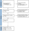
Pylephlebitis, defined as infective thrombophlebitis of the portal vein, is a rare condition with an incidence of 0.37-2.7 cases per 100,000 person-years, which can virtually complicate any intra-abdominal or pelvic infections that develop within areas drained by the portal venous circulation. The current systematic review aimed to investigate the etiology behind pylephlebitis in terms of pathogens involved and causative infective processes, and to report the most common symptoms at clinical presentation. We included 220 individuals derived from published cases between 1971 and 2022. Of these, 155 (70.5%) were male with a median age of 50 years. There were 27 (12.3%) patients under 18 years of age, 6 (2.7%) individuals younger than one year, and the youngest reported case was only 20 days old. The most frequently reported symptoms on admission were fever (75.5%) and abdominal pain (66.4%), with diverticulitis (26.5%) and acute appendicitis (22%) being the two most common causes. Pylephlebitis was caused by a single pathogen in 94 (42.8%) cases and polymicrobial in 60 (27.2%) cases. However, the responsible pathogen was not identified or not reported in 30% of the included patients. The most frequently isolated bacteria were Escherichia coli (25%), Bacteroides spp. (17%), and Streptococcus spp. (15%). The treatment of pylephlebitis consists initially of broad-spectrum antibiotics that should be tailored upon bacterial identification and continued for at least four to six weeks after symptom presentation. There is no recommendation for prescribing anticoagulants to all patients with pylephlebitis. However, they should be administered in patients with thrombosis progression on repeat imaging or persistent fever despite proper antibiotic therapy to increase the rates of thrombus resolution or decrease the overall mortality, which is approximately 14%.
Keywords: Bacteroides; Escherichia coli; Streptococcus; anticoagulant; appendicitis; diverticulitis; portal vein; portal vein thrombosis; pylephlebitis.
Conflict of interest statement
The authors declare no conflict of interest.
Figures


References
-
- Saxena R., Adolph M., Ziegler J.R., Murphy W., Rutecki G.W. Pylephlebitis: A case report and review of outcome in the antibiotic era. Am. J. Gastroenterol. 1996;91:1251–1253. - PubMed
-
- Takahashi H., Sakata I., Adachi Y. Treatment of portal vein septic thrombosis by infusion of antibiotics and an antifungal agent into portal vein and superior mesenteric artery: A case report. Hepatogastroenterology. 2003;50:1133–1135. - PubMed
-
- Mannaerts L., Bleeker-Rovers C.P., Koopman M., Punt C.J.A., Van Herpen C.M.L. Pylephlebitis after a duodenal ulcer in a patient with metastasised colon carcinoma treated with chemotherapy and bevacizumab: A case report. Neth. J. Med. 2009;67:69–71. - PubMed
Publication types
LinkOut - more resources
Full Text Sources
Miscellaneous

