. 2023 Jan 26;13(3):461.
doi: 10.3390/diagnostics13030461.

Diagnostic Utility of Double-Echo Steady-State (DESS) MRI for Fracture and Bone Marrow Edema Detection in Adolescent Lumbar Spondylolysis

Affiliations

Diagnostic Utility of Double-Echo Steady-State (DESS) MRI for Fracture and Bone Marrow Edema Detection in Adolescent Lumbar Spondylolysis

Atsushi Kitakado et al. Diagnostics (Basel). .

Abstract

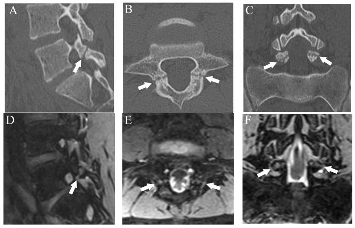
To evaluate the ability of double-echo steady-state (DESS) MRI to detect pars interarticularis fracture and bone marrow edema (BME) in spondylolysis, 500 lumber pars interarticularis from 50 consecutive patients (38 males and 12 females, mean age 14.2 ± 3.28 years) with spondylolysis who underwent both MRI and CT within 1 week were evaluated. All participants were young athletes who complained of lower back pain. Fractures were classified into four grades and CT was used as a reference; BME was evaluated in a binary manner and STIR was used as a reference. The diagnostic performance of fractures on DESS and T1WI, and BME on DESS was assessed by two radiologists independently. For fracture detection, DESS showed high diagnostic performance at a sensitivity of 94%, specificity of 99.5%, and accuracy of 98.8%, whereas T1WI showed lower sensitivity (70.1%). Fracture grading performed by DESS showed excellent agreement with CT grading (Kappa = 0.9). For BME, the sensitivity, specificity, and accuracy of DESS were 96.5%, 100%, and 99.6%, respectively. The inter-rater agreement of DESS for fracture and BME was 0.8 and 0.85, respectively. However, the inter-rater agreement for fracture on T1WI was 0.52. DESS had high diagnostic performance for fracture and BME in pars interarticularis. In conclusion, DESS had potential to detect all critical imaging findings in spondylolysis and may replace the role of CT.

Keywords: bone marrow edema; computed tomography; fracture; magnetic resonance imaging; pars interarticularis; spondylolysis.


Conflict of interest statement

The authors declare no conflict of interest.

Figures

Figure 1
Figure 1
Representative images of grade 1 fracture. The right pars interarticularis of L3 from a 14-year-old male. (A) Oblique-sagittal CT image showing faint attenuation in the cortex (arrow). (B) The faint attenuation corresponds to slight hyperintensity in the cortex on an oblique-sagittal DESS image (arrow). The window of the image was adjusted to enhance the faint finding. Note the BME in the pedicle (arrowhead). (C) Sagittal STIR image shows BME in the right pedicle of L3 (arrowhead).
Figure 2
Figure 2
Representative images of grade 2 fracture. The left pars interarticularis of L5 from a 16-year-old male. (A) Sagittal CT image showing an incomplete but clear fracture line starting from the caudal part of the pars interarticularis (arrow). (B) Sagittal DESS image showing a linear high signal that suggests the fracture line (arrow).
Figure 3
Figure 3
Representative images of grade 3 fracture. The right pars interarticularis of L5 from a 16-year-old male. (A) Sagittal CT image showing a complete fracture across the whole pars interarticularis (arrow). (B) Sagittal DESS image showing a linear high signal corresponding to the complete fracture (arrow).
Figure 4
Figure 4
Breakdown of sports activities. The number indicates number of patients.
Figure 5
Figure 5
Comparison of bilateral grade 3 fractures at L5 from an 11-year-old male in multiple planes between CT and DESS. (AC) CT shows complete fractures in L5 pars interarticularis on the right-sided sagittal, axial, and coronal plane, respectively (arrows). (DF) DESS shows comparable complete fracture on the right-sided sagittal, axial, and coronal plane, respectively (arrows).
Figure 6
Figure 6
Comparison of grade 2 fracture at the right pars interarticularis at L5 from a 14-year-old male in multiple planes between CT and DESS. (AC) CT shows grade 2 fracture on the right pars interarticularis on the right-sided sagittal, axial, and coronal plane, respectively (arrows). On the axial plane, fracture of the left side is also delineated (thin arrow). (DF) DESS shows grade 2 fractures comparable to CT on the right-sided sagittal, axial, and coronal plane, respectively (arrows). The sagittal and axial images delineate BME around the fracture (arrowhead), and the left pars interarticularis fracture is also delineated on the axial image (thin arrow).
Figure 7
Figure 7
The case that lower fracture grade was assigned by DESS than CT. (A) CT suggests an incomplete fracture which was consistent with grade 2 (arrow). (B) DESS shows a subtle high signal in the cortex of the pars interarticularis and evaluated as grade 1 (arrow). BME is also detected (arrowhead).

References

    1. Berger R.G., Doyle S.M. Spondylolysis 2019 update. Curr. Opin. Pediatr. 2019;31:61–68. doi: 10.1097/MOP.0000000000000706. - DOI - PubMed
    1. Micheli L.J., Wood R. Back pain in young athletes. Significant differences from adults in causes and patterns. Arch. Pediatr. Adolesc. Med. 1995;149:15–18. doi: 10.1001/archpedi.1995.02170130017004. - DOI - PubMed
    1. Logroscino G., Mazza O., Aulisa G., Pitta L., Pola E., Aulisa L. Spondylolysis and spondylolisthesis in the pediatric and adolescent population. Child’s Nerv. Syst. 2001;17:644–655. doi: 10.1007/s003810100495. - DOI - PubMed
    1. Fujii K., Katoh S., Sairyo K., Ikata T., Yasui N. Union of defects in the pars interarticularis of the lumbar spine in children and adolescents. The radiological outcome after conservative treatment. J. Bone Jt. Surg. Br. Vol. 2004;86:225–231. doi: 10.1302/0301-620X.86B2.14339. - DOI - PubMed
    1. Masci L., Pike J., Malara F., Phillips B., Bennell K., Brukner P. Use of the one-legged hyperextension test and magnetic resonance imaging in the diagnosis of active spondylolysis. Br. J. Sport. Med. 2006;40:940–946. doi: 10.1136/bjsm.2006.030023. - DOI - PMC - PubMed

LinkOut - more resources