Review
. 2023 Feb 3;13(3):563.
doi: 10.3390/diagnostics13030563.

Imaging Characteristics of Diffuse Idiopathic Skeletal Hyperostosis: More Than Just Spinal Bony Bridges

Affiliations
Review

Imaging Characteristics of Diffuse Idiopathic Skeletal Hyperostosis: More Than Just Spinal Bony Bridges

Iris Eshed. Diagnostics (Basel). .

Abstract

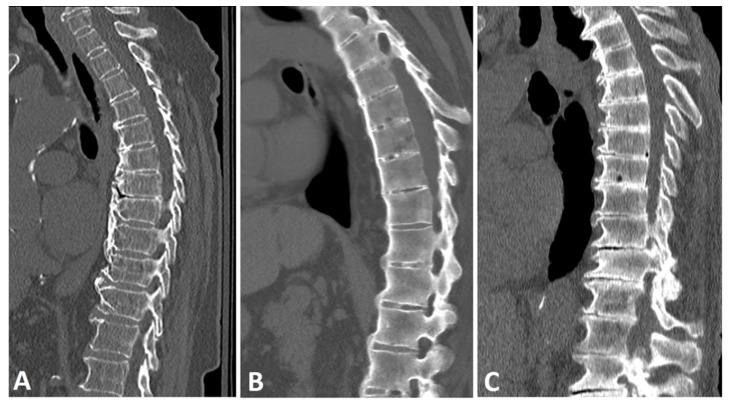
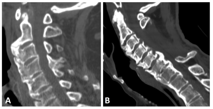
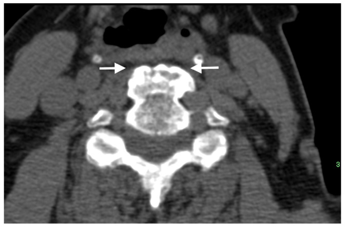
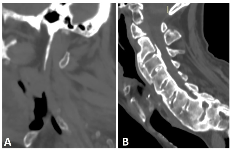
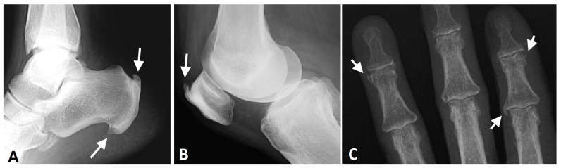
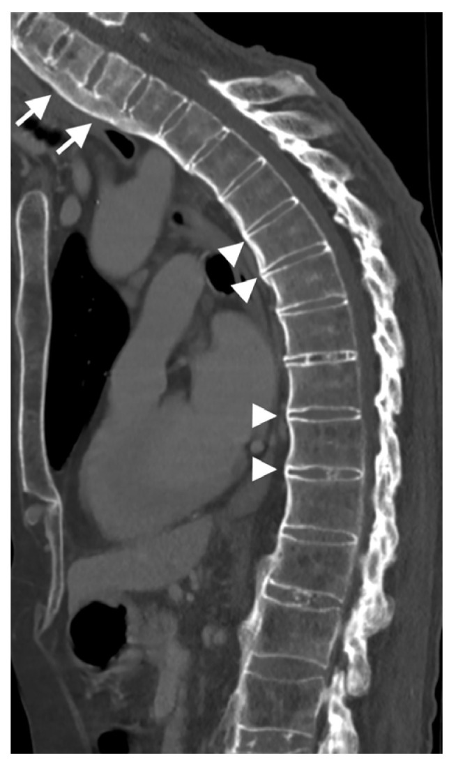
Diffuse idiopathic skeletal hyperostosis (DISH) is a systemic condition characterized by new bone formation and enthesopathies of the axial and peripheral skeleton. The pathogenesis of DISH is not well understood, and it is currently considered a non-inflammatory condition with an underlying metabolic derangement. Currently, DISH diagnosis relies on the Resnick and Niwayama criteria, which encompass end-stage disease with an already ankylotic spine. Imaging characterization of the axial and peripheral skeleton in DISH subjects may potentially help identify earlier diagnostic criteria and provide further data for deciphering the general pathogenesis of DISH and new bone formation. In the current review, we aim to summarize and characterize axial and peripheral imaging findings of the skeleton related to DISH, along with their clinical and pathogenetic relevance.

Keywords: CT; DISH; MRI; US; entheses; osteophytes; radiograph; sacroiliac joints; spine.


Conflict of interest statement

The author declares no conflict of interest.

Figures

Figure 1
Figure 1
Sagittal CT reconstructions of the thoracic spine in three different patients: (A) a patient with DISH with thick, flowing, horizontally oriented, coarse osteophytes; (B) a patient with AS with slender, vertically oriented syndesmophytes; and (C) a patient with degenerative disc disease with horizontally oriented, non-flowing osteophytes. Note that the disc space is relatively preserved in the DISH subject, but not in the patient with a degenerative spine.
Figure 2
Figure 2
Sagittal CT reconstruction images of two patients with thoracic (not shown) and cervical DISH. (A) Flowing chunky osteophytes are located anteriorly to the vertebrae, forming a “candle flame” or “parrot-beak” image. (B) Thick, not-yet-flowing anterior osteophytes and thick posterior osteophytes at the C5–6 level.
Figure 3
Figure 3
Axial CT image of a thick anterior osteophyte in the cervical spine of a patient with DISH that is impinging and causing narrowing of the adjacent esophagus (arrows).
Figure 4
Figure 4
Sagittal CT reconstructions of the cervical spine of two patients with thoracic DISH (not shown). (A) The elongated styloid process results from calcification and ossification of the stylohyoid ligament. (B) Flowing osteophytes characteristic of DISH accompanied by OPLL.
Figure 5
Figure 5
Axial CT images of two patients with thoracic DISH (not shown). (A) left-sided and (B) right-sided aortas with an anterior osteophyte located contralateral to the aorta’s position.
Figure 6
Figure 6
Sagittal CT reconstruction of the thoracic spine of a 78-year-old patient with thoracic DISH after low-energy trauma, showing an unstable extension-type fracture of the mid-thoracic spine. In the given slice, there are two vertically oriented bony bridges in the upper thoracic spine (arrowheads) that may lead to the misconception that this is a patient with ankylosing spondylitis; however, there are clearly many flowing osteophytes compatible with DISH in the lower part of the thoracic spine (arrows).
Figure 7
Figure 7
Axial CT images of the SIJs of two patients with DISH. (A) Characteristic extra-articular bridging osteophytes are seen anterior to the SIJs. (B) In addition to the ventral extra-articular osteophytes, clear ankylosis can be observed within the right and left SIJs.
Figure 8
Figure 8
AP radiograph of the pelvis of an 82-year-old female with thoracic DISH (not shown). Characteristic whiskering of the anterior superior iliac spine and ischial tuberosities (arrows) is present on both sides, resulting from enthesopathy in these regions.
Figure 9
Figure 9
Radiographs of the ankle, knee, and hand of a patient with thoracic DISH (not shown). (A) Lateral radiograph of the ankle, showing coarse, thick enthesophytes in the attachment of the Achilles tendon and plantar fascia to the calcaneus (arrows). (B) Lateral knee radiograph showing thick, coarse enthesophytes (arrow) in the attachment of the quadriceps tendon to the patella. (C) AP radiograph of the fingers, showing enthesophytes in the medial side of the 2nd metacarpophalangeal joint and 4th distal interphalangeal joint and the lateral side of the 2nd distal interphalangeal joint (arrows).
Figure 10
Figure 10
A sagittal CT reconstruction of the cervical and thoracic spine of a 60-year-old patient. There are thick-flowing osteophytes on the cervical spine (arrows), while thin slender vertical syndesmophytes (arrowheads) are in the thoracic spine.

References

    1. Resnick D., Niwayama G. Radiographic and pathologic features of spinal involvement in diffuse idiopathic skeletal hyperostosis (DISH) Radiology. 1976;119:559–568. doi: 10.1148/119.3.559. - DOI - PubMed
    1. Resnick D., Niwayama G. Entheses and enthesopathy. Anatomical, pathological, and radiological correlation. Radiology. 1983;146:1–9. doi: 10.1148/radiology.146.1.6849029. - DOI - PubMed
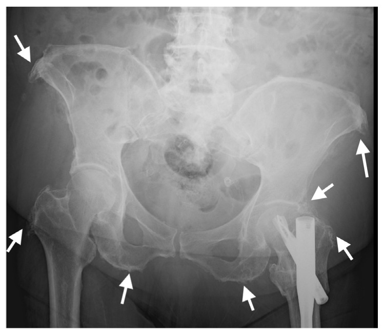
    1. Slonimsky E., Leibushor N., Aharoni D., Lidar M., Eshed I. Pelvic enthesopathy on CT is significantly more prevalent in patients with diffuse idiopathic skeletal hyperostosis (DISH) compared with matched control patients. Clin. Rheumatol. 2015;35:1823–1827. doi: 10.1007/s10067-015-3151-3. - DOI - PubMed
    1. Resnick D., Shaul S.R., Robins J.M. Diffuse idiopathic skeletal hyperostosis (DISH): Forestier’s disease with extraspinal manifestations. Radiology. 1975;115:513–524. doi: 10.1148/15.3.513. - DOI - PubMed
    1. Mader R., Novofestovski I., Adawi M., Lavi I. Metabolic syndrome and cardiovascular risk in patients with diffuse idiopathic skeletal hyperostosis. Semin. Arthritis Rheum. 2009;38:361–365. doi: 10.1016/j.semarthrit.2008.01.010. - DOI - PubMed

LinkOut - more resources