. 2023 Jan 19;12(3):372.
doi: 10.3390/cells12030372.

Aurora B SUMOylation Is Restricted to Centromeres in Early Mitosis and Requires RANBP2

Affiliations

Aurora B SUMOylation Is Restricted to Centromeres in Early Mitosis and Requires RANBP2

Erica Di Cesare et al. Cells. .

Abstract

Conjugation with the small ubiquitin-like modifier (SUMO) modulates protein interactions and localisation. The kinase Aurora B, a key regulator of mitosis, was previously identified as a SUMOylation target in vitro and in assays with overexpressed components. However, where and when this modification genuinely occurs in human cells was not ascertained. Here, we have developed intramolecular Proximity Ligation Assays (PLA) to visualise SUMO-conjugated Aurora B in human cells in situ. We visualised Aurora B-SUMO products at centromeres in prometaphase and metaphase, which declined from anaphase onwards and became virtually undetectable at cytokinesis. In the mitotic window in which Aurora B/SUMO products are abundant, Aurora B co-localised and interacted with NUP358/RANBP2, a nucleoporin with SUMO ligase and SUMO-stabilising activity. Indeed, in addition to the requirement for the previously identified PIAS3 SUMO ligase, we found that NUP358/RANBP2 is also implicated in Aurora B-SUMO PLA product formation and centromere localisation. In summary, SUMOylation marks a distinctive window of Aurora B functions at centromeres in prometaphase and metaphase while being dispensable for functions exerted in cytokinesis, and RANBP2 contributes to this control, adding a novel layer to modulation of Aurora B functions during mitosis.

Keywords: Aurora B; RANBP2; SUMOylation; in situ proximity ligation assay (isPLA); mitosis.


Conflict of interest statement

The authors declare no conflict of interest.

Figures
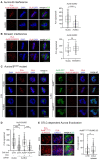
Figure 1
Figure 1
Aurora B-SUMO2/3 ligation products in prometaphase and metaphase HeLa cells. (A) Aurora B-SUMO2/3 PLA signals disappear in cells interfered for Aurora B, indicated as AurB(i). Control cultures were treated for RNA interference with neutral siRNAs targeting the firefly luciferase, indicated as GL2(i). The box plot represents the distribution of PLA signals in prometaphase and metaphase in control vs. Aurora B-interfered cultures (140 counted cells/group in 3 experiments); ****, extremely highly significant difference (p < 0.0001, Mann–Whitney test). (B) Aurora B-SUMO2/3 PLA signals persist in Borealin-interfered cells, which display extensive chromosome misalignment. The box plot on the right shows the abundance of Aurora B-SUMO 2/3 PLA signals/cell as described in (A) (60 counted cells/group, 2 experiments). ns, non-significant differences. (C) Mutation of K202 abolishes Aurora B PLA signals with SUMO2/3 peptides. Left: Aurora B-SUMO2/3 PLA products in control (CTR) HeLa cultures interfered with neutral (GL2) or with Aurora B-specific siRNAs: the latter abolished the PLA signals. Right: after interference to endogenous Aurora B, PLA assays depict SUMO2/3 products with dox-inducible Aurora B only in the cell line expressing wild-type kinase, but not Aurora BK202R mutant. (D) Quantification of Aurora B and SUMO2/3 PLA signals in the cell lines indicated in (C). + and − indicate dox addition. At least 60 cells per sample were counted (2 experiments). ns, non-significant difference; ****, p < 0.0001. (E) Aurora BphThr232-SUMO2/3 PLA products under conditions that increase merotelic attachments. The box plot quantifies Aurora BphThr232-SUMO2/3 PLA signals in controls (60) and STLC-released (86) cells (2 experiments): PLA signals increase in parallel with the increase in Aurora BphThr232 (**, p < 0.01). The Mann–Whitney test was used for all comparisons.
Figure 2
Figure 2
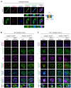
Distribution of Aurora B-SUMO2/3 PLA products in mitotic cells in situ. (A) Aurora B-SUMO2/3 PLA signals (red) co-localise with kinetochores (CREST, blue) in prometaphase and metaphase, and decrease abruptly in anaphase/telophase. (B) Localisation of active Aurora B, auto-phosphorylated at Thr232. (C) Canonical localisation of the total Aurora pool (red) at kinetochores (CREST, blue) and midbody (alpha-tubulin, green) during mitotic progression. Bar, 10 µm. (D) The schematics summarises the temporal distribution of Aurora B forms during mitotic progression.
Figure 3
Figure 3
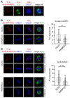
Aurora BK202R does not concentrate at centromeres. (A) Aurora BWT-GFP (top panel) and Aurora BK202R-GFP (bottom panel) in metaphase cells silenced for the endogenous Aurora B. In the blow-up (4×), Aurora BWT-GFP has a punctuated appearance bordered by CENP-A and, more externally, CENP-F (see schematics). Aurora BK202R-GFP (bottom panel) shows a more diffuse pattern over chromosomes. (B) Aurora BK202R fails to concentrate near CENP-A, independent of the absence (NOC) or presence (NOC release) of kinetochore attachments to microtubules. (C) Localisation of Aurora B, CENP-A and CENP-F, in kif11-inhibited cells (STLC) with unseparated poles and stretched kinetochores, or in STLC-released cells with stimulated Aurora B activity. Under all conditions, the K202R mutation impairs Aurora B concentration at centromeres, as can be appreciated in the magnification (bottom panels).
Figure 4
Figure 4
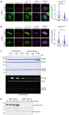
Impaired Aurora B and CENP-A phosphorylation at centromeres in the Aurora BK202R-expressing cell line. (A) After endogenous Aurora B silencing, Aurora BPh-Thr232 is depicted (red channel) at CREST-stained kinetochores in Aurora BWT-expressing metaphases, but substantially decreases in cells expressing Aurora BK202R mutant. Exogenous Aurora B proteins are visualised from the GFP signal. Data from three experiments in the wild-type vs. mutant Aurora B expressing cell lines are quantified in the bow plot (***, p < 0.001) (B) CENP-APh-Ser7 (red) is visible at metaphase centromeres in Aurora BWT- but decreases in Aurora BK202R-expressing cells. Data are quantified in the box plot (three experiments), (**, p < 0.01). (C) Aurora BK202R is not proficient for phosphorylation of NDC80 in vitro (left panel). After phosphorylation reactions with purified components at the indicated concentrations, ProQ Diamond staining visualises a dose-dependent increase in phosphorylated NDC80 with Aurora BWT/INCENP. No phosphorylated bands are seen with Aurora BK202R/INCENP (compare to the -INCENP, -ATP lanes). (D) Purified Aurora BWT, but not Aurora BK202R, is reactive to Aurora BPh-Thr232 antibody, indicating self-phosphorylation in vitro.
Figure 5
Figure 5
RANBP2 is required for Aurora B-SUMO2/3 PLA products. (A) RANBP2 and Aurora B form PLA products in mitotic cells. A representative late prometaphase is shown, in which PLA signals are juxtaposed to kinetochores (CREST, blue). (B) Borealin-SUMO2/3 PLA reactions in a control metaphase treated with neutral siRNAs (GL2) and, below, in a RANBP2-interfered cell. The box plot represents Borealin-SUMO2/3 PLA signals per cell in control (n, 55) and RANBP2-interfered (n, 65) metaphases in 2 experiments (**, p < 0.01). (C) Aurora B-SUMO2/3 PLA signals were analysed under the same conditions as above, in control (GL2) and RANBP2-interfered cells. Exemplifying prometaphases and metaphases are shown. The box plot represents Aurora B-SUMO2/3 PLA signals per cell in controls (n, 150) and RANBP2-interfered (n, 200) cells in 5 experiments; ****, highly significant difference (p < 0.0001).
Figure 6
Figure 6
RANBP2 silencing impairs Aurora B autophosphorylation at Thr232 (A) and phosphorylation of CENP-A at Ser7 (B) at centromeres. Experiments were repeated three times. The quantitative analysis of markers in control (interfered with neutral GL2 siRNAs) and RANBP2-interfered cultures is shown in the box plot and the statistical analysis is as follows: AurBphThr232 (n, 100 for GL2 and 107 for RANBP2), **** p < 0.0001; CENP-Aph-ser7 (n, 45 and 72 cells) **** p < 0.0001.

References

    1. Flotho A., Melchior F. Sumoylation: A regulatory protein modification in health and disease. Annu. Rev. Biochem. 2013;82:357–385. doi: 10.1146/annurev-biochem-061909-093311. - DOI - PubMed
    1. Varejão N., Lascorz J., Li Y., Reverter D. Molecular mechanisms in SUMO conjugation. Biochem. Soc. Trans. 2020;48:123–135. doi: 10.1042/BST20190357. - DOI - PubMed
    1. Vertegaal A.C.O. Signalling mechanisms and cellular functions of SUMO. Nat. Rev. Mol. Cell Biol. 2022;23:715–731. doi: 10.1038/s41580-022-00500-y. - DOI - PubMed
    1. Raman N., Nayak A., Muller S. The SUMO system: A master organizer of nuclear protein assemblies. Chromosoma. 2013;122:475–485. doi: 10.1007/s00412-013-0429-6. - DOI - PubMed
    1. Wotton D., Pemberton L.F., Merrill-Schools J. SUMO and Chromatin Remodeling. Adv. Exp. Med. Biol. 2017;963:35–50. - PubMed

Publication types

LinkOut - more resources