Review
. 2023 Jan 23;12(3):401.
doi: 10.3390/cells12030401.

Calcium/Calmodulin-Stimulated Protein Kinase II (CaMKII): Different Functional Outcomes from Activation, Depending on the Cellular Microenvironment

Affiliations
Review

Calcium/Calmodulin-Stimulated Protein Kinase II (CaMKII): Different Functional Outcomes from Activation, Depending on the Cellular Microenvironment

John A P Rostas et al. Cells. .

Abstract

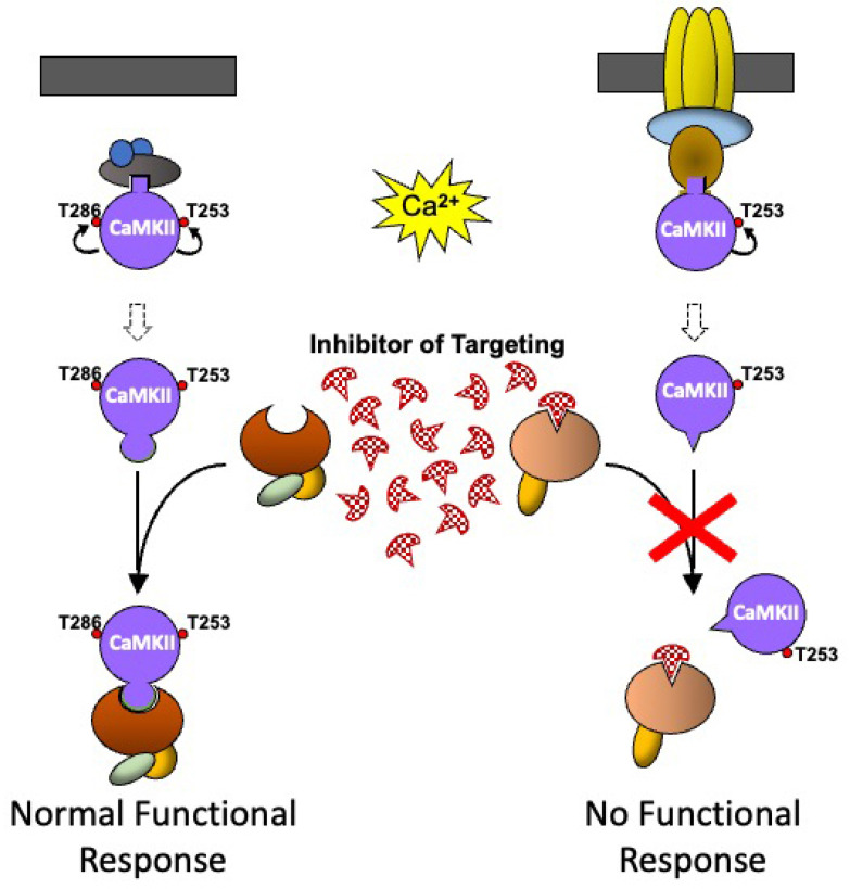
Calcium/calmodulin-stimulated protein kinase II (CaMKII) is a family of broad substrate specificity serine (Ser)/threonine (Thr) protein kinases widely expressed in many tissues that is capable of mediating diverse functional responses depending on its cellular and molecular microenvironment. This review briefly summarises current knowledge on the structure and regulation of CaMKII and focuses on how the molecular environment, and interaction with binding partner proteins, can produce different populations of CaMKII in different cells, or in different subcellular locations within the same cell, and how these different populations of CaMKII can produce diverse functional responses to activation following an increase in intracellular calcium concentration. This review also explores the possibility that identifying and characterising the molecular interactions responsible for the molecular targeting of CaMKII in different cells in vivo, and identifying the sites on CaMKII and/or the binding proteins through which these interactions occur, could lead to the development of highly selective inhibitors of specific CaMKII-mediated functional responses in specific cells that would not affect CaMKII-mediated responses in other cells. This may result in the development of new pharmacological agents with therapeutic potential for many clinical conditions.

Keywords: CaMKII; binding protein; calcium/calmodulin; molecular targeting; protein phosphorylation.


Conflict of interest statement

The authors declare no conflict of interest.

Figures

Figure 1
Figure 1
Schematic representations of the molecular structure of CaMKIIα: (A) CaMKII consists of a catalytic domain (orange), a regulatory domain (purple), which includes overlapping autoinhibitory and calmodulin (CaM) binding regions and multiple phosphorylation sites (yellow stars). The association domain (blue) is involved in the formation of CaMKII multimers. Splice sites (V1–4) are represented in orange/yellow. (B) Crystal structure (3SOa) of a single CaMKIIα subunit created using Chimera [44]. (C) Representation of the CaMKIIα holoenzyme structure, with the domains shown in the same colours as in (A), and showing the movement of the subunit and its catalytic domain following activation with calcium/calmodulin.
Figure 2
Figure 2
Targeting can produce localised functional outcomes by altering the kinase or adaptor activity of CaMKII. Coloured oblong shapes represent generic proteins with which CaMKII (purple disc) binds directly or indirectly. The small red disc represents the phosphorylation of a protein. See text for explanation of the sequence of events shown in the Figure.
Figure 3
Figure 3
A schematic representation of how CaMKII in different molecular environments can respond to stimuli differently. The binding partner with which CaMKII is associated can influence the functional outcomes that occur following a rise in intracellular calcium. Coloured oblong shapes represent generic proteins with which CaMKII (purple disc) binds directly or indirectly. Dark Grey bar represents a membrane. Geometric protrusions from CaMKII, and indents in the binding proteins represent binding sites for CaMKII. The small red disc represents the phosphorylation of a protein. See text for explanation of the sequence of events shown in the Figure.
Figure 4
Figure 4
A schematic diagram showing how stimulus-induced translocation of CaMKII between different binding proteins in a molecular environment can produce different functional responses to the activation of CaMKII by two sequential stimuli of the same type and how the influence of another signalling pathway can further modify the CaMKII response. The effects of two sequential stimulus-induced rises in calcium on non-phosphorylated CaMKII, when the surrounding binding partners are (A) also non-phosphorylated, or (B) phosphorylated. Coloured oblong shapes represent generic proteins with which CaMKII (purple disc) binds directly or indirectly. Dark grey bar represents a membrane. Geometric protrusions from CaMKII, and indents in the binding proteins represent binding sites for CaMKII. The small red disc represents the phosphorylation of a protein. See text for explanation of the sequence of events shown in the Figure.
Figure 5
Figure 5
A schematic representation of how pharmacological agents that inhibit CaMKII targeting could be used therapeutically. As CaMKII that is phosphorylated at different sites binds to different binding proteins to produce different functional responses, an inhibitor that specifically blocks the interaction of CaMKII with a particular binding partner will inhibit a specific functional response, while leaving all the other pools of CaMKII bound to other binding proteins, untouched, and able to produce their normal functional response. Coloured oblong shapes represent generic proteins with which CaMKII (purple disc) binds directly or indirectly. Dark grey bar represents a membrane. Geometric protrusions from CaMKII, and indents in the binding proteins represent binding sites for CaMKII. The small red disc represents the phosphorylation of a protein. See text for explanation of the sequence of events shown in the Figure.

References

    1. Miller S.G., Kennedy M.B. Distinct forebrain and cerebellar isozymes of type II Ca2+/calmodulin-dependent protein kinase associate differently with the postsynaptic density fraction. J. Biol. Chem. 1985;260:9039–9046. doi: 10.1016/S0021-9258(17)39454-1. - DOI - PubMed
    1. Giese K.P., Fedorov N.B., Filipkowski R.K., Silva A.J. Autophosphorylation at Thr286 of the alpha calcium-calmodulin kinase II in LTP and learning. Science. 1998;279:870–873. doi: 10.1126/science.279.5352.870. - DOI - PubMed
    1. Miller S., Yasuda M., Coats J.K., Jones Y., Martone M.E., Mayford M. Disruption of dendritic translation of CaMKIIalpha impairs stabilization of synaptic plasticity and memory consolidation. Neuron. 2002;36:507–519. doi: 10.1016/S0896-6273(02)00978-9. - DOI - PubMed
    1. Soderling T.R., Derkach V.A. Postsynaptic protein phosphorylation and LTP. Trends Neurosci. 2000;23:75–80. doi: 10.1016/S0166-2236(99)01490-3. - DOI - PubMed
    1. Taha S., Hanover J.L., Silva A.J., Stryker M.P. Autophosphorylation of alphaCaMKII is required for ocular dominance plasticity. Neuron. 2002;36:483–491. doi: 10.1016/S0896-6273(02)00966-2. - DOI - PubMed

MeSH terms

Substances

LinkOut - more resources