Review
. 2023 Jan 27;12(3):426.
doi: 10.3390/cells12030426.

Impact of 5-HT6 Receptor Subcellular Localization on Its Signaling and Its Pathophysiological Roles

Affiliations
Review

Impact of 5-HT6 Receptor Subcellular Localization on Its Signaling and Its Pathophysiological Roles

Séverine Chaumont-Dubel et al. Cells. .

Abstract

The serotonin (5-HT)6 receptor still raises particular interest given its unique spatio-temporal pattern of expression among the serotonin receptor subtypes. It is the only serotonin receptor specifically expressed in the central nervous system, where it is detected very early in embryonic life and modulates key neurodevelopmental processes, from neuronal migration to brain circuit refinement. Its predominant localization in the primary cilium of neurons and astrocytes is also unique among the serotonin receptor subtypes. Consistent with the high expression levels of the 5-HT6 receptor in brain regions involved in the control of cognitive processes, it is now well-established that the pharmacological inhibition of the receptor induces pro-cognitive effects in several paradigms of cognitive impairment in rodents, including models of neurodevelopmental psychiatric disorders and neurodegenerative diseases. The 5-HT6 receptor can engage several signaling pathways in addition to the canonical Gs signaling, but there is still uncertainty surrounding the signaling pathways that underly its modulation of cognition, as well as how the receptor's coupling is dependent on its cellular compartmentation. Here, we describe recent findings showing how the proper subcellular localization of the receptor is achieved, how this peculiar localization determines signaling pathways engaged by the receptor, and their pathophysiological influence.

Keywords: G-protein-coupled receptor; primary cilium; serotonin; signaling.


Conflict of interest statement

The authors declare no conflict of interest.

Figures

Figure 1
Figure 1
(A) Confocal images (20×) of 3D cultures (brain organoids) generated from ES cells expressing either cytosolic GFP (left) or 5-HT6R-GFP (right). The GFP or 5-HT6R are shown in green, nuclei are labeled with DAPI, and primary cilia are labeled with Arl13b (red). Scale bar 200 μm. (B) Confocal images (63×) of cells treated with either DMSO (no induction of the 5-HT6R expression) or with doxycycline (DOX, induction of the 5-HT6R expression) for 24 h, 7 days after starting differentiation. The 5-HT6R (yellow) is expressed in primary cilia, as shown by colocalization with the ciliary marker Arl13b (magenta). The inset on the merge image shows a 10× zoom on cilia labeled with Arl13b and GFP (Scale bar 20 μm). The histogram on the right shows that cilia length is significantly increased in cells expressing the 5-HT6R (DOX) when compared to DMSO-treated cells (6.70 ± 0.13 μm (n = 558) for DOX-treated cells vs. 4.76 ± 0.09 μm (n = 460) for DMSO-treated cells). **** p < 0.0001, unpaired t-test. Scale bar 20 μm.
Figure 2
Figure 2
The 5-HT6R possesses 2 cilia-targeting sequences (CTSs). When present, CTS1 (on the ic3 loop) interacts with the RABL2 GTPase, whereas CTS2 (on the C-terminal domain) interacts with TULP3. Both CTSs are required for normal cilia targeting (A). Cilia targeting is only partial when CTS2 is mutated, but paradoxically, interaction with TULP3 is increased rather than lost (B). When CTS1 is mutated, interaction with RABL2 is lost, and cilia targeting is diminished (C). Finally, mutating both CTSs results in a complete loss of ciliary localization of the receptor (D).
Figure 3
Figure 3
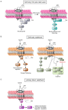
Spatio-temporal characteristics of the 5-HT6R signal transduction. Depending on its subcellular localization or developmental stage, the 5-HT6R can be coupled to different signaling pathways. (A) During the first post-natal week, the receptor is expressed at the membrane of the cell body. This relocation of the receptor allows for its coupling to Cdk5, which will phosphorylate the receptor and induce differentiation and neurite outgrowth through a Cdc42-dependent pathway. The receptor will subsequently couple to GPRIN1, to promote neurite elongation and branching through a Gs-cAMP-PKA-dependent signaling pathway. The interaction with Cdk5 and GPRIN1 is sequential; GPRIN1 cannot interact with the receptor phosphorylated by Cdk5 [39]. (B) At the adult stage, the somatic 5-HT6R can couple to Gs. Its association with different GIPs (G-protein-coupled receptor-interacting proteins), such as Fyn or JAB1, will modulate the receptor plasma membrane localization and its regulation of gene expression through the activation of the MAP kinase pathway. The receptor can also activate the mTOR pathway, which underlies its roles in cognition. Whether both signaling pathways can couple to receptors in the same conformational state remains to be determined. (C) The 5-HT6R is primarily located in the primary cilium, where it couples to the Gq-TRIO-RhoA pathway. Upon activation by serotonin from a nearby axon, the receptor in axo-ciliary synapse inhibits adducin-1 nuclear translocation, induces chromatin opening, and modulates epigenetic marks.

References

    1. Hewavitharana T., Wedegaertner P.B. Non-canonical signaling and localizations of heterotrimeric G proteins. Cell Signal. 2012;24:25–34. doi: 10.1016/j.cellsig.2011.08.014. - DOI - PMC - PubMed
    1. Weis W.I., Kobilka B.K. The Molecular Basis of G Protein-Coupled Receptor Activation. Annu. Rev. Biochem. 2018;87:897–919. doi: 10.1146/annurev-biochem-060614-033910. - DOI - PMC - PubMed
    1. Slosky L.M., Caron M.G., Barak L.S. Biased Allosteric Modulators: New Frontiers in GPCR Drug Discovery. Trends Pharm. Sci. 2021;42:283–299. doi: 10.1016/j.tips.2020.12.005. - DOI - PMC - PubMed
    1. Crilly S.E., Puthenveedu M.A. Compartmentalized GPCR Signaling from Intracellular Membranes. J. Membr. Biol. 2021;254:259–271. doi: 10.1007/s00232-020-00158-7. - DOI - PMC - PubMed
    1. Seifert R., Wenzel-Seifert K. Constitutive activity of G-protein-coupled receptors: Cause of disease and common property of wild-type receptors. Naunyn Schmiedebergs Arch Pharm. 2002;366:381–416. doi: 10.1007/s00210-002-0588-0. - DOI - PubMed

Publication types

LinkOut - more resources