. 2023 Feb 3;12(3):507.
doi: 10.3390/cells12030507.

Exploring the Interactions of Oncolytic Viral Therapy and Immunotherapy of Anti-CTLA-4 for Malignant Melanoma Mice Model

Affiliations

Exploring the Interactions of Oncolytic Viral Therapy and Immunotherapy of Anti-CTLA-4 for Malignant Melanoma Mice Model

Jui-Ling Yu et al. Cells. .

Abstract

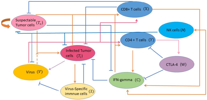
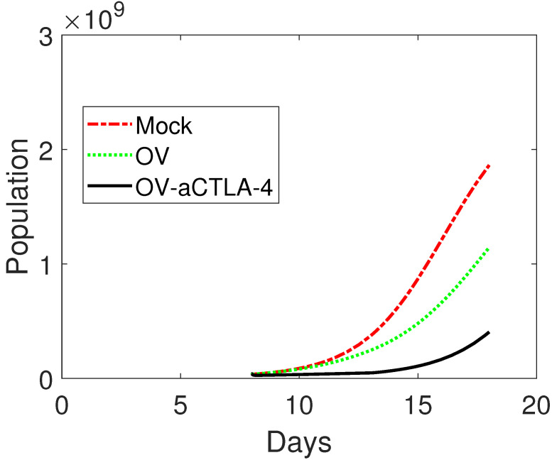
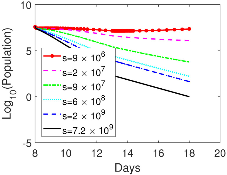
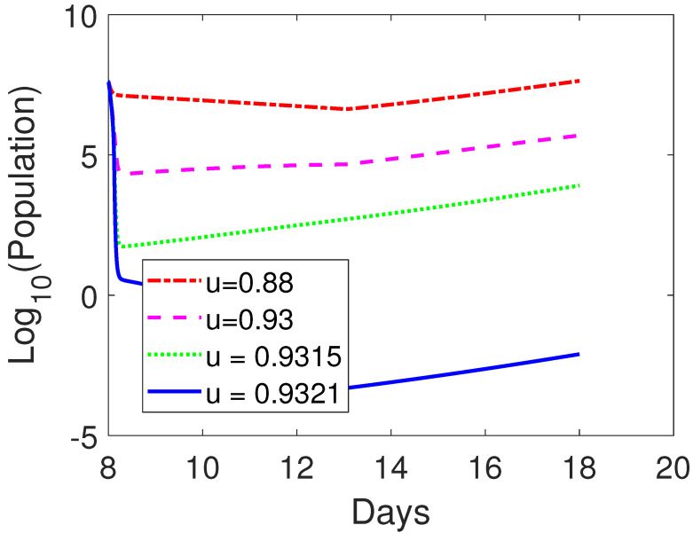
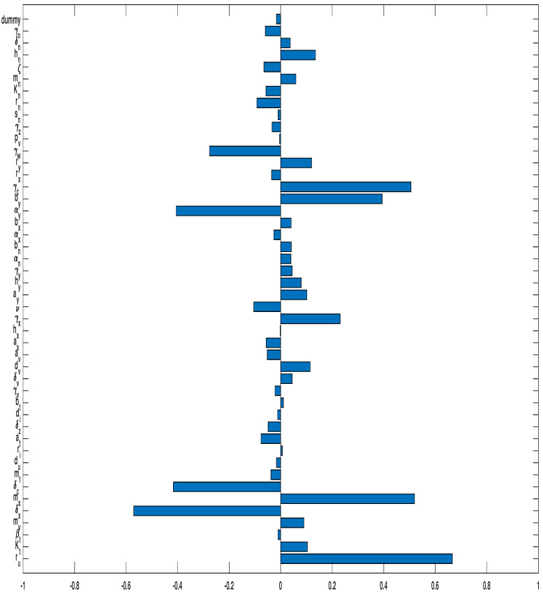
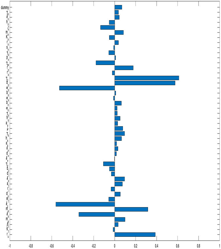
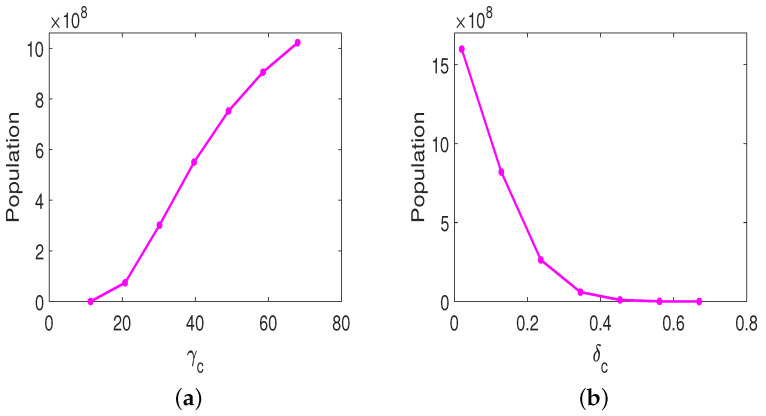
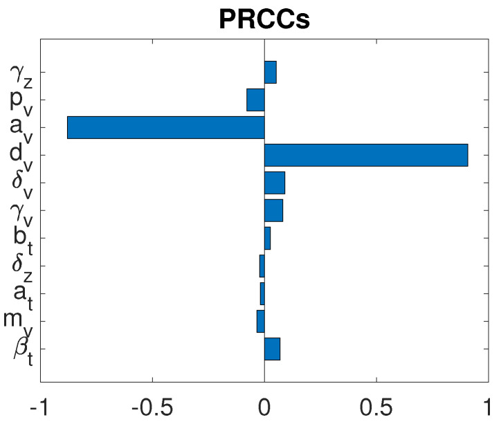
Oncolytic ability to direct target and lyse tumor cells makes oncolytic virus therapy (OVT) a promising approach to treating cancer. Despite its therapeutic potential to stimulate anti-tumor immune responses, it also has immunosuppressive effects. The efficacy of OVTs as monotherapies can be enhanced by appropriate adjuvant therapy such as anti-CTLA-4. In this paper, we propose a mathematical model to explore the interactions of combined therapy of oncolytic viruses and a checkpoint inhibitor, anti-CTLA-4. The model incorporates both the susceptible and infected tumor populations, natural killer cell population, virus population, tumor-specific immune populations, virus-specific immune populations, tumor suppressive cytokine IFN-g, and the effect of immune checkpoint inhibitor CTLA-4. In particular, we distinguish the tumor-specific immune abilities of CD8+ T, NK cells, and CD4+ T cells and describe the destructive ability of cytokine on tumor cells as well as the inhibitory capacity of CTLA-4 on various components. Our model is validated through the experimental results. We also investigate various dosing strategies to improve treatment outcomes. Our study reveals that tumor killing rate by cytokines, cytokine decay rate, and tumor growth rate play important roles on both the OVT monotherapy and the combination therapy. Moreover, parameters related to CD8+ T cell killing have a large impact on treatment outcomes with OVT alone, whereas parameters associated with IFN-g strongly influence treatment responses for the combined therapy. We also found that virus killing by NK cells may halt the desired spread of OVs and enhance the probability of tumor escape during the treatment. Our study reveals that it is the activation of host anti-tumor immune system responses rather than its direct destruction of the tumor cells plays a major biological function of the combined therapy.

Keywords: cytokines; immune checkpoint CTLA-4; mathematical modeling; melanoma; oncolytic virus therapy.


Conflict of interest statement

The authors declare no conflict of interest.

Figures

Figure 1
Figure 1
A schematic diagram depicts the interaction between oncolytic virus, immune cells, cytokines, and immune checkpoint with tumor cells. Uninfected tumor cells become infected by an oncolytic virus is presented. After successful viral penetration within the infected cells, infected cancer cells lyse and produce new infectious viral particles. Fragments from infected cancer cells stimulate anti-viral immune cells which subsequently kill infected cells and clear free virus. The anti-tumor immune cells (CD8+ T, NK cells, and CD4+ T cells) attack and destroy (direct/indirectly) both infected and uninfected cancer cells. T cells are also recruited by innate NK immune cells which become activated when they encounter the viruses. Cytokines activated by both susceptible and infected tumor cells, and secreted by activated T cells and NK cells, conduct the indirectly killing via activating macrophages, increasing phagocytosis of pathogen and tumor cells. Soluble proteins CTLA-4 are expressed by the activated CD4+ T cells and CD8+ T cells. It acts as a negative regulator of CD4+ T cell activation and prevents cytokine productions. NK cells are activated due to immunogenic cell death of infected tumor cells, leading to recruitment of NK cells. They are also activated in response to cytokines and vice versa. Finally, NK cells clear free viruses.
Figure 2
Figure 2
Comparison among numerically simulated dynamics of susceptible tumor cells for MOCK, OV, and OV-aCTLA-4 after treatments. The initial condition is (4×107,0,0,250,250,0,0,104). The treatment is administered on day 8 as the tumor reaches an average volume of 4×10⁷ cells and continues for 5 days. For OV only, the doses of oncolytic virus are s=2×10⁶. The same amount of oncolytic virus is applied to OV-aCTLA-4 treatment with 0.76 blockade rate of CTLA-4. Other parameter values are listed in Table 3.
Figure 3
Figure 3
Comparison among numerically simulated dynamics of susceptible tumor cells for OV-aCTLA-4 treatment on day 18 post-implementation with various amounts of oncolytic virus. The initial condition is as in Figure 2. The level of oncolytic virus is represented by s as indicated in the figure. The treatment started on day 8 post-implantation as the tumor size reaches 4×10⁷ and lasted for 5 days. The same CTLA-4 blocking rate as in Figure 2, u=0.76, is applied to OV-aCTLA-4 treatment. Other parameter values are listed in Table 3.
Figure 4
Figure 4
Comparison among numerically simulated dynamics of susceptible tumor cells for OV-aCTLA-4 treatment with various blockade rates of CTLA-4 on day 18 post-implementation. The initial condition is as in Figure 2. The various blockade rate of CTLA-4 is represented by u as in the figure. The therapy is administered on day 8 and holds for 5 days. The same dosages of oncolytic virus as in Figure 2, s=2×10⁶, is applied to OV-aCTLA-4 treatment. Other parameter values are given in Table 3.
Figure 5
Figure 5
PRCC of susceptible tumor size when the sole OVT is applied.
Figure 6
Figure 6
PRCC for the oncolytic virus parameters with anti-CTLA-4 against susceptible tumor cells. In all simulations, the tumor was treated with OVT and anti-CTLA-4.
Figure 7
Figure 7
Susceptible tumor size with combination therapy. The tumor size is plotted against (a) γc, the decay rate of tumor-suppressing cytokines; (b) δc, the tumor killing rate by cytokines.
Figure 8
Figure 8
GSA for the oncolytic virus related parameters under combination therapy.
Figure 9
Figure 9
Susceptible tumor size in oncolytic virus sensitivity analysis. The tumor size is plotted against (a) dv, the killing rate of virions by innate immune cells; (b) av, the proliferation rate of immune cells induced by oncolytic virus.

Similar articles

Cited by

References

    1. Aurelian L. Oncolytic virotherapy: The questions and the promise. Oncolytic Virother. 2013;2:19–29. doi: 10.2147/OV.S39609. - DOI - PMC - PubMed
    1. Seymour L.W., Fisher K.D. Oncolytic viruses: Finally delivering. Br. J. Cancer. 2016;114:357–361. doi: 10.1038/bjc.2015.481. - DOI - PMC - PubMed
    1. Chiocca E.A., Rabkin S.D. Oncolytic viruses and their application to cancer immunotherapy. Cancer Immunol. Res. 2014;2:295–300. doi: 10.1158/2326-6066.CIR-14-0015. - DOI - PMC - PubMed
    1. Dias J.D., Hemminki O., Diaconu I., Hirvinen M., Bonetti A., Guse K. Targeted cancer immunotherapy with oncolytic adenovirus coding for a fully human monoclonal antibody specific for CTLA-4. Gene Ther. 2012;19:988–998. doi: 10.1038/gt.2011.176. - DOI - PubMed
    1. Engeland C.E., Grossardt C., Veinalde R., Bossow S., Lutz D., Kaufmann J.K., Shevchenko I., Umansky V., Nettelbeck D.M., Weichert W., et al. CTLA-4 and PD-L1 Checkpoint Blockade Enhances Oncolytic Measles Virus Therapy. Mol. Ther. 2014;22:1949–1959. doi: 10.1038/mt.2014.160. - DOI - PMC - PubMed

Publication types