

Application of Deep Learning Model in the Sonographic Diagnosis of Uterine Adenomyosis
- PMID: 36767092
- PMCID: PMC9914280
- DOI: 10.3390/ijerph20031724
Application of Deep Learning Model in the Sonographic Diagnosis of Uterine Adenomyosis
Abstract
Background: This study aims to evaluate the diagnostic performance of Deep Learning (DL) machine for the detection of adenomyosis on uterine ultrasonographic images and compare it to intermediate ultrasound skilled trainees.
Methods: Prospective observational study were conducted between 1 and 30 April 2022. Transvaginal ultrasound (TVUS) diagnosis of adenomyosis was investigated by an experienced sonographer on 100 fertile-age patients. Videoclips of the uterine corpus were recorded and sequential ultrasound images were extracted. Intermediate ultrasound-skilled trainees and DL machine were asked to make a diagnosis reviewing uterine images. We evaluated and compared the accuracy, sensitivity, positive predictive value, F1-score, specificity and negative predictive value of the DL model and the trainees for adenomyosis diagnosis.
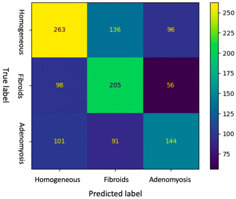
Results: Accuracy of DL and intermediate ultrasound-skilled trainees for the diagnosis of adenomyosis were 0.51 (95% CI, 0.48-0.54) and 0.70 (95% CI, 0.60-0.79), respectively. Sensitivity, specificity and F1-score of DL were 0.43 (95% CI, 0.38-0.48), 0.82 (95% CI, 0.79-0.85) and 0.46 (0.42-0.50), respectively, whereas intermediate ultrasound-skilled trainees had sensitivity of 0.72 (95% CI, 0.52-0.86), specificity of 0.69 (95% CI, 0.58-0.79) and F1-score of 0.55 (95% CI, 0.43-0.66).
Conclusions: In this preliminary study DL model showed a lower accuracy but a higher specificity in diagnosing adenomyosis on ultrasonographic images compared to intermediate-skilled trainees.
Keywords: adenomyosis; artificial intelligence; deep learning; endometriosis; trainee; ultrasound.
Conflict of interest statement
The authors declare no conflict of interest.
Figures
Similar articles
-
Question Mark Sign and Transvaginal Ultrasound Uterine Tenderness for the Diagnosis of Adenomyosis: A Prospective Validation.J Ultrasound Med. 2020 Jul;39(7):1405-1412. doi: 10.1002/jum.15237. Epub 2020 Feb 7. J Ultrasound Med. 2020. PMID: 32030800
-
Ultrasound diagnosis of endometriosis and adenomyosis: State of the art.Best Pract Res Clin Obstet Gynaecol. 2018 Aug;51:16-24. doi: 10.1016/j.bpobgyn.2018.01.013. Epub 2018 Feb 14. Best Pract Res Clin Obstet Gynaecol. 2018. PMID: 29506961 Review.
-
Transvaginal sonographic criteria for the diagnosis of adenomyosis based on histopathologic correlation.Taiwan J Obstet Gynecol. 2010 Mar;49(1):40-4. doi: 10.1016/S1028-4559(10)60007-1. Taiwan J Obstet Gynecol. 2010. PMID: 20466291
-
Comparison of Sensitivity and Specificity of Structured and Narrative Reports of Transvaginal Ultrasonogaphy for Adenomyosis.J Minim Invasive Gynecol. 2021 Jun;28(6):1216-1224. doi: 10.1016/j.jmig.2020.11.001. Epub 2020 Nov 15. J Minim Invasive Gynecol. 2021. PMID: 33207253
-
Role of transvaginal sonography and magnetic resonance imaging in the diagnosis of uterine adenomyosis.Fertil Steril. 2018 Mar;109(3):389-397. doi: 10.1016/j.fertnstert.2018.01.024. Fertil Steril. 2018. PMID: 29566851 Review.
Cited by
-
Deep learning based uterine fibroid detection in ultrasound images.BMC Med Imaging. 2024 Aug 19;24(1):218. doi: 10.1186/s12880-024-01389-z. BMC Med Imaging. 2024. PMID: 39160500 Free PMC article.
-
Metabolic syndrome score as an indicator in a predictive nomogram for lymph node metastasis in endometrial cancer.BMC Cancer. 2023 Jul 4;23(1):622. doi: 10.1186/s12885-023-11053-4. BMC Cancer. 2023. PMID: 37403054 Free PMC article.
-
Evolving the Era of 5D Ultrasound? A Systematic Literature Review on the Applications for Artificial Intelligence Ultrasound Imaging in Obstetrics and Gynecology.J Clin Med. 2023 Oct 29;12(21):6833. doi: 10.3390/jcm12216833. J Clin Med. 2023. PMID: 37959298 Free PMC article. Review.
-
Artificial Intelligence in the Management of Women with Endometriosis and Adenomyosis: Can Machines Ever Be Worse Than Humans?J Clin Med. 2024 May 16;13(10):2950. doi: 10.3390/jcm13102950. J Clin Med. 2024. PMID: 38792490 Free PMC article. Review.
-
Use of Automated Machine Learning for Classifying Hemoperitoneum on Ultrasonographic Images of Morrison's Pouch: A Multicenter Retrospective Study.J Clin Med. 2023 Jun 14;12(12):4043. doi: 10.3390/jcm12124043. J Clin Med. 2023. PMID: 37373736 Free PMC article.
References
-
- van den Bosch T., Dueholm M., Leone F.P.G., Valentin L., Rasmussen C.K., Votino A., Van Schoubroeck D., Landolfo C., Installé A.J., Guerriero S., et al. Terms, definitions and measurements to describe sonographic features of myometrium and uterine masses: A consensus opinion from the Morphological Uterus Sonographic Assessment (MUSA) group. Ultrasound Obstet. Gynecol. 2015;46:284–298. doi: 10.1002/uog.14806. - DOI - PubMed
-
- Exacoustos C., Morosetti G., Conway F., Camilli S., Martire F.G., Lazzeri L., Piccione E., Zupi E. New Sonographic Clas-sification of Adenomyosis: Do Type and Degree of Adenomyosis Correlate to Severity of Symptoms? J. Minim. Invasive Gynecol. 2020;27:1308–1315. doi: 10.1016/j.jmig.2019.09.788. - DOI - PubMed
Publication types
MeSH terms
LinkOut - more resources
Full Text Sources
Medical
Miscellaneous
