Review
. 2023 Jan 23;20(3):2088.
doi: 10.3390/ijerph20032088.

The Incremental Role of Multiorgan Point-of-Care Ultrasounds in the Emergency Setting

Affiliations
Review

The Incremental Role of Multiorgan Point-of-Care Ultrasounds in the Emergency Setting

Antonello D'Andrea et al. Int J Environ Res Public Health. .

Abstract

Point-of-care ultrasonography (POCUS) represents a goal-directed ultrasound examination performed by clinicians directly involved in patient healthcare. POCUS has been widely used in emergency departments, where US exams allow physicians to make quick diagnoses and to recognize early life-threatening conditions which require prompt interventions. Although initially meant for the real-time evaluation of cardiovascular and respiratory pathologies, its use has been extended to a wide range of clinical applications, such as screening for deep-vein thrombosis and trauma, abdominal ultrasonography of the right upper quadrant and appendix, and guidance for invasive procedures. Moreover, recently, bedside ultrasounds have been used to evaluate the fluid balance and to guide decongestive therapy in acutely decompensated heart failure. The aim of the present review was to discuss the most common applications of POCUS in the emergency setting.

Keywords: POCUS; VE x US; congestion; deep-vein thrombosis (DTV); echocardiography; emergency medicine (EM); lung ultrasound (LUS); point-of-care.


Conflict of interest statement

The authors declare no conflict of interest.

Figures

Figure 1
Figure 1
A-lines (arrow).
Figure 2
Figure 2
Lung point: “seashore sign” (left) turns into “stratosphere sign” (right) in M-mode view during deep inspiration.
Figure 3
Figure 3
Pleural effusion with lung region floating inside (arrow).
Figure 4
Figure 4
B-lines (left, indicated by arrow) and “B-pattern” (right).
Figure 5
Figure 5
“Tissue-like pattern” with air bronchogram inside (arrow).
Figure 6
Figure 6
Systolic bowing of interventricular septum towards the left ventricle (D-shape) is associated with pulmonary embolism and pulmonary hypertension.
Figure 7
Figure 7
Pericardial tamponade: end-diastolic right ventricular chamber collapse is associated with significant pericardial effusion.
Figure 8
Figure 8
Bubble test during pericardiocentesis: appearance of bubbles in the pericardium (arrow) indicates that the needle is correctly positioned in the pericardial space.
Figure 9
Figure 9
Ultrasonography of the right upper quadrant. A distended gallbladder with microlithiasis and large biliary sludge (left); multiple stones with posterior shadowing in the infundibulum (right).
Figure 10
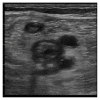
Figure 10
Ultrasonography of normal and abnormal appendixes. On the left, there are longitudinal and transverse USs of acute appendicitis showing thickened walls and diameter > 6 mm. On the right, there is a thinner normal appendix.
Figure 11
Figure 11
Pelvis and caliceal dilatation with proximal ureteral stone.
Figure 12
Figure 12
Intrarenal vein Doppler (above baseline) in a patient with acutely decompensated heart failure before (left) and after (right) decongestive therapy. The flow pattern appears monophasic/diastolic (L), becoming continuous as congestion reduces (R).
Figure 13
Figure 13
Compression ultrasound [Di Vilio A, Vergara A, Desiderio A, et al.: “A. Incremental value of compression ultrasound sonography in the emergency department. World J Crit Care Med. 2021 Sep 9;10(5):194–203].
Figure 14
Figure 14
The image shows the presence of an extensive thrombus with mixed echogenicity occupying the lumen of the femoral vein and the great saphenous vein.

References

    1. American College of Emergency Physicians ACEP emergency ultrasound guidelines 2001. Ann. Emerg. Med. 2001;38:470–481. doi: 10.1016/S0196-0644(01)70030-3. - DOI - PubMed
    1. Whitson M.R., Mayo P.H. Ultrasonography in the emergency department. Crit. Care. 2016;20:1–8. doi: 10.1186/s13054-016-1399-x. - DOI - PMC - PubMed
    1. Manson W.C., Kirksey M., Boublik J., Wu C.L., Haskins S.C. Focused assessment with sonography in trauma (FAST) for the regional anesthesiologist and pain specialist. Reg. Anesth. Pain Med. 2019;44:540–548. doi: 10.1136/rapm-2018-100312. - DOI - PubMed
    1. Bahner D., Blaivas M., Cohen H.L., Fox J.C., Hoffenberg S., Kendall J., Langer J., McGahan J.P., Sierzenski P., Tayal V.S., et al. AIUM Practice Guideline for the Performance of the Focused Assessment with Sonography for Trauma (FAST) Examination. J. Ultrasound Med. 2008;27:313–318. doi: 10.7863/jum.2008.27.2.313. - DOI - PubMed
    1. Lichtenstein D.A., Mezière G.A. Relevance of Lung Ultrasound in the Diagnosis of Acute Respiratory Failure: The BLUE Protocol. Chest. 2008;134:117–125. doi: 10.1378/chest.07-2800. - DOI - PMC - PubMed

LinkOut - more resources