. 2023 Jan 17;24(3):1821.
doi: 10.3390/ijms24031821.

A Newly Developed Chemically Defined Serum-Free Medium Suitable for Human Primary Keratinocyte Culture and Tissue Engineering Applications

Affiliations

A Newly Developed Chemically Defined Serum-Free Medium Suitable for Human Primary Keratinocyte Culture and Tissue Engineering Applications

Sergio Cortez Ghio et al. Int J Mol Sci. .

Abstract

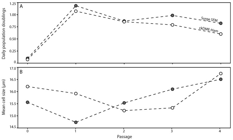
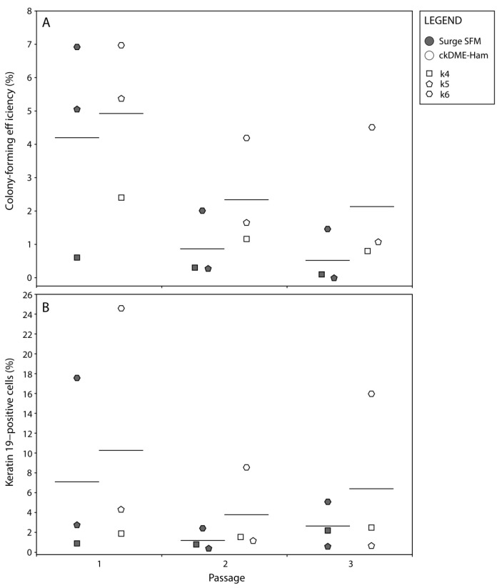
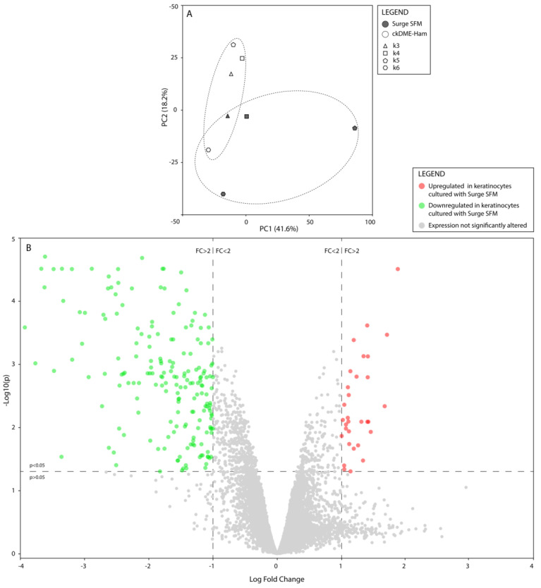
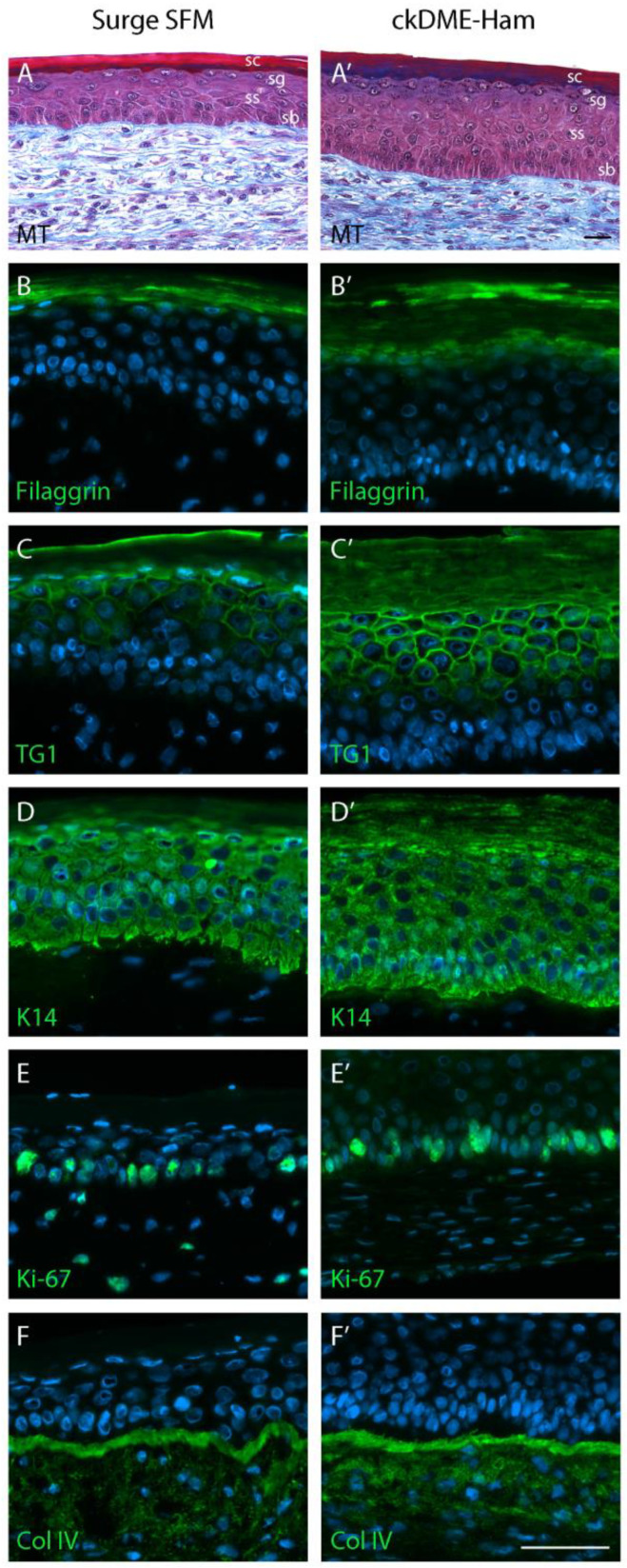
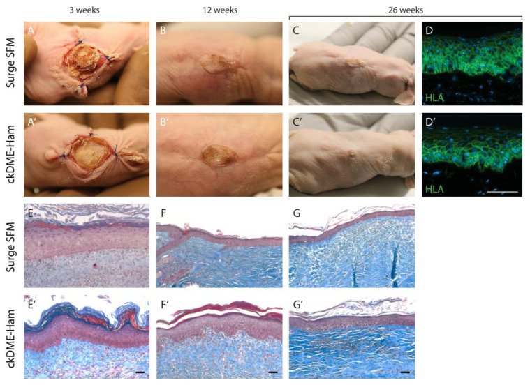
In our experience, keratinocytes cultured in feeder-free conditions and in commercially available defined and serum-free media cannot be as efficiently massively expanded as their counterparts grown in conventional bovine serum-containing medium, nor can they properly form a stratified epidermis in a skin substitute model. We thus tested a new chemically defined serum-free medium, which we developed for massive human primary keratinocyte expansion and skin substitute production. Our medium, named Surge Serum-Free Medium (Surge SFM), was developed to be used alongside a feeder layer. It supports the growth of keratinocytes freshly isolated from a skin biopsy and cryopreserved primary keratinocytes in cultured monolayers over multiple passages. We also show that keratin-19-positive epithelial stem cells are retained through serial passaging in Surge SFM cultures. Transcriptomic analyses suggest that gene expression is similar between keratinocytes cultured with either Surge SFM or the conventional serum-containing medium. Additionally, Surge SFM can be used to produce bilayered self-assembled skin substitutes histologically similar to those produced using serum-containing medium. Furthermore, these substitutes were grafted onto athymic mice and persisted for up to six months. In conclusion, our new chemically defined serum-free keratinocyte culture medium shows great promise for basic research and clinical applications.

Keywords: cell culture; defined medium; skin; stem cells; tissue engineering.


Conflict of interest statement

The authors declare no conflict of interest.

Figures

Figure 1
Figure 1
Line-connected dot plots of (A) the number of daily population doublings and (B) the mean cell size (μm) of k2 primary keratinocytes over five passages. Dot fill colors indicate the medium used to isolate and culture keratinocytes (gray: Surge SFM and white: ckDME-Ham). Dots are means of triplicates. Technical variability did not exceed 15%.
Figure 2
Figure 2
Dot plots of (A) the number of population doublings/day and (B) the mean cell size (μm) over two passages in culture of primary keratinocytes that were previously cryopreserved. Crossbars represent biological means. Dot fill colors indicate the medium used to culture keratinocytes (gray: Surge SFM and white: ckDME-Ham). Dot shapes identify keratinocyte populations (k3: triangle, k4: square, k5: pentagon, and k6: hexagon). Dots are means of triplicates. (C,C’) Phase contrast micrographs of k4 keratinocytes in P1 after 4 days of culture with either (C) Surge SFM or (C’) ckDME-Ham. Sharpness was increased 50% for visual clarity. Scale bar: 50 µm.
Figure 3
Figure 3
Dot plots of (A) keratinocytes’ holoclone (more than 4 mm in diameter) colony-forming efficiency (%) and (B) flow cytometry data for K19-positive cells (%) over three passages. Crossbars represent biological means. Dot fill colors indicate the medium used to culture keratinocytes (gray: Surge SFM and white: ckDME-Ham). Dot shapes identify keratinocyte populations (k4: square, k5: pentagon, and k6: hexagon). Dots are means of quintuplicates in (A) and means of triplicates in (B). Technical variability did not exceed 15%. Isotype controls did not exceed 0.1% K19-positive cells.
Figure 4
Figure 4
(A) Principal component analysis of normalized and filtered gene expression data. Dot fill colors indicate the medium used to culture keratinocytes (gray: Surge SFM and white: ckDME-Ham). Dot shapes identify keratinocyte populations (k3: triangle, k4: square, k5: pentagon, and k6: hexagon). Dotted ellipses represent both medium type clusters. (B) Volcano plot illustrating the results of the paired differential analysis of gene expression between keratinocytes cultured with Surge SFM and those cultured with ckDME-Ham. Dots represent individual genes. Gray genes’ expression was not significantly altered by culturing keratinocytes with Surge SFM rather than ckDME-Ham as opposed to colored genes whose expression was significantly (p < 0.05) modified by at least a 2-fold change in either direction (green: downregulated and red: upregulated).
Figure 5
Figure 5
Gene–gene and gene–function interaction network built around the two biological functions of interest, “Proliferation of keratinocytes” and “Differentiation of epithelial tissue”, using the gene expression differential analysis data.
Figure 6
Figure 6
Histology and immunostaining of the TES produced with (AF) Surge SFM or with (A’F’) ckDME-Ham. Epidermal layers are identified (sb: stratum basale, ss: stratum spinosum, sg: stratum granulosum, sc: stratum corneum). (A,A’) Masson’s trichrome staining. Immunofluorescence staining against (B,B’) filaggrin, (C,C’) transglutaminase 1 (TG1), (D,D’) keratin 14 (K14), (E,E’) Ki-67, and (F,F’) type IV collagen (Col IV). Cell nuclei were stained with Hoechst reagent. Scale bars: 50 µm.
Figure 7
Figure 7
Functional analysis of human TESs produced with either (AG) Surge SFM or (A’G’) ckDME-Ham. TESs were grafted on athymic mice. Macroscopic view at (A,A’) three weeks, (B,B’) 12 weeks, and (C,C’) 26 weeks after grafting. (D,D’) Immunofluorescence staining against human leucocyte antigen (HLA) 26 weeks after grafting the TESs. Cell nuclei were stained with Hoechst reagent. Masson’s trichrome staining at (E,E’) three weeks, (F,F’) 12 weeks, and (G,G’) 26 weeks after grafting. Scale bar: 50 µm.

References

    1. Green H. Cyclic AMP in relation to proliferation of the epidermal cell: A new view. Cell. 1978;15:801–811. doi: 10.1016/0092-8674(78)90265-9. - DOI - PubMed
    1. Green H., Rheinwald J., Sun T. Properties of an epithelial cell type in culture: The epidermal keratinocyte and its dependence on products of the fibroblast. Prog. Clin. Biol. Res. 1977;17:493–500. - PubMed
    1. Rheinwald J., Green H. Epidermal growth factor and the multiplication of cultured human epidermal keratinocytes. Nature. 1977;265:421–424. doi: 10.1038/265421a0. - DOI - PubMed
    1. Rheinwald J., Green H. Serial cultivation of strains of human epidemal keratinocytes: The formation keratinizing colonies from single cells. Cell. 1975;6:331–343. doi: 10.1016/S0092-8674(75)80001-8. - DOI - PubMed
    1. Cortez Ghio S., Cantin-Warren L., Guignard R., Larouche D., Germain L. Are the Effects of the Cholera Toxin and Isoproterenol on Human Keratinocytes’ Proliferative Potential Dependent on Whether They Are Co-Cultured with Human or Murine Fibroblast Feeder Layers? Int. J. Mol. Sci. 2018;19:2174. doi: 10.3390/ijms19082174. - DOI - PMC - PubMed

Substances

LinkOut - more resources