. 2023 Jan 17;24(3):1826.
doi: 10.3390/ijms24031826.

Identification of Pre-Renal and Intrinsic Acute Kidney Injury by Anamnestic and Biochemical Criteria: Distinct Association with Urinary Injury Biomarkers

Affiliations

Identification of Pre-Renal and Intrinsic Acute Kidney Injury by Anamnestic and Biochemical Criteria: Distinct Association with Urinary Injury Biomarkers

Sandra M Sancho-Martínez et al. Int J Mol Sci. .

Abstract

Acute kidney injury (AKI) is a syndrome of sudden renal excretory dysfunction with severe health consequences. AKI etiology influences prognosis, with pre-renal showing a more favorable evolution than intrinsic AKI. Because the international diagnostic criteria (i.e., based on plasma creatinine) provide no etiological distinction, anamnestic and additional biochemical criteria complement AKI diagnosis. Traditional, etiology-defining biochemical parameters, including the fractional excretion of sodium, the urinary-to-plasma creatinine ratio and the renal failure index are individually limited by confounding factors such as diuretics. To minimize distortion, we generated a composite biochemical criterion based on the congruency of at least two of the three biochemical ratios. Patients showing at least two ratios indicative of intrinsic AKI were classified within this category, and those with at least two pre-renal ratios were considered as pre-renal AKI patients. In this study, we demonstrate that the identification of intrinsic AKI by a collection of urinary injury biomarkers reflective of tubular damage, including NGAL and KIM-1, more closely and robustly coincide with the biochemical than with the anamnestic classification. Because there is no gold standard method for the etiological classification of AKI, the mutual reinforcement provided by the biochemical criterion and urinary biomarkers supports an etiological diagnosis based on objective diagnostic parameters.

Keywords: acute kidney injury; anamnesis; etiopathology; injury biomarkers; intrinsic; pre-renal.


Conflict of interest statement

The authors declare no conflict of interest.

Figures

Figure 1
Figure 1
Distribution of pre-renal and renal AKI patients according to the biochemical and anamnestic criteria.
Figure 2
Figure 2
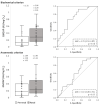
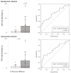
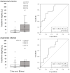
Urinary GM2AP levels of pre-renal and renal AKI patients following classification by biochemical and anamnestic criteria, shown as box plots (left panels) representing median values in arbitrary units (AU) of urinary GM2AP per mg urinary creatinine (Cru), and ROC curves (right panels). AUC: area under the ROC curve showing the pre-renal/renal classification efficacy of urinary GM2AP. The × in box plots represents the median value. **, p < 0.01.
Figure 3
Figure 3
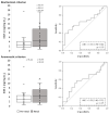
Urinary KIM-1 levels of pre-renal and renal AKI patients following classification by biochemical and anamnestic criteria, shown as box plots (left panels) representing median values in ng of urinary KIM-1 per mg urinary creatinine (Cru), and ROC curves (right panels). AUC: area under the ROC curve showing the pre-renal/renal classification efficacy of urinary KIM-1. The × in box plots represents the median value. *, p < 0.05.
Figure 4
Figure 4
Urinary NAG levels of pre-renal and renal AKI patients following classification by biochemical and anamnestic criteria, shown as box plots (left panels) representing median values in international units (IU) of urinary NAG per mg urinary creatinine (Cru), and ROC curves (right panels). AUC: area under the ROC curve showing the pre-renal/renal classification efficacy of urinary NAG. The × in box plots represents the median value. *, p < 0.05; ***, p < 0.001.
Figure 5
Figure 5
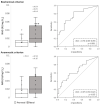
Urinary NGAL levels of pre-renal and renal AKI patients following classification by biochemical and anamnestic criteria, shown as box plots (left panels) representing median values in mg of urinary NGAL per mg urinary creatinine (Cru), and ROC curves (right panels). AUC: area under the ROC curve showing the pre-renal/renal classification efficacy of urinary NGAL. The × in box plots represents the median value. *, p < 0.05.
Figure 6
Figure 6
Urinary TCP1-eta levels of pre-renal and renal AKI patients following classification by biochemical and anamnestic criteria, shown as box plots (left panels) representing median values in arbitrary units (AU) of urinary TCP1-eta per mg urinary creatinine (Cru), and ROC curves (right panels). AUC: area under the ROC curve showing the pre-renal/renal classification efficacy of urinary TCP1-eta. The × in box plots represents the median value. *, p < 0.05.
Figure 7
Figure 7
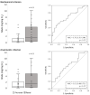
Urinary transferrin levels of pre-renal and renal AKI patients following classification by biochemical and anamnestic criteria, shown as box plots (left panels) representing median values in mg of urinary transferrin per mg urinary creatinine (Cru), and ROC curves (right panels). AUC: area under the ROC curve showing the pre-renal/renal classification efficacy of urinary transferrin. The × in box plots represents the median value. **, p < 0.01; ***, p < 0.001.
Figure 8
Figure 8
Summary of the etiological diagnostic capacity of urinary injury biomarkers (according to the area under the ROC curve) following pre-renal/renal classification by biochemical and anamnestic criteria. Color key: yellow, p < 0.05; orange, p < 0.01; green, p < 0.001; white, p > 0.05.

References

    1. Endre Z.H., Kellum J.A., Di Somma S., Doi K., Goldstein S.L., Koyner J.L., MacEdo E., Mehta R.L., Murray P.T. Differential diagnosis of AKI in clinical practice by functional and damage biomarkers: Workgroup statements from the tenth Acute Dialysis Quality Initiative Consensus Conference. Contrib. Nephrol. 2013;182:30–44. doi: 10.1159/000349964. - DOI - PubMed
    1. Sutherland S.M., Byrnes J.J., Kothari M., Longhurst C.A., Dutta S., Garcia P., Goldstein S.L. AKI in hospitalized children: Comparing the pRIFLE, AKIN, and KDIGO definitions. Clin. J. Am. Soc. Nephrol. 2015;10:554–561. doi: 10.2215/CJN.01900214. - DOI - PMC - PubMed
    1. Kerr M., Bedford M., Matthews B., O’donoghue D. The economic impact of acute kidney injury in England. Nephrol. Dial. Transplant. 2014;29:1362–1368. doi: 10.1093/ndt/gfu016. - DOI - PubMed
    1. Chertow G.M., Burdick E., Honour M., Bonventre J.V., Bates D.W. Acute kidney injury, mortality, length of stay, and costs in hospitalized patients. J. Am. Soc. Nephrol. 2005;16:3365–3370. doi: 10.1681/ASN.2004090740. - DOI - PubMed
    1. Vandijck D.M., Oeyen S., Decruyenaere J.M., Annemans L., Hoste E.A. Acute kidney injury, length of stay, and costs in patients hospitalized in the intensive care unit. Acta Clin. Belg. 2007;62:341–345. doi: 10.1179/acb.2007.077. - DOI - PubMed

LinkOut - more resources