. 2023 Jan 18;24(3):1886.
doi: 10.3390/ijms24031886.

Optimization and Technical Considerations for the Dye-Exclusion Protocol Used to Assess Blood-Brain Barrier Integrity in Adult Drosophila melanogaster

Affiliations

Optimization and Technical Considerations for the Dye-Exclusion Protocol Used to Assess Blood-Brain Barrier Integrity in Adult Drosophila melanogaster

Kesshni Bhasiin et al. Int J Mol Sci. .

Abstract

The blood-brain barrier (BBB) is a multicellular construct that regulates the diffusion and transport of metabolites, ions, toxins, and inflammatory mediators into and out of the central nervous system (CNS). Its integrity is essential for proper brain physiology, and its breakdown has been shown to contribute to neurological dysfunction. The BBB in vertebrates exists primarily through the coordination between endothelial cells, pericytes, and astrocytes, while invertebrates, which lack a vascularized circulatory system, typically have a barrier composed of glial cells that separate the CNS from humoral fluids. Notably, the invertebrate barrier is molecularly and functionally analogous to the vertebrate BBB, and the fruit fly, Drosophila melanogaster, is increasingly recognized as a useful model system in which to investigate barrier function. The most widely used technique to assess barrier function in the fly is the dye-exclusion assay, which involves monitoring the infiltration of a fluorescent-coupled dextran into the brain. In this study, we explore analytical and technical considerations of this procedure that yield a more reliable assessment of barrier function, and we validate our findings using a traumatic injury model. Together, we have identified parameters that optimize the dye-exclusion assay and provide an alternative framework for future studies examining barrier function in Drosophila.

Keywords: Drosophila melanogaster; barrier integrity; blood–brain barrier; dye-exclusion assay; traumatic injury.


Conflict of interest statement

The authors declare no conflict of interest.

Figures
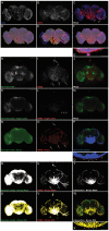
Figure 1
Figure 1
Different methods of assessing dye infiltration into the adult Drosophila brain. (AG) Representative maximum projection laser-scanning confocal images of a d10 w1118 female whole-mount brain injected with 10 kDa tetramethylrhodamine dextran (TMRD) on d9 showing: (A) DAPI, (B) TMRD, and (C) merged channels. White arrows point to structures outside the brain that have taken up TMRD, and white arrowheads point to TMRD at the glial barrier. (DG) NIS Elements software autodetection of (D) an overestimated ROI (shaded region), (E) an underestimated ROI (shaded region), and (F) manual correction of ROI to exclude structures outside the brain (arrows in (C)). Inset in (G) shows TMRD at the glial barrier (arrowheads) that is arbitrarily included in the ROI. (HJ) Representative maximum projection laser-scanning confocal images of a d10 w1118 female whole-mount brain injected with TMRD on d9 showing: (H) Neuropil (nc82) staining, (I) TMRD, and (J) merged channels. (K,L) Single z-plane from the same brain in (HJ) showing: (K) Neuropil (nc82) staining, (L) TMRD, and (M) merged channels. Arrowheads in (L) point to TMRD at the glial barrier. (NQ) NIS Elements binary analysis of a single z-slice image showing: (N) Neuropil (nc82) binary alone, (O) TMRD binary alone, and (P) nc82/TMRD intersection binary. Arrows in (O) show TMRD staining outside the brain that is included in the binary (O), but excluded from the intersection binary (P). Inset in (Q) shows TMRD at the blood–brain barrier (arrowheads) that is not included in the intersection binary. (RT) FIJI binary analysis of the same z-slice in (HJ) showing: (R) Neuropil (nc82) binary mask alone, (S) TMRD binary mask alone, and (T) nc82/TMRD intersection binary. Arrows in (S) show TMRD staining outside the brain that is included in the binary mask (S), but excluded from the intersection binary mask (T). (UX) FIJI autodetection of ROIs on (U) Neuropil (nc82) staining, (V) TMRD, and (W) nc82/TMRD intersection binary masks. Inset in (X) shows the intersection ROI overlaid over fluorescence at the glial barrier (arrowheads). Scale bar in (AF); (HP); (RW) = 100 µm. Scale bar in insets in (G,Q,X) = 20 µm.
Figure 2
Figure 2
Whole-body pre-fixation reduces baseline dye infiltration. (AC) Representative maximum projection laser-scanning confocal images of whole-mount brains from d10 w1118 females injected with 10 kDa TMRD on d9. Baseline TMRD infiltration in: (A) whole-body pre-fixation, (B) head pre-fixation with proboscis attached, and (C) head pre-fixation without proboscis. TMRD infiltration into the subesophageal zone (SEZ) was noted in both head pre-fixation conditions (arrows in (B,C)). (D) Quantification of normalized TMRD fluorescence infiltration across fixation conditions using nc82/TMRD intersection binary analysis. * p < 0.05; ** p < 0.01; Kruskal–Wallis test with Dunn’s multiple comparisons; n = 6–7; error bars are presented as mean ± SEM. Scale bar = 100 µm. Maximum projection images were generated from the middle ~75 µm region of the brain to highlight SEZ infiltration.
Figure 3
Figure 3
Whole-body pre-fixation reduces baseline TMRD fluorescence and reveals the effect of traumatic injury on dye infiltration. (A,C,F,H,K,M) Representative maximum projection laser-scanning confocal images of the TMRD channel and (B,D,G,I,L,N) single z-plane intersection binary of nc82 and TMRD signal, of whole-mount brains from d10 w1118 females injected with TMRD on d9. Brains were processed for fixation by: (AD) whole-body pre-fixation, (FI) head pre-fixation with proboscis attached, (KN) head pre-fixation without proboscis. Flies either did not experience a traumatic injury (No Hit) or were exposed to traumatic injury 2 h after dye injection (Hit). (E,J,O) Quantification of normalized TMRD fluorescence infiltration across fixation conditions using nc82/TMRD intersection binary analysis. * p < 0.05; n.s. = not significant. Mann–Whitney U-test (E,J), t-test (O); n = 6–9. Error bars are presented as mean ± SEM. Scale bar = 100 µm.

References

    1. Zlokovic B.V. The Blood-Brain Barrier in Health and Chronic Neurodegenerative Disorders. Neuron. 2008;57:178–201. doi: 10.1016/j.neuron.2008.01.003. - DOI - PubMed
    1. Alluri H., Wiggins-Dohlvik K., Davis M.L., Huang J.H., Tharakan B. Blood–Brain Barrier Dysfunction Following Traumatic Brain Injury. Metab. Brain Dis. 2015;30:1093–1104. doi: 10.1007/s11011-015-9651-7. - DOI - PubMed
    1. Sweeney M.D., Sagare A.P., Zlokovic B.V. Blood-Brain Barrier Breakdown in Alzheimer Disease and Other Neurodegenerative Disorders. Nat. Rev. Neurol. 2018;14:133–150. doi: 10.1038/nrneurol.2017.188. - DOI - PMC - PubMed
    1. Mason H.D., Johnson A.M., Mihelson N.A., Mastorakos P., McGavern D.B. Glia Limitans Superficialis Oxidation and Breakdown Promote Cortical Cell Death after Repetitive Head Injury. JCI Insight. 2021;6:e149229. doi: 10.1172/jci.insight.149229. - DOI - PMC - PubMed
    1. Carlson S.D., Juang J.L., Hilgers S.L., Garment M.B. Blood Barriers of the Insect. Annu. Rev. Entomol. 2000;45:151–174. doi: 10.1146/annurev.ento.45.1.151. - DOI - PubMed

LinkOut - more resources