Review
. 2023 Jan 19;24(3):1980.
doi: 10.3390/ijms24031980.

The Role of Different Types of microRNA in the Pathogenesis of Breast and Prostate Cancer

Affiliations
Review

The Role of Different Types of microRNA in the Pathogenesis of Breast and Prostate Cancer

Ekaterina A Sidorova et al. Int J Mol Sci. .

Abstract

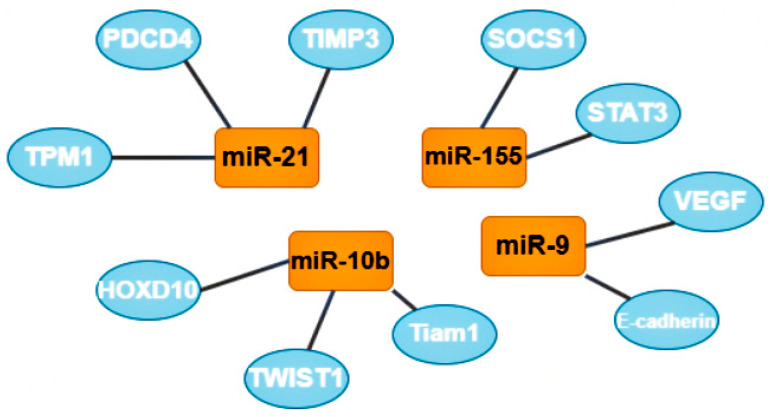
Micro ribonucleic acids (microRNAs or miRNAs) form a distinct subtype of non-coding RNA and are widely recognized as one of the most significant gene expression regulators in mammalian cells. Mechanistically, the regulation occurs through microRNA binding with its response elements in the 3'-untranslated region of target messenger RNAs (mRNAs), resulting in the post-transcriptional silencing of genes, expressing target mRNAs. Compared to small interfering RNAs, microRNAs have more complex regulatory patterns, making them suitable for fine-tuning gene expressions in different tissues. Dysregulation of microRNAs is well known as one of the causative factors in malignant cell growth. Today, there are numerous data points regarding microRNAs in different cancer transcriptomes, the specificity of microRNA expression changes in various tissues, and the predictive value of specific microRNAs as cancer biomarkers. Breast cancer (BCa) is the most common cancer in women worldwide and seriously impairs patients' physical health. Its incidence has been predicted to rise further. Mounting evidence indicates that microRNAs play key roles in tumorigenesis and development. Prostate cancer (PCa) is one of the most commonly diagnosed cancers in men. Different microRNAs play an important role in PCa. Early diagnosis of BCa and PCa using microRNAs is very useful for improving individual outcomes in the framework of predictive, preventive, and personalized (3P) medicine, thereby reducing the economic burden. This article reviews the roles of different types of microRNA in BCa and PCa progression.

Keywords: 3P medicine; breast cancer; metastatic disease; microRNA; molecular mechanisms; non-coding RNA; prostate cancer; tumor biomarkers.


Conflict of interest statement

The authors declare no conflict of interest.

Figures

Figure 1
Figure 1
Metastasis and tumor invasion. Tumor cells at the primary tumor sites leave their primary site by dint of invasion into the surrounding stroma (stage 1: invasion). Then they penetrate (stage 2: intravasation) and migrate through the blood or lymph vessels into the circulation (stage 3: dissemination). Some of tumor cells survive and migrate to the target organ parenchyma (tropic sites of metastasis), which is the “pre-metastatic niche” (stage 4: extravasation). Finally, cells adapt and proliferate to form metastasis with the subsequent formation of a clinically obvious tumor (stage 5: colonization/proliferation at the tropic sites). Myeloid cells, such as tumor-associated macrophages (TAMs), can promote metastatic spread through blood and lymphatic vessels. Transforming growth factor-β (TGF-β), produced by TAMs, and cancer-associated fibroblasts (CAFs) are regulators of the epithelial–mesenchymal transition and metastasis. In the pre-metastatic niche, natural killer cells (NK cells) and T cells inhibit tumor growth.
Figure 2
Figure 2
Regulatory patterns of mRNA and microRNA interaction. MicroRNAs bind to the mRNA MRE complex in 3’-untranslated regions, resulting in the post-transcriptional silencing of genes. The mode of silencing in RISC depends whenever mRNA and microRNA are completely or partially complementary. In cases where molecules are perfectly complementary, the RISC simply cleaves the target mRNA. In cases where molecules are no perfectly complementary, the RISC binds to the MRE only by the heptameric 5’seed region of the guide microRNA, leading to acceleration of mRNA decay. This happens due to deadenylation or interruption translation at its different stages. RISC—the RNA-induced silencing complex, MRE—microRNA response element.
Figure 3
Figure 3
The role of microRNA-153 in carcinogenesis. MicroRNA-153 is involved in epithelial-mesenchymal transition (EMT)-associated signaling pathways that stimulate tumorigenesis, cancer progression, and metastasis. A mesenchymal–epithelial transition (MET) is a reversible biological process that involves the transition from mesenchymal cells to polarized cells called epithelia.
Figure 4
Figure 4
The most studied oncomirs and their target genes. MiR-21 has 3 common target genes: tropomyosin 1 (TPM1), programmed cell death 4 (PDCD4), and metallopeptidase inhibitor 3 (TIMP3). MiR-10b also has 3 target genes, including homeobox D10 (HOXD10), twist-related protein 1 (TWIST1) and T cell lymphoma invasion and metastasis-inducing protein 1 (Tiam 1). Mir-9 target genes are vascular endothelial growth factors (VEGF) and E-cadherin (also known as CDH1). MiR-155 has 2 target genes: suppressor of cytokine signaling 1 (SOCS1) and signal transducer and activator of transcription 3 (STAT3).
Figure 5
Figure 5
The most studied oncosuppressors and their target genes. MiR-145 has 3 target genes: neuroblastoma RAS viral oncogene homolog (N-RAS), MUC1, and vascular endothelial growth factors (VEGF). Let-7 family target genes: radixin (RDX), Harvey rat sarcoma viral oncogene homolog (H-RAS), high-mobility group AT-hook 2 (HGMA2), serine/threonine-protein kinase (PAK1), diaphanous-related formin 2 (DIAPH2), and integrin subunit beta (ITGB). MiR-205 and miR-200 have 2 mutual target genes: zinc finger E-box binding homeobox 1 and 2 (ZEB1, ZEB2).
Figure 6
Figure 6
Main pathways in which miR-99a inhibits in cancer cells. microRNA-99a inhibits FGFR3 on the cancer cells. Inhibited FGFR3 cannot activate downstream cascade pathways: PI3K/AKT and RAS/RAF/MEK/MAPK. For the PI3K/AKT signaling pathway, FGFR3 phosphate FRS2 inhibits GRB2. This inhibited molecular PI3K, which inhibits mTOR and as a result, reduces proliferation and transcription in the nucleus. As for the RAS/RAF/MEK/MAPK pathway, inhibited GRB2 inactivate SOS, RAS, RAF, MEK, and MAPK, and eventually increase proliferation and transcription. FGFR2 (CD332)—fibroblast growth factor receptor 2, FRS2—fibroblast growth factor receptor substrate 2, GRB2—growth factor receptor-bound protein 2, PI3K—phosphoinositide 3-kinase, AKT—RAC-alpha serine/threonine-protein kinase, protein kinase B alpha, RAS—rat sarcoma virus kinase, RAF (rapidly accelerated fibrosarcoma)—proto-oncogene serine/threonine-protein kinase, mTOR—mammalian target of rapamycin kinase, MEK (MAP2K, MAPKK)—mitogen-activated protein kinase, MAPK—mitogen-activated protein kinase, 4E-BP1—eukaryotic translation initiation factor 4E-binding protein 1, S6K1—ribosomal protein S6 kinase beta-1.
Figure 7
Figure 7
Epithelial-mesenchymal transition (EMT) and clinical significance of partial EMT and full EMT. Cells undergoing partial EMT have an ability to migrate and colonize lungs. Cell in full EMT do not colonize lungs. Therapy resistance is characteristic of both partial EMT and full EMT.
Figure 8
Figure 8
Mechanism of increasing apoptosis of cancer cells when miR-4728 does not inhibit NOXA. NOXA displaces BAK1 from MCL-1 as they have a similar conformation. The MCL-1/NOXA complex activates BCL-2, which activates the apoptosis of cancer cells. NOXA—NADPH oxidase activator, MCL-1—mantle cell lymphoma 1 protein, BCL-1—B cell lymphoma 1 protein, BCL-2—B cell lymphoma 2 protein, BAK1—BCL-2 homologous antagonist killer.
Figure 9
Figure 9
Suppressing effect of miR-4728 in cancer cells. MiR-4728 inhibited NOXA, thus BAK1 and BIM/BCL2L11 can combine to MCL-1. This complex inhibits BCL-2 and inactivates cell apoptosis, therefore, trastuzumab cannot impact on cells with HER2 expression. NOXA—NADPH oxidase activator, MCL-1—mantle cell lymphoma-1, BCL-2—B cell lymphoma 2 protein, BAK1—BCL-2 homologous antagonist killer, HER2—human epidermal growth factor receptor 2, BIM/BCL2L11—Bcl-2 interacting mediator of cell death (commonly called Bcl-2-like protein 11).

References

    1. Sung H., Ferlay J., Siegel R.L., Laversanne M., Soerjomataram I., Jemal A., Bray F. Global Cancer Statistics 2020: GLOBOCAN Estimates of Incidence and Mortality Worldwide for 36 Cancers in 185 Countries. CA Cancer J. Clin. 2021;71:209–249. doi: 10.3322/caac.21660. - DOI - PubMed
    1. Tran N.L., Nagle R.B., Cress A.E., Heimark R.L. N-Cadherin Expression in Human Prostate Carcinoma Cell Lines: An Epithelial-Mesenchymal Transformation Mediating Adhesion with Stromal Cells. Am. J. Pathol. 1999;155:787–798. doi: 10.1016/S0002-9440(10)65177-2. - DOI - PMC - PubMed
    1. Pakzad R., Mohammadian-Hafshejani A., Ghoncheh M., Pakzad I., Salehiniya H. The incidence and mortality of prostate cancer and its relationship with development in Asia. Prostate Int. 2015;3:135–140. doi: 10.1016/j.prnil.2015.09.001. - DOI - PMC - PubMed
    1. Grant C.M., Kyprianou N. Epithelial mesenchymal transition (EMT) in prostate growth and tumor progression. Transl. Androl. Urol. 2013;2:202–211. doi: 10.3978/j.issn.2223-4683.2013.09.04. - DOI - PMC - PubMed
    1. Mazurakova A., Koklesova L., Samec M., Kudela E., Kajo K., Skuciova V., Csizmár S.H., Mestanova V., Pec M., Adamkov M., et al. Anti-breast cancer effects of phytochemicals: Primary, secondary, and tertiary care. EPMA J. 2022;13:315–334. doi: 10.1007/s13167-022-00277-2. - DOI - PMC - PubMed