Review
. 2023 Jan 19;24(3):1998.
doi: 10.3390/ijms24031998.

Morphological and Functional Remodeling of Vascular Endothelium in Cardiovascular Diseases

Affiliations
Review

Morphological and Functional Remodeling of Vascular Endothelium in Cardiovascular Diseases

Ghassan Bkaily et al. Int J Mol Sci. .

Abstract

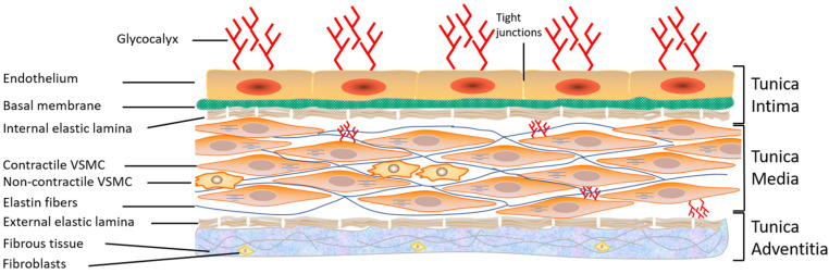
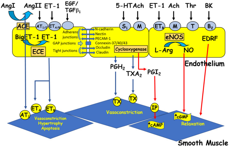
The vascular endothelium plays a vital role during embryogenesis and aging and is a cell monolayer that lines the blood vessels. The immune system recognizes the endothelium as its own. Therefore, an abnormality of the endothelium exposes the tissues to the immune system and provokes inflammation and vascular diseases such as atherosclerosis. Its secretory role allows it to release vasoconstrictors and vasorelaxants as well as cardio-modulatory factors that maintain the proper functioning of the circulatory system. The sealing of the monolayer provided by adhesion molecules plays an important role in cardiovascular physiology and pathology.

Keywords: adhesion molecules; atherosclerosis; calcium; endothelium; endothelium dysfunction; endothelium pathology; endothelium physiology; endothelium released factors; endothelium remodeling; hypertension; ion transporters.


Conflict of interest statement

The authors declare that the research was conducted in the absence of any commercial or financial relationships that could be construed as a potential conflict of interest.

Figures

Figure 1
Figure 1
Structure of the vascular wall. Schematic representation showing the three layers of the vascular wall: tunica intima, tunica media, and tunica adventitia, as well as the components of each layer. VSMC: vascular smooth muscle cells (from Bkaily et al., 2021 [2]).
Figure 2
Figure 2
The endothelium produces vasoactive factors that cause either relaxation or contraction of the vascular smooth muscle. Ang I and II: angiotensin I and II, ACE: angiotensin-converting enzyme, Ach: acetylcholine, BK: bradykinin, cAMP/cGMP: cyclic adenosine/guanosine monophosphate, ECE: endothelin-converting enzyme, EDRF: endothelium-derived relaxing factor, ET-1: endothelin-1, 5HT: 5-hydroxytryptamine (serotonin), L-Arg: L-arginine, NO: nitric oxide, NOS: nitric oxide synthase, PGH2: prostaglandin H2, PGI2: prostacyclin, TGFβ1: transforming growth factor beta 1, Thr: thrombin, and TXA2: thromboxane A2. Circles represent receptors (AT: angiotensin receptor, B: bradykinin receptor, ET: endothelin receptor, M: muscarinic receptor, IP: purinergic receptor, S: serotonin receptor, T: thrombin receptor, and TX: thromboxane receptor).
Figure 3
Figure 3
Current to voltage (I/V) relationship curve (A) and open probability/voltage relationship (B) of the voltage-dependent steady-state R-type Ca2+ channel in human aortic VSMCs recorded using the patch clamp technique (modified from Bkaily et al. 1997) [44].
Figure 4
Figure 4
Increase in the volume of endocardial endothelial cells (EECs) in young and old hereditary cardiomyopathic hamsters (HCMHs). (AD) Freshly isolated and cultured EECs from 10-week-old normal hamsters (NH) (A), 10-week-old HCMH (B), 32-week-old NH (C), 32-week-old HCMH, and (D) show an increase in the volume (in µm3) of EECs in HCMHs compared to those of age-matched NHs. In panels A–D, the white scale bar is in µm (modified from Jacques and Bkaily 2019) [104].
Figure 5
Figure 5
Modulation of cytosolic and nuclear calcium levels of human VSMCs by physical contact with human VECs. Quantitative 3D confocal microscopy images showing the distribution of cytosolic and nuclear Ca2+ in hVSMCs in pure culture of hVSMCs without contact (A) and in contact with other hVSMCs (B), as well as in co-culture with hVECs (C). The pseudo-color scale represents the level of fluorescence intensity of the Fluo-3–Ca2+ complex from 0 (black, absence of fluorescence) to 255 (white, maximum fluorescence) in panels A, B, and C. The white scale bar value is in micrometers (modified from Hassan et al., 2018) [105].
Figure 6
Figure 6
Activation of immune cells induces the release of factors that modulate the function and junctions between VECs, which permits the infiltration of activated neutrophils/eosinophils, and polymorphonuclear cells (PMN). The remodeling of the vascular endothelium induces the secretion of factors that induce either relaxation or contraction of VSMCs. The activated monocytes/macrophages and VECs’ released factors promote the proliferation of contractile and non-contractile VSMCs that characterize atherosclerosis (modified from Bkaily, 1994) [27].

References

    1. Mann M.J., Gibbons G.H., Tsao P.S., von der Leyen H.E., Cooke J.P., Buitrago R., Kernoff R., Dzau V.J. Cell cycle inhibition preserves endothelial function in genetically engineered rabbit vein grafts. J. Clin. Investig. 1997;99:1295–1301. doi: 10.1172/JCI119288. - DOI - PMC - PubMed
    1. Bkaily G., Abou Abdallah N., Simon Y., Jazzar A., Jacques D. Vascular smooth muscle remodeling in health and disease. Can. J. Physiol. Pharmacol. 2021;99:171–178. doi: 10.1139/cjpp-2020-0399. - DOI - PubMed
    1. Kotlyarov S. Immune Function of Endothelial Cells: Evolutionary Aspects, Molecular Biology and Role in Atherogenesis. Int. J. Mol. Sci. 2022;23:9770. doi: 10.3390/ijms23179770. - DOI - PMC - PubMed
    1. Florey L. The endothelial cell. Br. Med. J. 1966;2:487–490. doi: 10.1136/bmj.2.5512.487. - DOI - PMC - PubMed
    1. Burnett J.C., Jr. Coronary endothelial dysfunction in the hypertensive patient: From myocardial ischemia to heart failure. J. Hum. Hypertens. 1997;11:45–49. doi: 10.1038/sj.jhh.1000400. - DOI - PubMed

MeSH terms