. 2023 Jan 20;24(3):2118.
doi: 10.3390/ijms24032118.

Yet Another Similarity between Mitochondrial and Bacterial Ribosomal Small Subunit Biogenesis Obtained by Structural Characterization of RbfA from S. aureus

Affiliations

Yet Another Similarity between Mitochondrial and Bacterial Ribosomal Small Subunit Biogenesis Obtained by Structural Characterization of RbfA from S. aureus

Aydar G Bikmullin et al. Int J Mol Sci. .

Abstract

Ribosome biogenesis is a complex and highly accurate conservative process of ribosomal subunit maturation followed by association. Subunit maturation comprises sequential stages of ribosomal RNA and proteins' folding, modification and binding, with the involvement of numerous RNAses, helicases, GTPases, chaperones, RNA, protein-modifying enzymes, and assembly factors. One such assembly factor involved in bacterial 30S subunit maturation is ribosomal binding factor A (RbfA). In this study, we present the crystal (determined at 2.2 Å resolution) and NMR structures of RbfA as well as the 2.9 Å resolution cryo-EM reconstruction of the 30S-RbfA complex from Staphylococcus aureus (S. aureus). Additionally, we show that the manner of RbfA action on the small ribosomal subunit during its maturation is shared between bacteria and mitochondria. The obtained results clarify the function of RbfA in the 30S maturation process and its role in ribosome functioning in general. Furthermore, given that S. aureus is a serious human pathogen, this study provides an additional prospect to develop antimicrobials targeting bacterial pathogens.

Keywords: 30S biogenesis; RbfA; Staphylococcus aureus; bacterial ribosomal proteins; ribosome; translation.


Conflict of interest statement

The authors declare no conflict of interest.

Figures

Figure 1
Figure 1
The structure of S. aureus RbfA: (a) Crystal structure; (b) solution NMR structure; (c) comparison of crystal (yellow) and NMR (red) structures (Cα RMSD = 1.76 Å); (d) electrostatic surface of S. aureus RbfA (at pH 7.5); (e) comparisons of RbfA structure from S. aureus with its homologs from T. thermophilus (Cα RMSD = 0.82 Å), H. influenzae (Cα RMSD = 0.82 Å), and M. pneumoniae (Cα RMSD = 3.32 Å). The homologs’ structures are colored gray.
Figure 2
Figure 2
The 2.9 Å cryo-EM density map of the S. aureus 30S–RbfA complex (gray) and its comparison with the map of the free S. aureus 30S (EMD-3624, orange). The RbfA extra density is colored green. h—head and b—a body of the 30S.
Figure 3
Figure 3
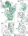
Comparison of 16S rRNA in the mature 30S (PDB ID: 5ND8, green) and 30S–RbfA complex (in this study, blue) from S. aureus: (a) overall view and (b) the head displacement; (c,d) the conformational changes of helices h28 and h44. The rest of the 16S rRNA is colored white.
Figure 4
Figure 4
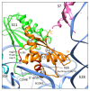
The binding site of RbfA (orange) on the 30S subunit. The RbfA contacting region with 16S rRNA (hkh motif and α1/β1) is colored red. The S7, S11, S18 proteins are colored pink, green, and cyan, respectively. The 16S rRNA is colored blue.
Figure 5
Figure 5
Structure comparisons of S. aureus RbfA (crystal—yellow, red—NMR, and grey—AlphaFold prediction) and M. musculus mtRBFA (inward conformation without C- and N-extensions—green). Cα RMSD NMR/AlphaFold = 1.893 Å and Cα RMSD mtRBFA/AlphaFold = 1.123 Å.
Figure 6
Figure 6
The cryo-EM structure of S. aureus RbfA: (a) overall view and (b) comparison of the cryo-EM structure (orange) with the crystal (yellow, Cα RMSD = 0.69 Å) and NMR (red, Cα RMSD = 1.91 Å) structures.
Figure 7
Figure 7
The models and densities of the RbfA C-terminus. (a) M. musculus (PDB ID: 7PNU, EMDB-13552); (b) S. aureus (this study); and (c) E. coli (PDB ID: 7BOH, EMD-12243).
Figure 8
Figure 8
Comparison of bacterial and mitochondrial small subunits bound with RbfA/mtRBFA (PDB ID: 7PNU): (a) the localization of RbfA/mtRBFA on small subunits (the rRNAs are colored blue and ribosomal proteins are colored grey) and (b) the interactions between RbfA/S7 and mtRBFA/mtS7 proteins.

References

    1. Crick F. Central dogma of molecular biology. Nature. 1970;227:561–563. doi: 10.1038/227561a0. - DOI - PubMed
    1. Melnikov S., Ben-Shem A., Garreau de Loubresse N., Jenner L., Yusupova G., Yusupov M. One core, two shells: Bacterial and eukaryotic ribosomes. Nat. Struct. Mol. Biol. 2012;19:560–567. doi: 10.1038/nsmb.2313. - DOI - PubMed
    1. Narla A., Ebert B.L. Ribosomopathies: Human disorders of ribosome dysfunction. Blood. 2010;115:3196–3205. doi: 10.1182/blood-2009-10-178129. - DOI - PMC - PubMed
    1. Wilson D.N. Ribosome-targeting antibiotics and mechanisms of bacterial resistance. Nat. Rev. Microbiol. 2014;12:35–48. doi: 10.1038/nrmicro3155. - DOI - PubMed
    1. Kang J., Brajanovski N., Chan K.T., Xuan J., Pearson R.B., Sanij E. Ribosomal proteins and human diseases: Molecular mechanisms and targeted therapy. Signal Transduct. Target. Ther. 2021;6:323. doi: 10.1038/s41392-021-00728-8. - DOI - PMC - PubMed

MeSH terms